. 2015 Jan;125(1):258-62.
doi: 10.1172/JCI78473. Epub 2014 Dec 1.

Exome sequencing reveals MCM8 mutation underlies ovarian failure and chromosomal instability

Exome sequencing reveals MCM8 mutation underlies ovarian failure and chromosomal instability

Saleh AlAsiri et al. J Clin Invest. 2015 Jan.

Abstract

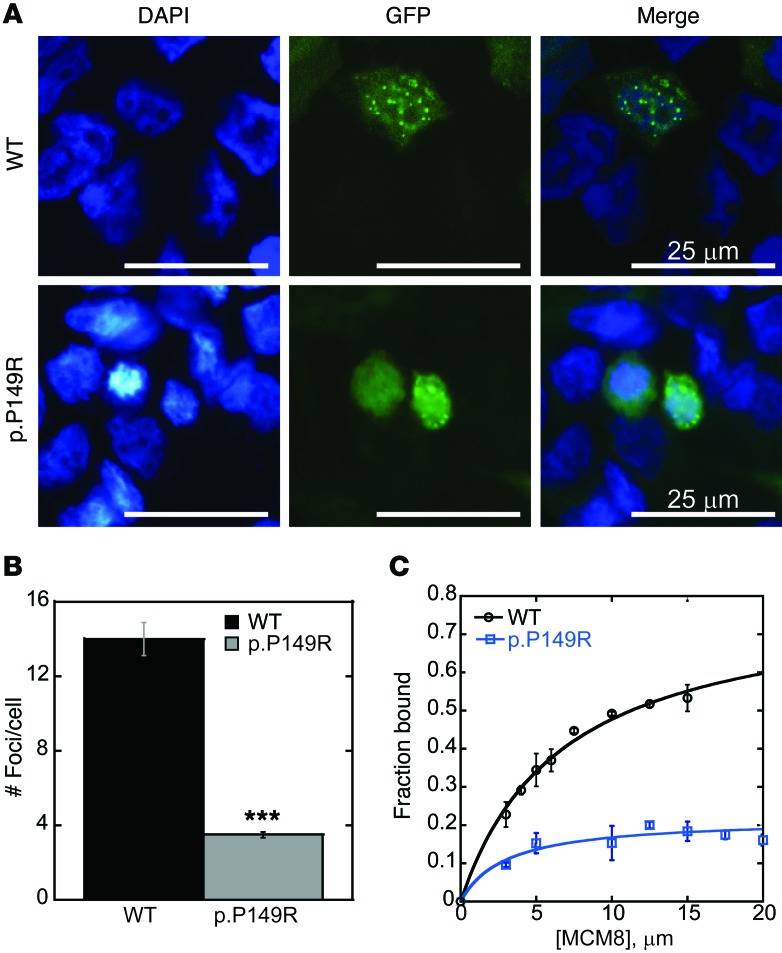
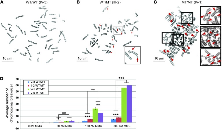
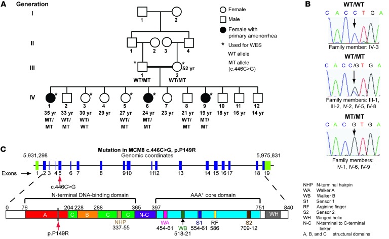
Premature ovarian failure (POF) is a genetically and phenotypically heterogeneous disorder that includes individuals with manifestations ranging from primary amenorrhea to loss of menstrual function prior to age 40. POF presents as hypergonadotropic hypogonadism and can be part of a syndrome or occur in isolation. Here, we studied 3 sisters with primary amenorrhea, hypothyroidism, and hypergonadotropic hypogonadism. The sisters were born to parents who are first cousins. SNP analysis and whole-exome sequencing revealed the presence of a pathogenic variant of the minichromosome maintenance 8 gene (MCM8, c.446C>G; p.P149R) located within a region of homozygosity that was present in the affected daughters but not in their unaffected sisters. Because MCM8 participates in homologous recombination and dsDNA break repair, we tested fibroblasts from the affected sisters for hypersensitivity to chromosomal breaks. Compared with fibroblasts from unaffected daughters, chromosomal break repair was deficient in fibroblasts from the affected individuals, likely due to inhibited recruitment of MCM8 p.P149R to sites of DNA damage. Our study identifies an autosomal recessive disorder caused by an MCM8 mutation that manifests with endocrine dysfunction and genomic instability.


Figures

Figure 3
Figure 3. MCM8 mutation disrupts MCM8 foci formation and DNA binding.
(A) MCM8 c.446C>G (p.P149R) mutation inhibits MCM8 foci formation at sites of DNA damage. HEK293T cells were transfected with wild-type MCM8-GFP (WT) or mutant MCM8-GFP (p.P149R; shown in green) and treated with 300 nM MMC for 6 hours. Nuclei were counterstained with DAPI (blue). Foci formed in 293T cells expressing wild-type MCM8, but not in cells expressing mutant MCM8. Four independent experiments for transfection of MCM8-GFP (WT vs. p.P149R) coupled with DNA damage were performed. Representative confocal images are shown. (B) Twenty representative cells per condition were quantified for the number of damage-induced nuclear foci. A 2-tailed t test (assuming unequal variance) revealed a statistically significant difference (***P < 0.001) between wild-type (14 ± 0.9 foci/cell) and c.446C>G-expressing (3.5 ± 0.2 foci/cell) cells. Error bars represent SEM. (C) MCM8 c.446C>G (p.P149R) inhibited DNA binding by EMSA. Wild-type MCM8 or mutant MCM8 (p.P149R) protein was bound to a random 46 nt ssDNA oligonucleotide. Gels were imaged, and quantification of the fraction of band shift was performed. Data were fit to a single-site binding model defined by ΔF[MCM8]/KD + [MCM8], where F is the fraction bound and KD is the dissociation constant. Mutant MCM8 (p.P149R, blue squares) showed a significant reduction in DNA binding affinity for ssDNA at each concentration when compared with that of wild-type MCM8 (WT, black circles). Each point is the average of 3 replicates. Error bars represent SD.
Figure 2
Figure 2. MCM8 mutation impairs DNA break repair.
Cells from homozygous MCM8 c.446C>G individuals have an impaired ability to repair dsDNA breaks induced by MMC. A total of 4 experimental groups were treated with MMC for each sample. Representative metaphase cells treated with 300 nM MMC are shown from (A) a healthy and fertile individual with the MCM8 WT/WT genotype (family member IV-3), (B) an unaffected WT/MT genotype individual (family member III-2), and (C) an affected female homozygous for the MCM8 c.446C>G pathogenic variant (MT/MT genotype; family member IV-1). Arrows point to chromosomal breaks. Original magnification, ×63/1.4 for chromosomal spreads and ×4 for inserts from the spreads. Scale bars: 10 μm. (D) At least 10 metaphase cells were used to calculate the total number of chromosomal breaks per cell for each MMC concentration. The number of chromosomal breaks counted was limited to 60 per cell, and those with more were indicated as 60+ breaks per cell. Error bars represent SEM. *P < 0.01, **P < 0.001, and ***P < 0.0001 by 2-tailed t test.
Figure 1
Figure 1. Pedigree of a family with 3 daughters afflicted by premature ovarian failure and homozygous for the MCM8 c.446C>G variant.
(A) Family members are designated by Arabic numerals. Horizontal lines between individuals represent marriage. Double horizontal lines indicate consanguinity in a marriage. Vertical lines represent lineage. Below each individual, the individual’s current age (if known) and MCM8 genotype are provided. (B) Sanger sequencing was used to validate genotypes, and representative chromatograms are shown. Individuals who are heterozygous for the c.446C>G MCM8 variant show overlapping C and G peaks (middle graph). Individuals homozygous for the c.446C>G MCM8 variant have a single G peak (bottom graph). (C) MCM8 is encoded on chromosome 20: 5,931,298-5,975,831 (NCBI37/hg19), and the c.446C>G variant in exon 5 is shown (red arrow). Full boxes represent exons (blue denotes coding sequences; green denotes noncoding sequences), and introns are indicated by lines. MCM8 consists of an N-terminal DNA-binding domain and a AAA+ core domain. The c.446C>G substitution caused a change in the amino acid sequence p.P149R within the predicted DNA-binding domain (red arrow). All domains are color coded with the homology model (Supplemental Figure 3).

Comment in

  • Reproductive aging and MCM8/9.
    Yatsenko SA, Rajkovic A. Yatsenko SA, et al. Oncotarget. 2015 Jun 30;6(18):15750-1. doi: 10.18632/oncotarget.4589. Oncotarget. 2015. PMID: 26119146 Free PMC article. No abstract available.

References

    1. Nelson LM. Clinical practice. Primary ovarian insufficiency. N Engl J Med. 2009;360(6):606–614. doi: 10.1056/NEJMcp0808697. - DOI - PMC - PubMed
    1. Wood MA, Rajkovic A. Genomic markers of ovarian reserve. Semin Reprod Med. 2013;31(6):399–415. doi: 10.1055/s-0033-1356476. - DOI - PMC - PubMed
    1. Vijayraghavan S, Schwacha A. The eukaryotic Mcm2-7 replicative helicase. Subcell Biochem. 2012;62:113–134. doi: 10.1007/978-94-007-4572-8_7. - DOI - PubMed
    1. Gozuacik D, et al. Identification and functional characterization of a new member of the human Mcm protein family: hMcm8. Nucleic Acids Res. 2003;31(2):570–579. doi: 10.1093/nar/gkg136. - DOI - PMC - PubMed
    1. Lutzmann M, et al. MCM8- and MCM9-deficient mice reveal gametogenesis defects and genome instability due to impaired homologous recombination. Mol Cell. 2012;47(4):523–534. doi: 10.1016/j.molcel.2012.05.048. - DOI - PubMed

Publication types

Substances