. 2015 Jan 2;14(1):434-46.
doi: 10.1021/pr501174p. Epub 2014 Dec 9.

Glycomic analysis of membrane glycoproteins with bisecting glycosylation from ovarian cancer tissues reveals novel structures and functions

Affiliations

Glycomic analysis of membrane glycoproteins with bisecting glycosylation from ovarian cancer tissues reveals novel structures and functions

Heba Allam et al. J Proteome Res. .

Abstract

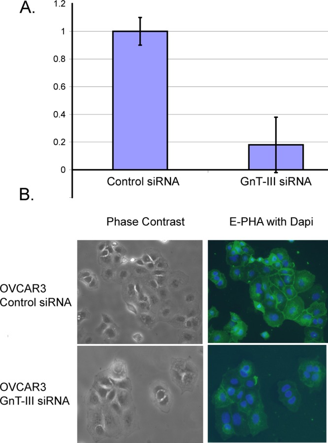
Biomarkers capable of detecting and targeting epithelial ovarian cancer cells for diagnostics and therapeutics would be extremely valuable. Ovarian cancer is the deadliest reproductive malignancy among women in the U.S., killing over 14 000 women each year. Both the lack of presenting symptoms and high mortality rates illustrate the need for earlier diagnosis and improved treatment of this disease. The glycosyltransferase enzyme GnT-III encoded by the Mgat3 gene is responsible for the addition of GlcNAc (N-acetylglucosamine) to form bisecting N-linked glycan structures. GnT-III mRNA expression is amplified in ovarian cancer tissues compared with normal ovarian tissue. We use a lectin capture strategy coupled to nano-ESI-RPLC-MS/MS to isolate and identify the membrane glycoproteins and unique glycan structures associated with GnT-III amplification in human ovarian cancer tissues. Our data illustrate that the majority of membrane glycoproteins with bisecting glycosylation are common to both serous and endometrioid histological subtypes of ovarian cancer, and several have been reported to participate in signaling pathways such as Notch, Wnt, and TGFβ.

Keywords: biomarkers; bisecting N-linked glycans; glycoproteins; human tissue; mass spectrometry; ovarian cancer.


Figures

Figure 1
Figure 1
E-PHA binds bisecting glycans in a GnT-III dependent manner. (A) Real-time PCR measurement of GnT-III mRNA levels in OVCAR3 human ovarian cancer cells expressing control siRNA or GnT-III siRNA. GnT-III levels were normalized to RPL4 and the control siRNA level was set to 1.0 for comparison; error bars represent the mean ± SE for triplicate measurements. (B) E-PHA staining of glycoproteins with bisecting glycosylation in OVCAR3 cells; nuclei are stained with DAPI, magnification 20×.
Figure 2
Figure 2
(A) Schematic flow of glycoproteomic experiments. Membrane proteins were analyzed by nanoflow-ESI–RPLC–MS/MS before E-PHA capture to verify equivalent protein quality and quantity. Biotinylated E-PHA was added to membrane extracted proteins, and glycoproteins with bisecting glycans were captured using streptavidin magnetic resin prior to nanoflow-ESI–RPLC–MS/MS. Membrane glycoproteins enriched in tumor tissues relative to nonmalignant tissues by E-PHA capture by at least 1.5 times or 150% by spectral counts were identified and are listed in Table 1. (B) Venn diagram showing the percentage of glycoproteins in Table 1 from endometrioid only (13% purple), serous only (6% blue), or shared (81% blue/purple). (C) Functional annotation of E-PHA enriched membrane glycoproteins using DAVID 2009.
Figure 3
Figure 3
Prevalence of unique bisecting N-linked glycan structures in ovarian carcinoma tissues. PNGaseF released N-linked glycan structures from glycoproteins enriched by E-PHA capture. Glycans were released from pooled E-PHA captured glycoproteins from endometrioid (n = 3), serous (n = 3), or total membrane proteins from nonmalignant control surface epithelial cells (n = 1). TIM profile was filtered with neutral loss of HexNAc and bisecting N-linked glycan structures were identified by MSn analysis manually. Peaks labeled M7, M8, and M9 indicate high mannose type N-linked glycans.
Figure 4
Figure 4
Bisecting N-linked glycan structures identified from human endometrioid and serous EOC. Bisecting structures identified from endometrioid and serous ovarian cancer tissues by MSn analysis are summarized. Represented glycan structures are in accordance with the guidelines from the Consortium for Functional Glycomics (CFG): blue square, N-acetylglucosamine (GlcNAc); green circle, mannose (Man); yellow circle, galactose (Gal); red triangle, fucose (Fuc).
Figure 5
Figure 5
Protein transport is impaired in GnT-III siRNA cells. (A) OVCAR3 control siRNA cells and GnT-III siRNA cells were serum-starved prior to incubation with fluorescein-EGF (50 ng/mL) for 1 h on ice. Cells were placed in a 37 °C incubator for 30 min prior to removal of EGF that was not internalized by an acid wash. Cells were fixed and stained using an EEA1 monoclonal antibody. Nuclei are stained with DAPI, magnification 40×.
Figure 6
Figure 6
Protein transport and response to EGF is restored with mouse GnT-III expression. (A) OVCAR3 cells expressing GnT-III siRNA constitutively were transiently transfected with mouse HA-GnT-III (gift from Pamela Stanley). Cells expressing mouse HA-GnT-III show positive E-PHA staining and HA antibody staining indicating expression and activity in human cells. Nuclei are stained with DAPI, magnification 20×. (B) Transport of EGF fluorescein-bound receptors is rescued by mouse HA-GnT-III expression. OVCAR3 GnT-III siRNA cells were transiently transfected with mouse HA-GnT-III (top panel) or empty vector (bottom panel). Cells were serum-starved for 2 h 1 day after transfection prior to incubation with fluorescein-EGF, as described for Figure 4. (C) OVCAR3 GnT-III siRNA cells were transiently transfected with vector-only or mouse HA-GnT-III prior to plating into 96-well plates. Cells were serum-starved and incubated with vehicle only or EGF 35 ng/mL for 24 h. Cell proliferation was measured using CellTiter Aqueous One (Promega) reagent. Error bars represent the ± SEM from three separate experiments.

References

    1. Bhaumik M.; Seldin M. F.; Stanley P. Cloning and chromosomal mapping of the mouse Mgat3 gene encoding N-acetylglucosaminyltransferase III. Gene 1995, 1642295–300. - PubMed
    1. Koles K.; et al. Identification of N-glycosylated proteins from the central nervous system of Drosophila melanogaster. Glycobiology 2007, 17121388–1403. - PubMed
    1. Abbott K. L.; et al. Targeted glycoproteomic identification of biomarkers for human breast carcinoma. J. Proteome Res. 2008, 741470–1480. - PMC - PubMed
    1. Tabb D. L.; Fernando C. G.; Chambers M. C. MyriMatch: highly accurate tandem mass spectral peptide identification by multivariate hypergeometric analysis. J. Proteome Res. 2007, 62654–661. - PMC - PubMed
    1. Ma Z. Q.; et al. IDPicker 2.0: Improved protein assembly with high discrimination peptide identification filtering. J. Proteome Res. 2009, 883872–3881. - PMC - PubMed

Publication types

Substances