. 2014 Nov 15;22(22):6387-91.
doi: 10.1016/j.bmc.2014.09.050. Epub 2014 Oct 2.

A bicyclic peptide scaffold promotes phosphotyrosine mimicry and cellular uptake

Affiliations

A bicyclic peptide scaffold promotes phosphotyrosine mimicry and cellular uptake

Justin S Quartararo et al. Bioorg Med Chem. .

Abstract

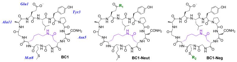
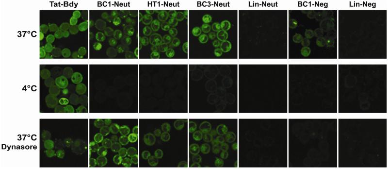
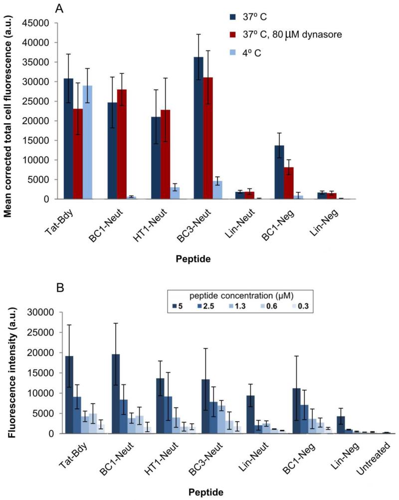
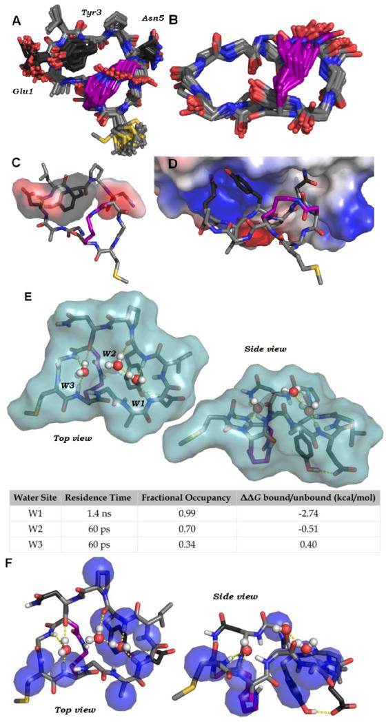
While peptides are promising as probes and therapeutics, targeting intracellular proteins will require greater understanding of highly structured, cell-internalized scaffolds. We recently reported BC1, an 11-residue bicyclic peptide that inhibits the Src homology 2 (SH2) domain of growth factor receptor-bound protein 2 (Grb2). In this work, we describe the unique structural and cell uptake properties of BC1 and similar cyclic and bicyclic scaffolds. These constrained scaffolds are taken up by mammalian cells despite their net neutral or negative charges, while unconstrained analogs are not. The mechanism of uptake is shown to be energy-dependent and endocytic, but distinct from that of Tat. The solution structure of BC1 was investigated by NMR and MD simulations, which revealed discrete water-binding sites on BC1 that reduce exposure of backbone amides to bulk water. This represents an original and potentially general strategy for promoting cell uptake.

Keywords: Bicyclic peptide; Cell uptake; Cyclic peptide; Molecular dynamics; NMR structure; Phosphotyrosine.


Figures

Figure 1
Figure 1
Chemical structures of the BC1 scaffold and BODIPY-FL-labeled analogs. R1 is the adduct of hydrazine-functionalized BODIPY-FL, and R2 is the adduct of maleimide-functionalized BODIPY-FL. Fluorescein-labeled peptides were also prepared and tested to rule out dye-dependent effects on cell uptake. HT1-Neut and HT1-Neg are head-to-tail cyclic analogs of BC1-Neut and BC1-Neg, and Lin-Neut and Lin-Neg are linear analogs of BC1-Neut and BC1-Neg. See Supplementary Information for complete structural and synthesis details.
Figure 2
Figure 2
Analysis of peptide internalization by confocal fluorescence microscopy. Mammalian breast epithelial cancer cells (MDA-MB-453) were grown to 95% confluence in a 24-well glass-bottom plate and incubated with 10 μM fluorescently-labeled peptide in PBS for 1 hour at 37°C or 4°C. Low-temperature conditions were used to demonstrate the extent to which the uptake process was energy-dependent. Alternatively, cells were pre-treated with 80 μM dynasore (a dynamin inhibitor that prevents clathrin-mediated endocytosis) for 30 min at 37° C prior to incubation with dye-labeled peptides at the same temperature. Cells were washed thoroughly in PBS prior to imaging by confocal microscopy. While peptides were incubated in PBS, we also verified their stability in buffered human serum as described previously (Supplementary Figure S3).
Figure 3
Figure 3
Quantification of cell fluorescence. (A) Corrected total cell fluorescence (CTCF) of individual cells for each experimental condition tested. Images collected by confocal fluorescence microscopy were background-subtracted using the ImageJ software suite to compute CTCF values for individual cells. The mean CTCF of 5 representative cells within each image is plotted. Error bars denote standard deviation. (B) Quantitation of cellular internalization by direct fluorometry. MDA-MB-453 cells were seeded in a 96 well plate at 105 cells/ml and grown for several hours prior to incubation with the indicated concentrations of peptide for 1 hour at 37° C in PBS. Cells were then washed and lysed in 1% Triton x-100 prior to fluorometric analysis at 485 nm excitation, 535 nm emission. Supplementary Figure S8 shows the same data but with a narrower y-axis.
Figure 4
Figure 4
Solution structure of BC1. (A) NMR-derived structural model of BC1. 90 NOE-derived distance constraints were used in two-stages simulated annealing and energy minimization using CNSolve. The 30 lowest-energy structures are shown. Carbons are shown in light gray, side chain carbons of the binding epitope are shown in dark grey, and side chain carbons of the cross-link are shown in purple. (B) The atoms of the backbone and cross-link, with all other side chain atoms omitted. Backbone RMSD is 0.33 Å. (C) A representative structure of BC1 showing the putative binding epitope (transparent surface). A pTyr-mimicking epitope is formed by Gly1 and Tyr3. Pro4 organizes a turn that positions Asn5 on the same face as Glu1 and Tyr3, consistent with established binding modes for peptide ligands of Grb2-SH2. (D) A representative solution structure of BC1 was docked into the phosphotyrosine-binding site of the Grb2-SH2 domain (PDB ID: 1JYR) using Molecular Operating Environment (Chemical Computing Group) with the OPLS-AA forcefield. The protein is depicted as an electrostatic potential surface. Following energy minimization, Glu1 and Tyr3 form similar interactions with the Grb2-SH2 domain as pTyr-containing peptides. Asn5 is able to access a known specificity pocket. (E) A 100 ns molecular dynamics simulation of BC1 in explicit water revealed three distinct water-binding pockets formed by backbone carbonyls and amide protons. Surface represents the Van der Waals surface of BC1. The table shows approximate residence time, fractional occupancy (percentage of the simulation steps in which a water occupied the pocket) and calculated δδG of binding for each water. (F) Representative NMR structure of BC1 showing heavy atoms (blue spheres) that have protons with cross-relaxation peaks with water protons. For these protons, the water cross-peak in the ROESY was opposite in sign to the same cross-peak in the NOESY, which is characteristic of direct magnetization transfer to water (Supplementary Figure S11).,

References

    1. Bock JE, Gavenonis J, Kritzer JA. ACS Chem. Biol. 2013;8:488–499. - PMC - PubMed
    1. Walensky LD, Bird GH. J. Med. Chem. 2014 doi 10.1021/jm4011675. - PMC - PubMed
    1. Duchardt F, Fotin-Mleczek M, Schwarz H, Fischer R, Brock R. Traffic. 2007;8:848–866. - PubMed
    1. Frank AO, Vangamudi B, Feldkamp MD, Souza-Fagundes EM, Luzwick JW, Cortez D, Olejniczak ET, Waterson AG, Rossanese OW, Chazin WJ, Fesik SW. J. Med. Chem. 2014;57:2455–2461. - PMC - PubMed
    1. Wallbrecher R, Depré L, Verdurmen WPR, Bovée-Geurts PH, van Duinkerken RH, Zekveld MJ, Timmerman P, Brock R. Bioconjug. Chem. 2014;25:955. - PubMed

Publication types