. 2015 Mar;132(5):546-58.
doi: 10.1111/jnc.13002. Epub 2015 Jan 29.

Sigma receptor 1 activation attenuates release of inflammatory cytokines MIP1γ, MIP2, MIP3α, and IL12 (p40/p70) by retinal Müller glial cells

Affiliations

Sigma receptor 1 activation attenuates release of inflammatory cytokines MIP1γ, MIP2, MIP3α, and IL12 (p40/p70) by retinal Müller glial cells

Arul Shanmugam et al. J Neurochem. 2015 Mar.

Abstract

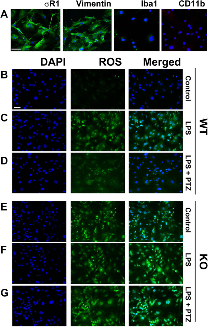
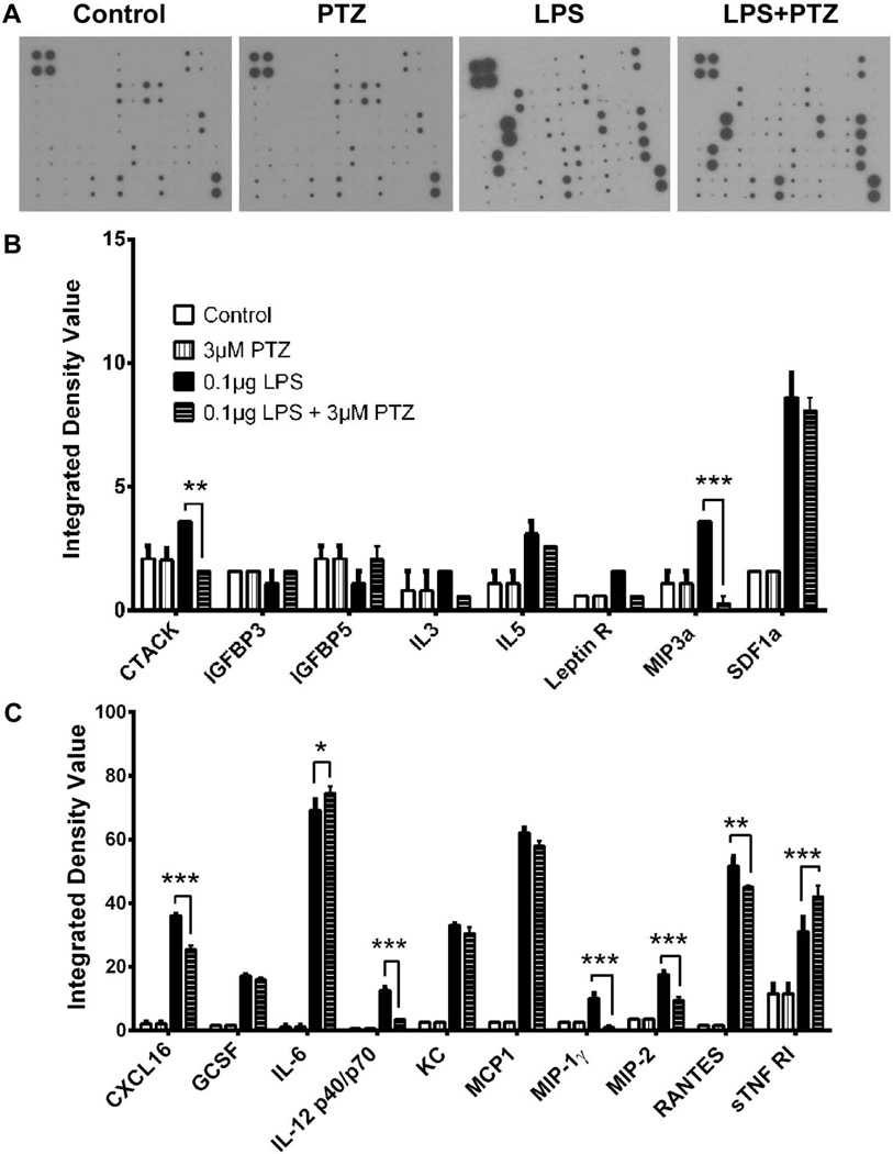
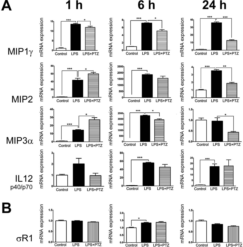
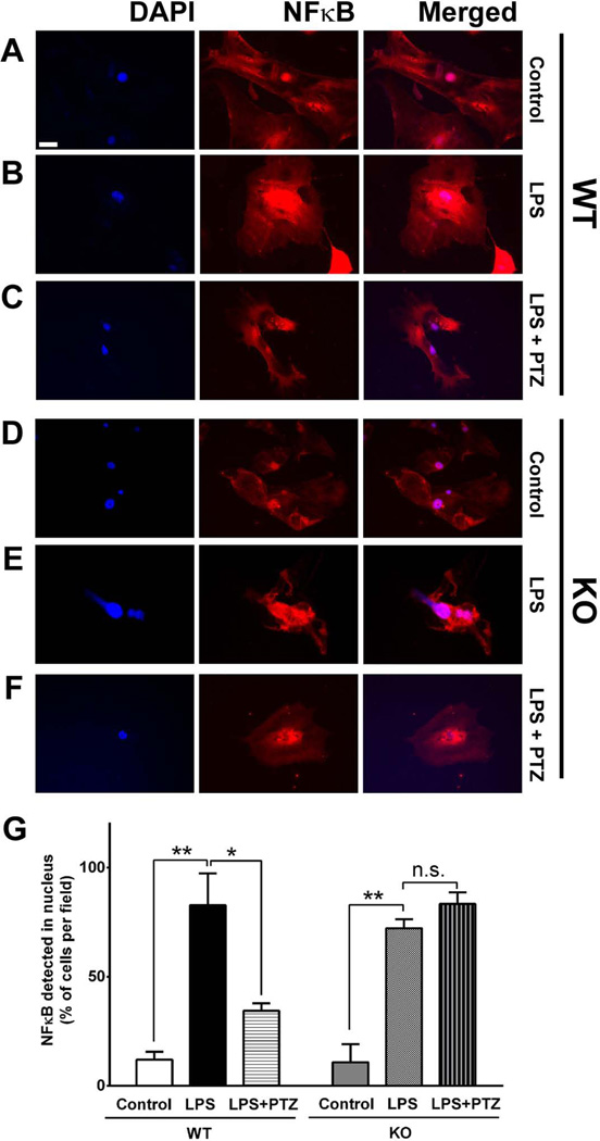
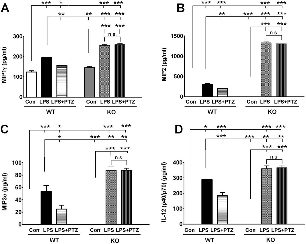
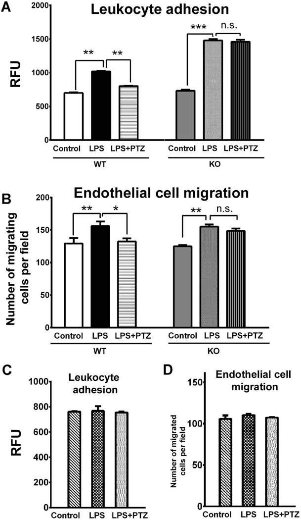
The high-affinity sigma receptor 1 (σR1) ligand (+)-pentazocine ((+)-PTZ) affords profound retinal neuroprotection in vitro and in vivo by a yet-unknown mechanism. A common feature of retinal disease is Müller cell reactive gliosis, which includes cytokine release. Here, we investigated whether lipopolysaccharide (LPS) stimulates cytokine release by primary mouse Müller cells and whether (+)-PTZ alters release. Using a highly sensitive inflammatory antibody array we observed significant release of macrophage inflammatory proteins (MIP1γ, MIP2, MIP3α) and interleukin-12 (IL12 (p40/p70)) in LPS-treated cells compared to controls, and a significant decrease in secretion upon (+)-PTZ treatment. Müller cells from σR1 knockout mice demonstrated increased MIP1γ, MIP2, MIP3α and IL12 (p40/p70) secretion when exposed to LPS compared to LPS-stimulated WT cells. We investigated whether cytokine secretion was accompanied by cytosolic-to-nuclear NFκB translocation and whether endothelial cell adhesion/migration was altered by released cytokines. Cells exposed to LPS demonstrated increased NFκB nuclear location, which was reduced significantly in (+)-PTZ-treated cells. Media conditioned by LPS-stimulated-Müller cells induced leukocyte-endothelial cell adhesion and endothelial cell migration, which was attenuated by (+)-PTZ treatment. The findings suggest that release of certain inflammatory cytokines by Müller cells can be attenuated by σR1 ligands providing insights into the retinal neuroprotective role of this receptor.

Keywords: (+)-pentazocine; cytokine array; mouse; radial glial cell; retinal degeneration; retinal disease.


Figures

Fig 1
Fig 1. Examination of σR1, vimentin and microglial marker expression and ROS production in primary mouse Müller cells
(A) Immunolabeling of primary Müller cells with antibodies against σR1 (green florescence); vimentin (green fluorescence); Iba1 (green fluorescence); and CD11b (red fluorescence). Nuclei were stained with DAPI (blue). Panels B – G: Fluorescent detection of ROS (green florescence), nuclei were stained with DAPI (blue florescence) in σR1+/+ (wildtype, WT) cells (B-D) and σR1−/− (knockout, KO) cells (E-G). Müller cells receiving no LPS exposure (control, B and E)); Müller cells exposed 24 h to LPS [0.1 µg/ml] (C and F); Müller cells exposed 24 h to LPS [0.1 µg/ml] pre-and co-treated with (+)-PTZ [3µM] (D and G). Calibration bar = 100 µM.
Fig 2
Fig 2. Detection of cytokines secreted by primary mouse Müller cells in response to LPS and (+)-PTZ
(A) Representative arrays used to assess cytokines secreted by primary Müller cells that received no treatment (control), were treated with (+)-PTZ [3µM], were exposed 24 h to LPS [0.1 µg/ml] (LPS) or were exposed 24 h to LPS [0.1 µg/ml] and were pre-and co-treated with (+)-PTZ [3µM] (LPS+PTZ). Spots on each array represent individual cytokines. The upper four spots on the left corner of each array are internal standards provided with the array. Spot intensities were quantified densitometrically and were compared to the density of the internal standards yielding an integrated density value (IDV) for each cytokine. Of the 62 cytokines that can be detected using the array, (B) and (C) provide quantitative data for the cytokines whose secretion was detected in Müller cells (*p<0.05, ** p<0.01, *** p<0.001).
Fig 3
Fig 3. Analysis of genes encoding MIP1γ, MIP2, MIP3α, IL12 (p40/p70) and σR1 in Müller cells treated with LPS and (+)-PTZ
mRNA was isolated from mouse Müller cells that received no treatment (control), were exposed 1, 6, or 24 h to LPS [0.1 µg/ml] (LPS) or were exposed 1, 6, or 24 h to LPS [0.1 µg/ml] plus pre-and co-treated with (+)-PTZ [3µM] (LPS+PTZ); qRT-PCR was performed to analyze the expression of (A) MIP1γ, MIP2, MIP3α, IL12 p40, (B) σR1 using primer pairs listed in Table 1 Gene levels were expressed relative to gene expression for control (non-LPS treated) condition (arbitrarily assigned a value of 1) and were normalized to GAPDH. Each experiment was performed in triplicate (*p<0.05, ** p<0.01, *** p<0.001).
Fig 4
Fig 4. Immunocytochemical analysis of LPS-induced NFκB translocation
Immunofluorescent labeling of σR1+/+ (wildtype, WT) and σR1−/− (knockout, KO) mouse Müller cells that (A, C) received no treatment (control), (B, E) were exposed 15 min to LPS [0.1 µg/ml] (LPS), or (C, F) were exposed 15 min to LPS [0.1 µg/ml] plus pre-and co-treated with (+)-PTZ [3µM] (LPS+PTZ) with an antibody against NFκB (p65), detected with fluorescent dye (red); nuclei stained with DAPI (blue). (G) Quantitation of the data: ten fields were imaged per well and cells in which NFκB was located in the nucleus were counted, these data were expressed as a ratio to the total number of cells in the field. The experiments were performed in triplicate and values expressed as percentage of cells per field that localized NF-κB to the nucleus (*p<0.05, ** p<0.01, n.s. = not significant). Calibration bar = 100 µM.
Fig 5
Fig 5. ELISA detection of MIP1γ, MIP2, MIP3α, and IL12 (p40/p70) secretion by WT and σR1−/− Müller cells
Müller cells harvested from either σR1+/+ (wildtype, WT) or σR1−/− (knockout, KO) mice were incubated with or without LPS [1.0 µg/ml] for 24 h in the presence/absence of (+)-PTZ [3µM] (LPS+PTZ) following which the conditioned media was collected and subjected to ELISA to quantify secretion of (A) MIP1γ, (B) MIP2, (C) MIP3α,and (D) IL12 (p40/p70). Data are presented as pg/ml. Each experiment was performed in triplicate (*p<0.05, ** p<0.01, *** p<0.001, n.s. = not significant).
Fig 6
Fig 6. Assessment of leukocyte adhesion and endothelial cell migration as a consequence of LPS stimulation of Müller cells
Media conditioned by either σR1+/+ (wildtype, WT) or σR1−/− (knockout, KO) Müller cells that received no treatment (control), were exposed 24 h to LPS [0.1 µg/ml] (LPS) or were exposed 24 h to LPS [0.1 µg/ml] and were pre-and co-treated with (+)-PTZ [3µM] (LPS+PTZ) was used to evaluate: (A) Effects on the adhesion of leukocytes to endothelial cells. The graph depicts level of fluorescence of the lysate detected at 480 nm/520 nm; values were plotted as relative fluorescence units (RFU). (B) Extent of migration of HUVEC cells through a porous filter. The graph depicts the number of cells that had migrated through the porous filter detected by immunofluorescence. Data are expressed as mean ± S.E.M. Each experiment was performed in triplicate; *p<0.05, ** p<0.01, n.s. = not significant. The effects on leukocyte adhesion (C) or endothelial cell migration (D) when exposed to serum-free media (control), or media containing LPS, or media containing LPS and (+)-PTZ were investigated in the same manner as described for the conditioned media studies. There were no significant effects on leukocyte adhesion or endothelial cell migration when incubated under these conditions.

References

    1. Aderka D, Engelmann H, Maor Y, Brakebusch C, Wallach D. Stabilization of the bioactivity of tumor necrosis factor by its soluble receptors. J Exp Med. 1992;175:323–329. - PMC - PubMed
    1. Armstrong DA, Major JA, Chudyk A, Hamilton TA. Neutrophil chemoattractant genes KC and MIP-2 are expressed in different cell populations at sites of surgical injury. J Leukoc Biol. 2004;75:641–648. - PubMed
    1. Aydar E, Palmer CP, Klyachko VA, Jackson MB. The sigma receptor as a ligand-regulated auxiliary potassium channel subunit. Neuron. 2002;34:399–410. - PubMed
    1. Biedermann T, Kneilling M, Mailhammer R, Maier K, Sander CA, Kollias G, Kunkel SL, Hültner L, Röcken M. Mast cells control neutrophil recruitment during T cell-mediated delayed-type hypersensitivity reactions through tumor necrosis factor and macrophage inflammatory protein 2. J Exp Med. 2000;192:1441–1452. - PMC - PubMed
    1. Behensky AA, Yasny IE, Shuster AM, Seredenin SB, Petrov AV, Cuevas J. Stimulation of sigma receptors with afobazole blocks activation of microglia and reduces toxicity caused by amyloid-β25–35. J Pharmacol Exp Ther. 2013;347:458–467. - PubMed

Publication types

MeSH terms

Associated data

LinkOut - more resources