. 2014 Nov 6;95(5):565-78.
doi: 10.1016/j.ajhg.2014.10.006. Epub 2014 Nov 6.

Dosage changes of a segment at 17p13.1 lead to intellectual disability and microcephaly as a result of complex genetic interaction of multiple genes

Affiliations

Dosage changes of a segment at 17p13.1 lead to intellectual disability and microcephaly as a result of complex genetic interaction of multiple genes

Claudia M B Carvalho et al. Am J Hum Genet. .

Abstract

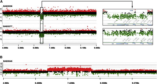
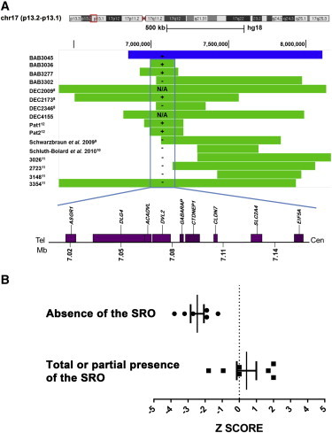
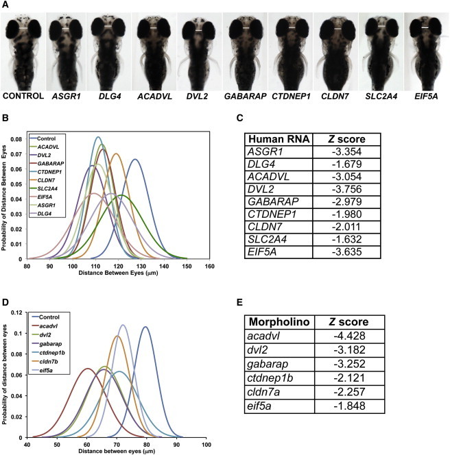
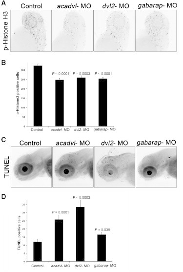
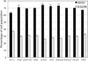
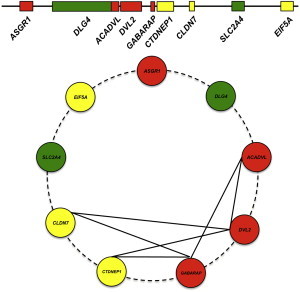
The 17p13.1 microdeletion syndrome is a recently described genomic disorder with a core clinical phenotype of intellectual disability, poor to absent speech, dysmorphic features, and a constellation of more variable clinical features, most prominently microcephaly. We identified five subjects with copy-number variants (CNVs) on 17p13.1 for whom we performed detailed clinical and molecular studies. Breakpoint mapping and retrospective analysis of published cases refined the smallest region of overlap (SRO) for microcephaly to a genomic interval containing nine genes. Dissection of this phenotype in zebrafish embryos revealed a complex genetic architecture: dosage perturbation of four genes (ASGR1, ACADVL, DVL2, and GABARAP) impeded neurodevelopment and decreased dosage of the same loci caused a reduced mitotic index in vitro. Moreover, epistatic analyses in vivo showed that dosage perturbations of discrete gene pairings induce microcephaly. Taken together, these studies support a model in which concomitant dosage perturbation of multiple genes within the CNV drive the microcephaly and possibly other neurodevelopmental phenotypes associated with rearrangements in the 17p13.1 SRO.


Figures

Figure 1
Figure 1
High-Resolution aCGH Plot for Individuals BAB3036, BAB3277, and BAB3045 (A) Individuals with small deletions of 17p13.1 region. Black box delimits the smallest region of overlap deleted in subjects with microcephaly. (B) Individual with triplication involving 17p13.1 region. CNVs were detected by oligonucleotide probes for which the mean normalized log2 (Cy5/Cy3) ratio of the CGH signal reached mean thresholds of −1 (indicating a heterozygous deletion [CN = 1]), 0.6 (indicating a duplication [CN = 3]), or 1.0 (indicating a triplication [CN = 4]).
Figure 2
Figure 2
Representative Summary of CNVs Involving 17p13.1 (A) Top: Individuals with deletions are represented by green rectangles and the individual with a triplication is represented by blue rectangle. The graphical normalized data for each individual was obtained by applying the most distal and proximal oligonucleotide genomic probe coordinates to the custom track at UCSC Genome Browser website. Positions are given relative to build hg18. Vertical blue rectangle delimits the smallest region of overlap (SRO) for the microcephaly phenotype in this cohort. Plus sign indicates presence of absolute microcephaly; minus sign indicates absence of absolute microcephaly (refer to Table 2 for Z score values). N/A indicates information not available. Bottom: Dosage-sensitive region associated with small head size (157 kb) chr17: 6,996,378–7,152,828 (hg18). (B) Graph of Z scores of head circumferences in subjects with 17p13.1 deletions. Black dots: individual Z score of subjects who harbor CNVs encompassing entirely predefined SRO of 157 kb. Black squares: individual Z scores of subjects who harbor CNVs that do not encompass predefined SRO or encompass it partially. All measurements are plotted as age- and sex-matched Z scores. Bars indicate mean and 95% confidence intervals.
Figure 3
Figure 3
Delimited 17p13.1 Region Harbors Dosage-Sensitive Genes as Experimentally Assayed in Zebrafish (A) Overexpression of capped human mRNA corresponding to nine loci in the 17p13.1 region in zebrafish embryos leads to microcephaly at 4 dpf. Representative dorsal images of zebrafish embryos injected with the indicated human mRNA scored for microcephaly at 4 dpf. (B) Graph represents the probability distribution curve of distance between eyes in zebrafish embryos upon overexpression injection of individual mRNAs. (C) List of individual capped human mRNAs and corresponding Z score measured as distance between eyes in zebrafish at 4 dpf (B). (D) Graph represents the probability distribution curve of distance between eyes in zebrafish embryos injected with splice blocker morpholinos of the indicated genes and scored for microcephaly at 4 dpf. (E) The corresponding Z scores of the probability distribution curves in (D).
Figure 4
Figure 4
Loss of the Primary Drivers of Microcephaly Lead to Reduced Cell Division in Zebrafish (A) Zebrafish embryos were injected with the morpholinos as indicated and stained for phospho-histone H3 at 2 dpf. Representative images are shown for control and morpholino suppression. The dashed lines in the controls indicate the regions used for calculating the number of histone-positive cells. (B) Graph represents average and standard error of histone-positive cells in each sample from 20 embryos. (C) Representative images of TUNEL staining of 3 dpf zebrafish embryos either uninjected or injected with 3 ng of acadvl, dvl2, or gabarap MO. (D) Graph represents the average number of TUNEL-positive cells and standard error for each condition.
Figure 5
Figure 5
Loss of the Primary Drivers Result in Decreased S/G2/M Population Neuro2A Cells Neuro2A cells were transfected with a pool of four different siRNAs targeting Asgr1 and compared with cells transfected with scrambled siRNA (Scr si). The remaining seven genes were suppressed with a combination of five shRNAs in pLKO.1 vector for each gene and compared against the control cells transfected with scrambled shRNA (pLKO.1 scr). Three days after transfection, cells were harvested, fixed, permeabilized, and stained with propidium iodide (PI). Cell cycle analysis was carried by flow cytometry and the average percentage of G0/G1 and S/G2/M population of each condition from three experiments is represented and the error bars indicate the standard deviation. ∗∗p < 0.005.
Figure 6
Figure 6
Schematic Model for Dosage-Sensitive Region at 17p13.1 Associated with Small Head Size in Human Top: Model representing loci that leads to severe (red), moderate (yellow), and no scored phenotype (green). Bottom: Summary of the binary genetic interactions tested between loci for microcephaly in zebrafish embryos.

References

    1. Bi W., Sapir T., Shchelochkov O.A., Zhang F., Withers M.A., Hunter J.V., Levy T., Shinder V., Peiffer D.A., Gunderson K.L. Increased LIS1 expression affects human and mouse brain development. Nat. Genet. 2009;41:168–177. - PMC - PubMed
    1. vanTuinen P., Dobyns W.B., Rich D.C., Summers K.M., Robinson T.J., Nakamura Y., Ledbetter D.H. Molecular detection of microscopic and submicroscopic deletions associated with Miller-Dieker syndrome. Am. J. Hum. Genet. 1988;43:587–596. - PMC - PubMed
    1. Lupski J.R., de Oca-Luna R.M., Slaugenhaupt S., Pentao L., Guzzetta V., Trask B.J., Saucedo-Cardenas O., Barker D.F., Killian J.M., Garcia C.A. DNA duplication associated with Charcot-Marie-Tooth disease type 1A. Cell. 1991;66:219–232. - PubMed
    1. Chance P.F., Alderson M.K., Leppig K.A., Lensch M.W., Matsunami N., Smith B., Swanson P.D., Odelberg S.J., Disteche C.M., Bird T.D. DNA deletion associated with hereditary neuropathy with liability to pressure palsies. Cell. 1993;72:143–151. - PubMed
    1. Potocki L., Bi W., Treadwell-Deering D., Carvalho C.M., Eifert A., Friedman E.M., Glaze D., Krull K., Lee J.A., Lewis R.A. Characterization of Potocki-Lupski syndrome (dup(17)(p11.2p11.2)) and delineation of a dosage-sensitive critical interval that can convey an autism phenotype. Am. J. Hum. Genet. 2007;80:633–649. - PMC - PubMed

Publication types

MeSH terms

Supplementary concepts