Case Reports
. 2014 Nov 6;95(5):611-21.
doi: 10.1016/j.ajhg.2014.10.007. Epub 2014 Nov 6.

Mutations in SNX14 cause a distinctive autosomal-recessive cerebellar ataxia and intellectual disability syndrome

Affiliations
Case Reports

Mutations in SNX14 cause a distinctive autosomal-recessive cerebellar ataxia and intellectual disability syndrome

Anna C Thomas et al. Am J Hum Genet. .

Erratum in

  • Am J Hum Genet. 2015 Jun 4;96(6):1008-9

Abstract

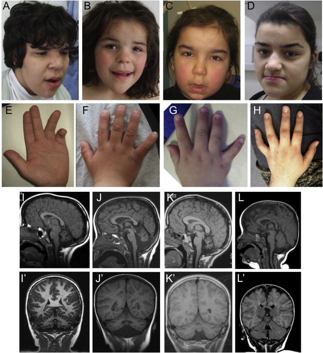
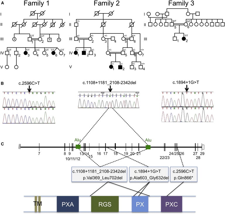
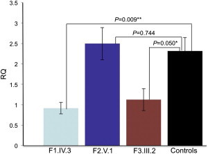
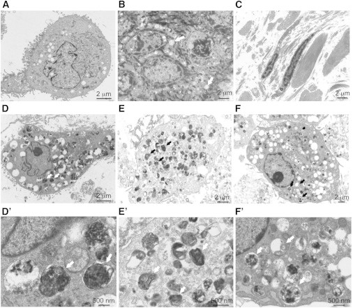
Intellectual disability and cerebellar atrophy occur together in a large number of genetic conditions and are frequently associated with microcephaly and/or epilepsy. Here we report the identification of causal mutations in Sorting Nexin 14 (SNX14) found in seven affected individuals from three unrelated consanguineous families who presented with recessively inherited moderate-severe intellectual disability, cerebellar ataxia, early-onset cerebellar atrophy, sensorineural hearing loss, and the distinctive association of progressively coarsening facial features, relative macrocephaly, and the absence of seizures. We used homozygosity mapping and whole-exome sequencing to identify a homozygous nonsense mutation and an in-frame multiexon deletion in two families. A homozygous splice site mutation was identified by Sanger sequencing of SNX14 in a third family, selected purely by phenotypic similarity. This discovery confirms that these characteristic features represent a distinct and recognizable syndrome. SNX14 encodes a cellular protein containing Phox (PX) and regulator of G protein signaling (RGS) domains. Weighted gene coexpression network analysis predicts that SNX14 is highly coexpressed with genes involved in cellular protein metabolism and vesicle-mediated transport. All three mutations either directly affected the PX domain or diminished SNX14 levels, implicating a loss of normal cellular function. This manifested as increased cytoplasmic vacuolation as observed in cultured fibroblasts. Our findings indicate an essential role for SNX14 in neural development and function, particularly in development and maturation of the cerebellum.


Figures

Figure 1
Figure 1
Phenotype of Affected Individuals from the Three Families Presented Photographs and brain MRI scans from family 1 individual IV.3 aged 19 years (A) and 22 years (E); family 1 individual IV.6 aged 6 years (B, F) and 7 years (I, I′); family 2 individual V.1 aged 4.5 years (C, G, J, J′); family 3 individual III.2 aged 22 years (D, H) and 10 years (K, K′); and family 2 individual V.2 aged 9 months (L, L′). Notice the similar facial features mainly characterized by broad face, fullness of the upper eyelid, broad nasal base and slight underdevelopment of the alae, broad and long philtrum, thick lower lip vermillion, and fifth finger brachycamptodactyly. In the first years of life, no neuroradiological anomalies were observed in affected individuals, as depicted here by the normal T1-weighted mid-sagittal (L) and coronal (L′) MRI sections, which have not yet been repeated for this individual. MRI images performed for other individuals during infancy are unavailable but were reported to be normal. At older ages, affected individuals have a small cerebellum with thin folia and enlarged fissures, suggestive of global cerebellar atrophy as shown here in the T1-weighted MRI images from three of the children, one from each family (mid-sagittal sections I–K; coronal sections I′–K′). The pons appear small but in comparison are well preserved.
Figure 2
Figure 2
Identification of SNX14 Mutations in Three Affected Families (A) Pedigrees of the three families showing genotypes in tested individuals. (B) Sequence traces for families 1 and 3 show point mutations in genomic DNA (top trace, mutant; bottom trace, wild-type). For family 2, the sequence trace spans the deletion breakpoint, in genomic DNA, and indicates the location of the breakpoints within two Alu repeats in the schematic diagram of the SNX14 locus shown in (C) (see also Figure S1 for further details). (C) Schematic representation of part of the SNX14 genomic locus (top) and the SNX14 protein (bottom) indicating the location and effect of the mutations detected in the three families. The protein consists of two predicted transmembrane domains (TM) at the N terminus, followed by the PXA domain, RGS domain, conserved PX phosphoinositide binding domain, and PXC domain situated toward the C terminus. The deletion in family 2 is predicted to remove the RGS and PX domains, whereas the splice site mutation in family 3 removes part of the PX domain.
Figure 3
Figure 3
SNX14 RNA Expression in Fibroblasts from Affected and Control Individuals qRT-PCR showing relative quantification (RQ ΔΔCT method) of SNX14 expression in affected fibroblasts samples from each family compared with controls. Both GAPDH and HPRT1 were used as combined endogenous controls, with RQ calculated using StepOne analysis software v.2.1 (Life Technologies). Each experiment was biologically replicated three times with each sample analyzed in triplicate. Using a two-tailed t test, family 1 individual IV.3 shows a 60% decrease in expression compared to combined controls, where fibroblasts from five different control individuals have been analyzed (N = 5), ∗∗p = 0.009. Family 3 individual III.2 shows a 51% drop in expression compared to controls, p = 0.05. However, family 2 individual V.1 shows no significant difference in expression compared to controls, p = 0.74. Error bars correspond to mean ± SEM.
Figure 4
Figure 4
Electron Micrographs of Skin Biopsy and Cultured Fibroblasts (A) Control cultured fibroblast at 800× magnification. (B and C) Skin section from family 3 individual III.2 at 800× magnification. (B) In the epidermis, there was mild hyperkeratosis with keratinocytes showing increased vacuolation, containing fine nonspecific granular material (arrows). (C) In the dermis, collagen and elastic tissue had a normal appearance and distribution with infrequent vacuoles in the fibroblasts. (D–F) Cultured fibroblasts from family 1 individual IV.6, family 2 individual V.1, and family 3 individual III.2, respectively, at 800× magnification. The cells showed numerous cytoplasmic vacuoles, often containing dense staining material suggestive of lipid degeneration. Same cells at 3,000× magnification shown in (D′)–(F′). Vacuoles contained granular material or multilamellar bodies or were empty (see white and black arrows for examples).

References

    1. Ezgu F., Krejci P., Li S., de Sousa C., Graham J.M., Jr., Hansmann I., He W., Porpora K., Wand D., Wertelecki W. Phenotype-genotype correlations in patients with Marinesco-Sjögren syndrome. Clin. Genet. 2014;86:74–84. - PubMed
    1. Strehle E.M. Sialic acid storage disease and related disorders. Genet. Test. 2003;7:113–121. - PubMed
    1. Waterham H.R., Ebberink M.S. Genetics and molecular basis of human peroxisome biogenesis disorders. Biochim. Biophys. Acta. 2012;1822:1430–1441. - PubMed
    1. Matthijs G., Schollen E., Pardon E., Veiga-Da-Cunha M., Jaeken J., Cassiman J.J., Van Schaftingen E. Mutations in PMM2, a phosphomannomutase gene on chromosome 16p13, in carbohydrate-deficient glycoprotein type I syndrome (Jaeken syndrome) Nat. Genet. 1997;16:88–92. - PubMed
    1. Philip N., Chabrol B., Lossi A.M., Cardoso C., Guerrini R., Dobyns W.B., Raybaud C., Villard L. Mutations in the oligophrenin-1 gene (OPHN1) cause X linked congenital cerebellar hypoplasia. J. Med. Genet. 2003;40:441–446. - PMC - PubMed

Publication types

Supplementary concepts

LinkOut - more resources