Case Reports
. 2014 Nov 6;95(5):622-32.
doi: 10.1016/j.ajhg.2014.10.008. Epub 2014 Nov 6.

Mutations in CKAP2L, the human homolog of the mouse Radmis gene, cause Filippi syndrome

Affiliations
Case Reports

Mutations in CKAP2L, the human homolog of the mouse Radmis gene, cause Filippi syndrome

Muhammad Sajid Hussain et al. Am J Hum Genet. .

Abstract

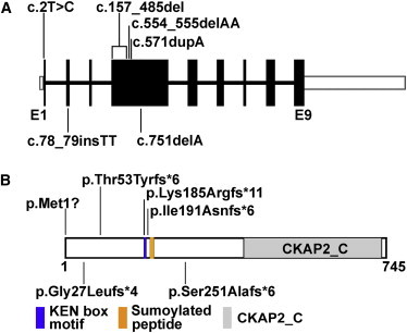
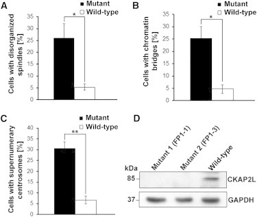
Filippi syndrome is a rare, presumably autosomal-recessive disorder characterized by microcephaly, pre- and postnatal growth failure, syndactyly, and distinctive facial features, including a broad nasal bridge and underdeveloped alae nasi. Some affected individuals have intellectual disability, seizures, undescended testicles in males, and teeth and hair abnormalities. We performed homozygosity mapping and whole-exome sequencing in a Sardinian family with two affected children and identified a homozygous frameshift mutation, c.571dupA (p.Ile191Asnfs(∗)6), in CKAP2L, encoding the protein cytoskeleton-associated protein 2-like (CKAP2L). The function of this protein was unknown until it was rediscovered in mice as Radmis (radial fiber and mitotic spindle) and shown to play a pivotal role in cell division of neural progenitors. Sanger sequencing of CKAP2L in a further eight unrelated individuals with clinical features consistent with Filippi syndrome revealed biallelic mutations in four subjects. In contrast to wild-type lymphoblastoid cell lines (LCLs), dividing LCLs established from the individuals homozygous for the c.571dupA mutation did not show CKAP2L at the spindle poles. Furthermore, in cells from the affected individuals, we observed an increase in the number of disorganized spindle microtubules owing to multipolar configurations and defects in chromosome segregation. The observed cellular phenotypes are in keeping with data from in vitro and in vivo knockdown studies performed in human cells and mice, respectively. Our findings show that loss-of-function mutations in CKAP2L are a major cause of Filippi syndrome.


Figures

Figure 1
Figure 1
Clinical Presentation of Filippi-Syndrome-Affected Individuals with Mutations in CKAP2L Affected individual FP1-1 and his brother, FP1-3, are shown at the age of 3 years and 5 months. FP1-1 was reported previously as the first child born to an Italian family. Note the facial abnormalities and cutaneous syndactyly of the fingers and toes. Although the clinical presentation is very similar, the broad nasal bridge and underdeveloped alae nasi seem to be more pronounced in the younger brother (FP1-3), whereas the cutaneous syndactyly of the fingers is more severe in the older brother (FP1-1). In the bottom row, affected individual FP5-1 is shown at the age of 16 years. He is the first child of double first cousins of Pakistani origin and was reported previously at the age of 5 years and 4 months. Earlier in his life, he underwent a surgical intervention to release his cutaneous syndactyly of the third and fourth fingers of both hands. Parents provided written consent for the publication of photographs of their children.
Figure 2
Figure 2
Position of CKAP2L Mutations Identified in Filippi-Syndrome-Affected Families (A) Genomic structure of human CKAP2L. The nine exons of CKAP2L are displayed by boxes, which are drawn to scale. The filled boxes represent the open reading frame, and the open boxes represent the UTRs of the transcript. The introns are drawn as connecting lines of arbitrary length. Vertical lines indicate the positions of the mutations. Heterozygous mutations are shown below the schematic, whereas all homozygous mutations are shown above. (B) Domain organization of CKAP2L as predicted by the NCBI Conserved Domain Database. A peptide of 329 amino acids (residues 415–734) was recognized as cytoskeleton-associated protein 2 C terminus (CKAP2_C). The position of a KEN box motif (Lys-Glu-Asn) is highlighted in blue (residues 185–187). KEN box motifs are known to be a target sequence of APC/C protein CDH1 to initiate proteasomal degradation. The position of a nonconsensus sumoylated peptide identified by mass spectrometry is shown in yellow (residues 198–206).
Figure 3
Figure 3
Subcellular Localization of CKAP2L in Wild-Type LCLs Wild-type LCLs were cultivated in RPMI-1640 (GIBCO, Life Technologies) supplemented with 10% fetal bovine serum and antibiotics (penicillin and streptomycin). For analysis of spindle microtubules, the cells were incubated with tubulin-stabilization buffer before and after blocking with PBG. Primary antibodies specific to CKAP2L (NBP1-83450, Novus Biologicals, 1:300; green), γ-tubulin (GTU-88, Sigma-Aldrich, 1:300; red), and α-tubulin (YL 1/2, 1:20; turquoise) were used. Secondary antibodies Alexa Fluor 488 donkey anti-rabbit IgG (A21206, Invitrogen), Alexa Fluor 568 goat anti-mouse IgG (A11004, Invitrogen), and Alexa Fluor 647 goat anti-rat IgG (A21247, Invitrogen) were used as appropriate, and DAPI (D9564, Sigma) was used for DNA detection. CKAP2L was detected at the spindle pole from prometaphase to telophase. Interphase cells did not show any CKAP2L staining. Scale bars represent 5 μm.
Figure 4
Figure 4
Analysis of CKAP2L Localization, Spindle Morphology, Centrosomes, and DNA in LCLs from Affected Individuals Carrying the c.571dupA Mutation Confocal-microscopy images of an affected individual’s LCLs, stained for CKAP2L (green), α-tubulin (turquoise), and γ-tubulin (red) and stained with DAPI (blue). Immunoreactivity of CKAP2L was not observed at the spindles or the spindle poles of dividing cells. Defective spindles (metaphase), supernumerary centrosomes (metaphase), and abnormal chromatin bridges (inset) between separating daughter nuclei during cytokinesis accompanied the absence of CKAP2L. Scale bars represent 5 μm.
Figure 5
Figure 5
Statistical Analysis of Abnormalities of Spindles, Centrosomes, and DNA in LCLs from Affected Individuals (A) Statistical data of disorganized spindle microtubules observed in wild-type and mutant LCLs from three different experiments (n > 50 cells per experiment). Error bars represent the SEM. p value = 0.0314 (Student’s t test). (B) Statistical data of chromosome lagging observed between the daughter nuclei during cytokinesis in wild-type and mutant LCLs. A total of 150 cells during mitosis from three different experiments (n = 50 cells per experiment) were analyzed. Error bars represent the SEM. p value = 0.0143 (Student’s t test). (C) Statistical data of supernumerary centrosomes observed during mitosis in wild-type and mutant LCLs from three different experiments (n > 50 cells per experiment). Error bars represent the SEM. p value = 0.0021 (Student’s t test). (D) A representative immunoblot with cell lysates from control and mutant LCLs. Wild-type LCLs and affected individuals’ LCLs were harvested by centrifugation and lysed in ice-cold RIPA buffer (R0278, Sigma-Aldrich) supplemented with a proteinase-inhibitor cocktail (Sigma). After centrifugation, proteins in the supernatant were denatured at 95°C for 5 min in SDS sample buffer. Proteins were separated by 4%–12% SDS-PAGE (EC-890, National Diagnostics) and blotted onto nitrocellulose membrane (PROTRANR, Germany). Protein detection was performed with goat polyclonal anti-CKAP2L as the primary antibody (sc-242448, Santa Cruz Biotechnology) and GAPDH (G9295, Sigma-Aldrich) as the loading control. Anti-goat IgG peroxidase conjugate (A9452, Sigma) was used as the secondary antibody, and then the blots were developed with an enhanced chemiluminescence system. CKAP2L was not detected in lysates from mutant LCLs, whereas a predicted 84 kDa band was detected in wild-type lysates. GAPDH was used as a loading control.
Figure 6
Figure 6
Detection of CKAP2L in the Murine Embryonic Limb Buds (A and B) A transverse section of an E10.5 whole mouse embryo coimmunostained for CKAP2L (affinity-purified rabbit polyclonal antibody, 1:10,000; green, B) and the mitosis-specific marker phosphohistone H3 (mouse monoclonal IgG1, clone 6G3, Cell Signaling Technology, 1:1,000; red, A). Nuclei were counterstained with TOPRO-3 (Life Technologies; blue). Robust immunoreactivity of CKAP2L was observed in the neural progenitor cells throughout the neural tube and the myotome (double arrows in B). The scale bar represents 100 μm. Abbreviations are as follows: nt, neural tube; and lb, forelimb bud. Asterisks indicate the dorsal aorta and vein. (C) Higher magnification of the forelimb bud around the apical ectodermal ridge, stained for CKAP2L (green) and phosphohistone H3 (red). The immunoreactivity of CKAP2L was significantly lower in the connective tissues, including the limb buds, than in the neural tube, but close inspection clearly disclosed CKAP2L staining at the mitotic spindles (arrows) of dividing mesenchymal cells, which were loosely distributed in the developing limb bud. Note that the section shown is different from that in (A). The scale bar represents 75 μm. The following abbreviation is used: epi, epithelial ectoderm. (D) Individual dividing cells in anaphase (left panel) and metaphase (right panel) within the limb bud mesenchyme. CKAP2L localized to the mitotic spindle and spindle poles of mitotic cells. The scale bar represents 50 μm. (E) CKAP2L staining in an E12.5 forelimb bud. CKAP2L immunoreactivity (green) was detected in the dividing cells (arrows) at the boundary region between the protruding cartilage and the surrounding mesenchyme. These CKAP2L-positive cells might include the mitotic mesenchymal cells and the dividing chondrocyte progenitors. In addition, CKAP2L was detected in the fine processes of myogenic progenitor cells, which delaminated from the ventrolateral lips of the dermomyotome and migrated to give rise to limb muscles. TOPRO-3-stained nuclei are in blue. The scale bar represents 25 μm. The following abbreviation is used: ctl, developing cartilage.

References

    1. Meinecke P. Short stature, microcephaly, characteristic face, syndactyly and mental retardation: the Filippi syndrome. Report on a second family. Genet. Couns. 1993;4:147–151. - PubMed
    1. Filippi G. Unusual facial appearance, microcephaly, growth and mental retardation, and syndactyly. A new syndrome? Am. J. Med. Genet. 1985;22:821–824. - PubMed
    1. Orrico A., Hayek G. An additional case of craniodigital syndrome: variable expression of the Filippi syndrome? Clin. Genet. 1997;52:177–179. - PubMed
    1. Woods C.G., Crouchman M., Huson S.M. Three sibs with phalangeal anomalies, microcephaly, severe mental retardation, and neurological abnormalities. J. Med. Genet. 1992;29:500–502. - PMC - PubMed
    1. Walpole I.R., Parry T., Goldblatt J. Expanding the phenotype of Filippi syndrome: a report of three cases. Clin. Dysmorphol. 1999;8:235–240. - PubMed

Publication types

MeSH terms

Supplementary concepts