. 2014 Nov 4;20(5):910-918.
doi: 10.1016/j.cmet.2014.10.001. Epub 2014 Nov 4.

The LXR-Idol axis differentially regulates plasma LDL levels in primates and mice

Affiliations

The LXR-Idol axis differentially regulates plasma LDL levels in primates and mice

Cynthia Hong et al. Cell Metab. .

Abstract

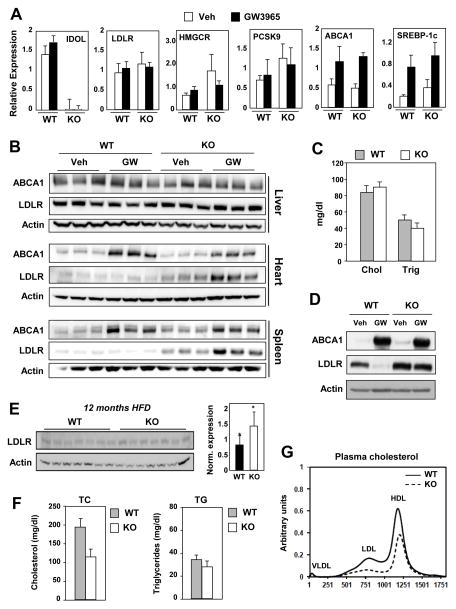
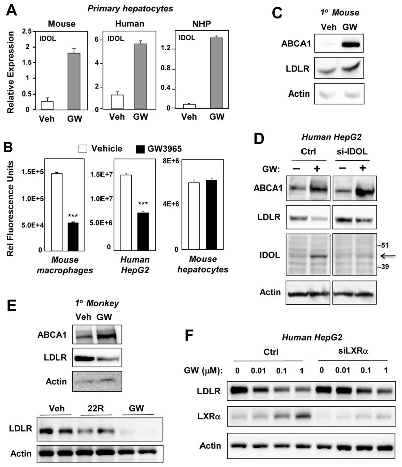
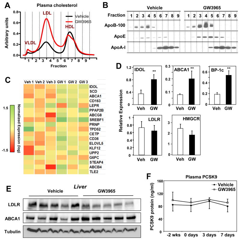
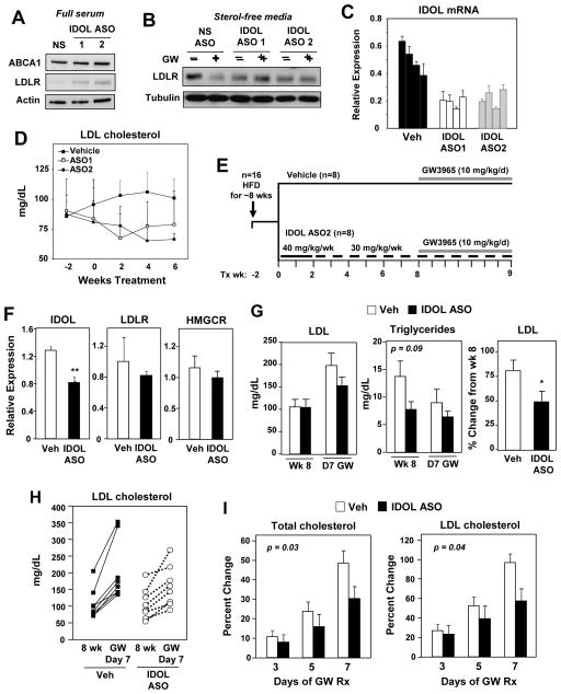
The LXR-regulated E3 ubiquitin ligase IDOL controls LDLR receptor stability independent of SREBP and PCSK9, but its relevance to plasma lipid levels is unknown. Here we demonstrate that the effects of the LXR-IDOL axis are both tissue and species specific. In mice, LXR agonist induces Idol transcript levels in peripheral tissues but not in liver, and does not change plasma LDL levels. Accordingly, Idol-deficient mice exhibit elevated LDLR protein levels in peripheral tissues, but not in the liver. By contrast, LXR activation in cynomolgus monkeys induces hepatic IDOL expression, reduces LDLR protein levels, and raises plasma LDL levels. Knockdown of IDOL in monkeys with an antisense oligonucleotide blunts the effect of LXR agonist on LDL levels. These results implicate IDOL as a modulator of plasma lipid levels in primates and support further investigation into IDOL inhibition as a potential strategy for LDL lowering in humans.


Figures

Figure 1
Figure 1. IDOL controls peripheral LDLR expression in mice
WT and Idol-null mice were treated with the LXR agonist GW3965 at 40 mg/kg for 3 days (n = 3–4/group). (A) Hepatic gene expression was quantified by real-time PCR and normalized to 36B4. (B) Protein lysates from liver, heart, and spleen were analyzed by immunoblotting with antibodies against ABCA1, LDLR, and β-Actin. (n = 3–4 mice/treatment) (C) Total serum cholesterol and triglyceride levels for chow-fed animals. (D) Immunoblot analysis of protein extracts from thioglycollate-elicited peritoneal macrophages isolated from WT and Idol-null mice and treated with GW3965 (1 μM). Immunoblot shown is representative of three independent experiments. (E) WT and Idol-null mice were fed western diet for 12 months. Protein lysates from liver were analyzed by immunoblotting and quantified using ImageJ (n=7 mice/genotype). Significance was determined by student t-test (* p < 0.05). (F) Total serum cholesterol and triglyceride levels for the animals shown in E. (G) Plasma lipoprotein cholesterol distribution for the animals shown in E. Error bars represent SEM.
Figure 2
Figure 2. Species-specific regulation of hepatic LDLR protein expression by the LXR–IDOL pathway
Primary hepatocytes from mice, humans, and nonhuman primates were serum-starved and treated with GW3965 (1 μM) for 24 h. (A) Regulation of IDOL mRNA expression by LXR agonists. (B) Quantification of cellular uptake of DiI-LDL uptake in the presence or absence of GW3965. Cell-associated fluorescence was measured with a Typhoon Instrument. n=3. ***P< 0.001. (C,D) Immunoblot analysis of protein lysates from primary mouse hepatocytes (C) or human HepG2 cells (D) treated with GW3965 (1 μM) for 24 h. (E) Immunoblotting of protein lysates from cynomolgus monkey hepatocytes treated with GW3965 (1 μM) or 22R-hydroxycholesterol (2.5 μM) for 24 h. (F) Immunoblot analysis of protein lysates from control or LXRα-siRNA-transfected HepG2 cells treated for 24 h with increasing doses of GW3965. Data are representative of three independent experiments. Error bars represent SEM.
Figure 3
Figure 3. LXR activation in nonhuman primates reduces hepatic LDLR protein and raises plasma LDL levels
(A) Plasma lipoprotein cholesterol distribution of cynomolgus monkeys treated with vehicle or GW3965 (10 mg/kg for 7 days) (n = 5 pooled samples/group). (B) Plasma apolipoprotein (apoB-100, apoE, and apoA-I) distributions for animals shown in A. (C) Transcriptional profiling of mRNA from the livers of cynomolgus monkeys treated for 2 days with vehicle or GW3965. Tissues were obtained 6 h after the last dose and transcript levels were analyzed with Affymetrix arrays. (D) Hepatic gene expression from the animals in D as determined by real-time PCR; BP-1C = SREBP-1c (n=5/group; ** p < 0.01). (E) Protein lysates of the liver from cynomolgus monkeys treated with vehicle or GW3965 (10 mg/kg for 7 days). Tissues were obtained 24 h after the final dose; immunoblots were performed for LDLR, ABCA1, and tubulin. (F) ELISA of plasma levels of PCSK9 in cynomolgus monkeys treated with vehicle or GW3965 (10 mg/kg for 7 days) (n = 5/group). Error bars represent SEM
Figure 4
Figure 4. IDOL knockdown modulates hepatic LDLR expression in nonhuman primates
(A,B) Immunoblot analysis of HepG2 cells transfected with control or IDOL ASOs. (A) Cells were cultured in full serum. (B) Cells were cultured in sterol-deficient media containing 5 μM simvastatin plus 100 μM mevalonic and treated for 24 h with GW3965 (1 μM). Data are representative of three independent experiments. (C) Activity of IDOL ASOs in vivo. Cynomolgus monkeys were treated for 8 weeks with vehicle or IDOL ASOs, and hepatic IDOL expression levels were quantified by real-time PCR. Each bar represents an individual animal. (D) Plasma LDL cholesterol was measured at the designated time points for animals shown in C. (E) Schematic showing the experimental design for evaluating the effects of IDOL ASO treatment on the response to LXR activation in monkeys. (F) Liver gene expression determined by real-time PCR (n = 8/group). *** p < 0.001. (G) Average plasma LDL cholesterol level (Left) and average triglyceride level (Middle) before and after GW3965 treatment. (Right) Percent change in LDL cholesterol levels (average for group) after GW3965 treatment. (H) Plasma LDL levels of each nonhuman primate at week 8 and day 7 of GW3965 treatment. (I) Percent change in total and LDL cholesterol levels over the course of the entire GW3965 treatment period. Two-way ANOVA was used to assess statistical significance. Error bars represent SEM.

References

    1. Abifadel M, Varret M, Rabes JP, Allard D, Ouguerram K, Devillers M, Cruaud C, Benjannet S, Wickham L, Erlich D, et al. Mutations in PCSK9 cause autosomal dominant hypercholesterolemia. Nat Genet. 2003;34:154–156. - PubMed
    1. Agellon LB, Walsh A, Hayek T, Moulin P, Jiang XC, Shelanski SA, Breslow JL, Tall AR. Reduced high density lipoprotein cholesterol in human cholesteryl ester transfer protein transgenic mice. J Biol Chem. 1991;266:10796–10801. - PubMed
    1. Brown MS, Goldstein JL. A receptor-mediated pathway for cholesterol homeostasis. Science. 1986;232:34–47. - PubMed
    1. Brown MS, Goldstein JL. The SREBP pathway: regulation of cholesterol metabolism by proteolysis of a membrane-bound transcription factor. Cell. 1997;89:331–340. - PubMed
    1. Calkin AC, Goult BT, Zhang L, Fairall L, Hong C, Schwabe JW, Tontonoz P. FERM-dependent E3 ligase recognition is a conserved mechanism for targeted degradation of lipoprotein receptors. Proc Natl Acad Sci U S A. 2011;108:20107–20112. - PMC - PubMed

Publication types