Review
. 2015 Jan;21(1):24-33.
doi: 10.1016/j.molmed.2014.10.009. Epub 2014 Oct 30.

Human cancer immunotherapy with antibodies to the PD-1 and PD-L1 pathway

Affiliations
Review

Human cancer immunotherapy with antibodies to the PD-1 and PD-L1 pathway

Kim C Ohaegbulam et al. Trends Mol Med. 2015 Jan.

Abstract

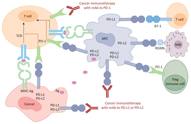
The programmed death 1 (PD-1) receptor and its ligands programmed death ligand 1 (PD-L1) and PD-L2, members of the CD28 and B7 families, play critical roles in T cell coinhibition and exhaustion. Overexpression of PD-L1 and PD-1 on tumor cells and tumor-infiltrating lymphocytes, respectively, correlates with poor disease outcome in some human cancers. Monoclonal antibodies (mAbs) blockading the PD-1/PD-L1 pathway have been developed for cancer immunotherapy via enhancing T cell functions. Clinical trials with mAbs to PD-1 and PD-L1 have shown impressive response rates in patients, particularly for melanoma, non-small-cell lung cancer (NSCLC), renal cell carcinoma (RCC), and bladder cancer. Further studies are needed to dissect the mechanisms of variable response rate, to identify biomarkers for clinical response, to develop small-molecule inhibitors, and to combine these treatments with other therapies.

Keywords: PD-1; PD-L1; PD-L2; human cancer; immunotherapy; monoclonal antibody.


Figures

Figure 1
Figure 1. Crystal structures of the PD-1/PD-L1 and PD-1/PD-L2 complexes
(A) Overlay of the crystal structures of human PD-1 (PDB Code 3RRQ) and the mouse PD-1/human PD-L1 complex (3BIK). (B) Overlay of human PD-1 with the mouse PD-1/PD-L2 complex (3BP5). Pink: human PD-1, green: mouse PD-1, yellow: human PD-L1, grey: mouse PD-L2
Figure 2
Figure 2. Human cancer immunotherapy with anti-PD-1 and anti-PD-L1/L2 antibodies
Antigen-presenting cells (APC) take up antigens (Ag) released from cancer cells and present to T cells. Cancer cells can also present Ag to activated T cells in the context of MHC. Upon T cell activation, PD-1 receptors are expressed on T cells and inhibit immune responses by engagement of PD-L1 and PD-L2 ligands on APC and PD-L1 on cancer cells. Therefore, monoclonal antibody (mAb)-mediated specific blockade of the PD-1/PD-L1/PD-L2 pathway can enhance anti-tumor immunity. In addition to binding to PD-1, PD-L1 and PD-L2 also bind B7-1 and repulsive guidance molecule B, respectively. In addition to T cells and APC, PD-1 and PD-L1 can be induced on other immune cells.

Similar articles

Cited by

References

    1. Keir ME, et al. PD-1 and its ligands in tolerance and immunity. Annu Rev Immunol. 2008;26:677–704. - PMC - PubMed
    1. Ishida Y, et al. Induced expression of PD-1, a novel member of the immunoglobulin gene superfamily, upon programmed cell death. EMBO J. 1992;11:3887–3895. - PMC - PubMed
    1. Barber DL, et al. Restoring function in exhausted CD8 T cells during chronic viral infection. Nature. 2006;439:682–687. - PubMed
    1. Wherry EJ. T cell exhaustion. Nat Immunol. 2011;12:492–499. - PubMed
    1. Hofmeyer KA, et al. The PD-1/PD-L1 (B7-H1) pathway in chronic infection-induced cytotoxic T lymphocyte exhaustion. Journal of biomedicine & biotechnology. 2011;2011:451694. - PMC - PubMed

Publication types