. 2015 Jan:36:66-79.
doi: 10.1016/j.biomaterials.2014.08.012. Epub 2014 Oct 13.

The ultrastructure of fibronectin fibers pulled from a protein monolayer at the air-liquid interface and the mechanism of the sheet-to-fiber transition

Affiliations

The ultrastructure of fibronectin fibers pulled from a protein monolayer at the air-liquid interface and the mechanism of the sheet-to-fiber transition

Maria Mitsi et al. Biomaterials. 2015 Jan.

Abstract

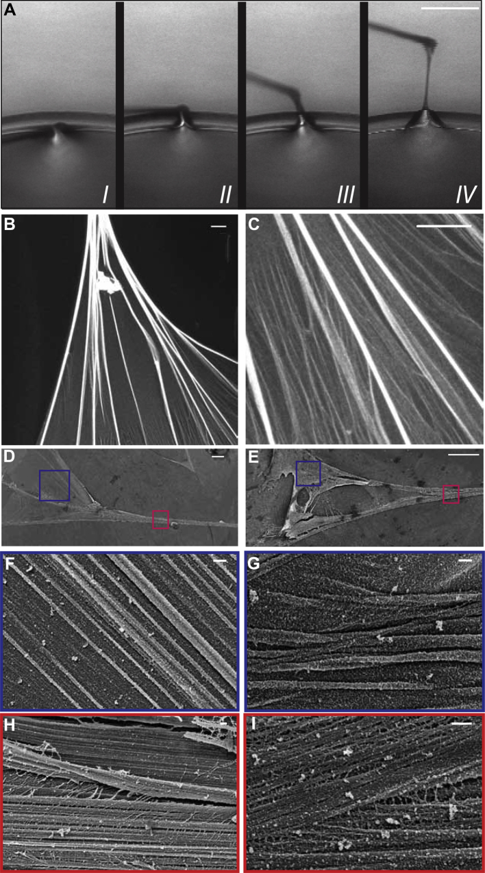
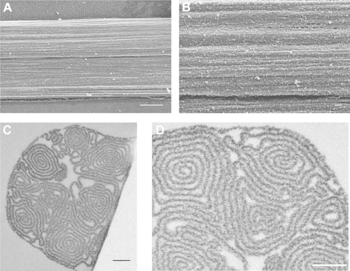
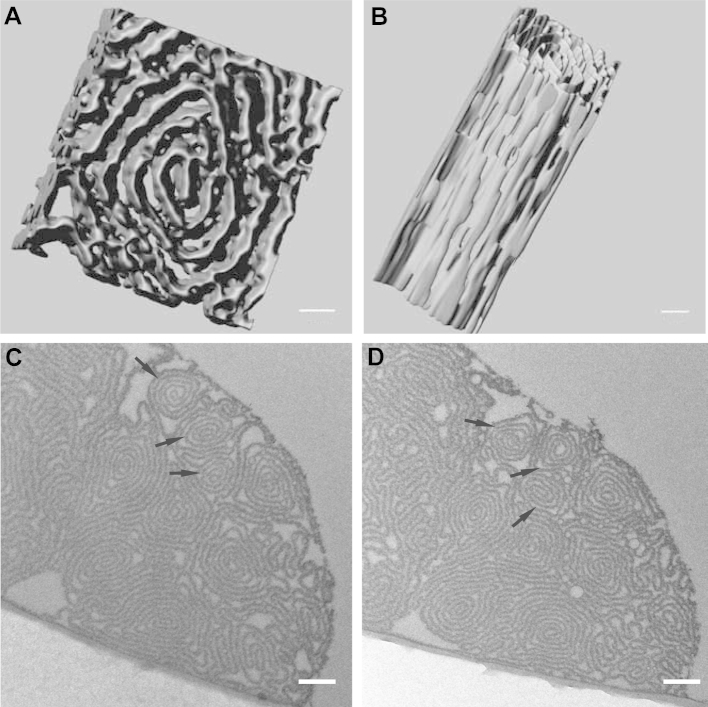
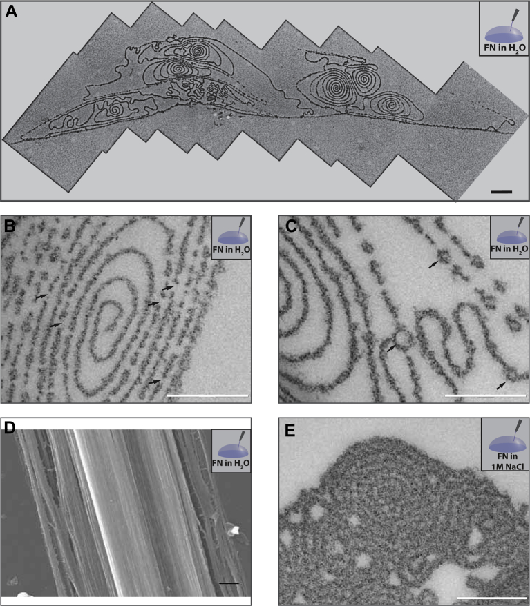
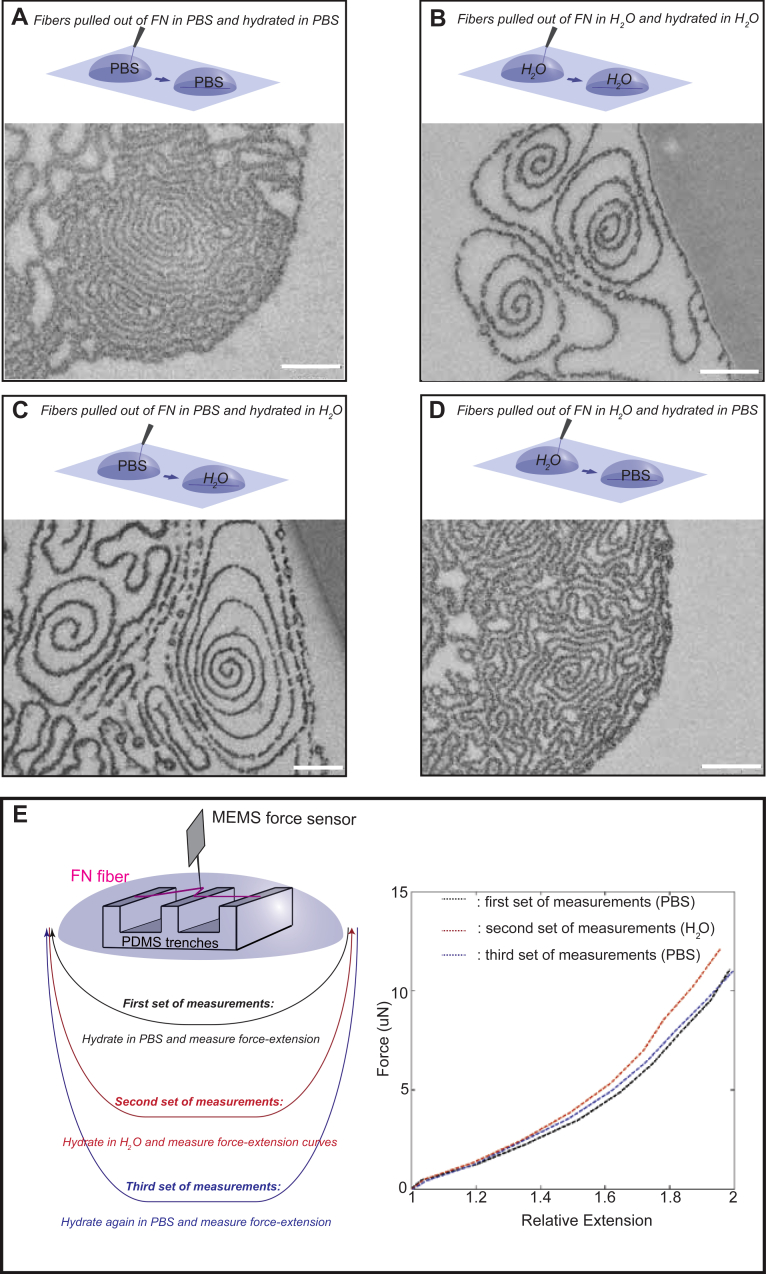
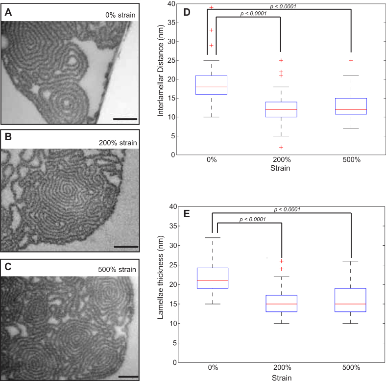
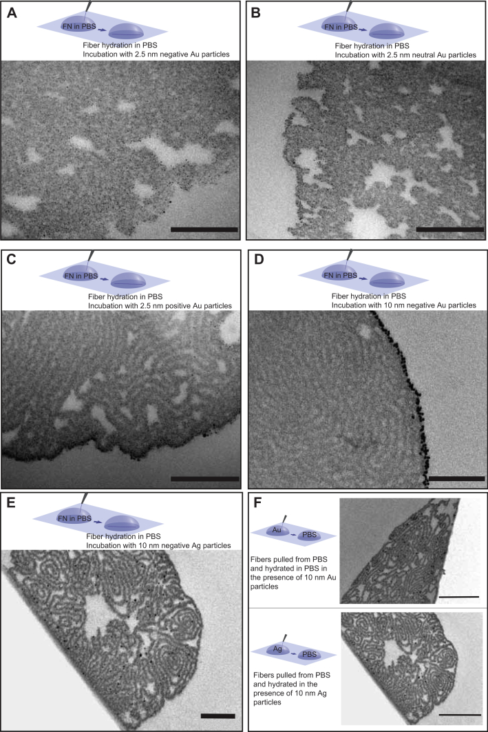
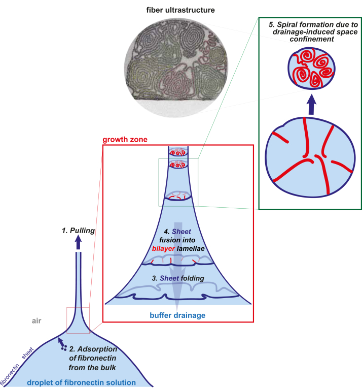
Fibronectin is a globular protein that circulates in the blood and undergoes fibrillogenesis if stretched or under other partially denaturing conditions, even in the absence of cells. Stretch assays made by pulling fibers from droplets of solutions containing high concentrations of fibronectin have previously been introduced in mechanobiology, particularly to ask how bacteria and cells exploit the stretching of fibronectin fibers within extracellular matrix to mechano-regulate its chemical display. Our electron microscopy analysis of their ultrastructure now reveals that the manually pulled fibronectin fibers are composed of densely packed lamellar spirals, whose interlamellar distances are dictated by ion-tunable electrostatic interactions. Our findings suggest that fibrillogenesis proceeds via an irreversible sheet-to-fiber transition as the fibronectin sheet formed at the air-liquid interface of the droplet is pulled off by a sharp tip. This far from equilibrium process is driven by the externally applied force, interfacial surface tension, shear-induced fibronectin self-association, and capillary force-induced buffer drainage. The ultrastructural characterization is then contrasted with previous FRET studies that characterized the molecular strain within these manually pulled fibers. Particularly relevant for stretch-dependent binding studies is the finding that the interior fiber surfaces are accessible to nanoparticles smaller than 10 nm. In summary, our study discovers the underpinning mechanism by which highly hierarchically structured fibers can be generated with unique mechanical and mechano-chemical properties, a concept that might be extended to other bio- or biomimetic polymers.

Keywords: Electron microscopy; Fibrillogenesis; Fibronectin; Kinetically trapped supramolecular system; Monolayer-to-lamella transition; Sheet-to-fiber transition.


Figures

Fig. 1
Fig. 1
Fibronetin fibers can be generated by pulling their monolayers away from the air-liquid interface. Fibers were pulled out of a 0.4 mg/ml fibronectin solution in PBS with a constant speed of 8 μm/s, using the tip of a MEMS force sensor (A). Similar fibers, pulled with a pipet tip out of a 0.4 mg/ml fibronectin (labeled with Alexa488 (B, C)) or unlabeled (D–J)) solution in PBS, deposited on the substrate and the fan-like region that connects the fiber to the droplet of the fibronectin solution was imaged by confocal microscopy (B, C; 63× oil objective) or by cryo-SEM, following high pressure freezing, freeze substitution and freeze drying of the specimen (D–I). The regions highlighted by the blue and red boxes in D and E are shown at higher magnification at F, G and H, I respectively. Scale bar: 200 μm for A, 10 μm for B, C, 20 μm for D, E and 200 nm for F–I.
Fig. 2
Fig. 2
Manually pulled fibronectin fibers show a lamellar organization. Manually pulled fibers were fixed by high pressure freezing, freeze dried and imaged in the SEM at −120 °C (A, B) or were chemically fixed with glutaraldehyde, ethanol dehydrated, embedded in Epon, ultrathin sectioned (50 nm) and observed by TEM (C, D). Scale bar: 1 μm for A, 100 nm for B and 200 nm for C and D.
Fig. 3
Fig. 3
Three dimensional ultrastructure of manually pulled fibronectin fibers. Fibers were pulled out of a 0.4 mg/ml fibronectin solution in PBS, chemically fixed with glutaraldehyde, ethanol dehydrated and embedded in Epon. 50 nm thick serial sections were cut from the blocks and were observed under the TEM. The images were processed as described for Fig. 2, and the segmented images were used for a 3D reconstruction. The reconstruction of one spiral domain from 11 serial sections is shown (A, B). In C and D, sequential sections 10 μm apart from a single fiber are shown. Scale bars: 20 nm for A, B and 500 nm for C, D.
Fig. 4
Fig. 4
The internal structure of fibers is stabilized by repulsive electrostatic interactions. Fibers pulled out of a 0.4 mg/ml fibronectin solution in distilled H2O (A,B,C,D) or 1 m NaCl (E) were chemically fixed with glutaraldehyde, ethanol dehydrated and embedded in Epon, ultrathin sectioned (50 nm) and observed by TEM (A, B, C, E) or high pressure frozen, freeze substituted and freeze dried and imaged in the SEM at −120 °C (D). For A, several images were required to capture the entire fiber cross section, and are shown stitched together. Scale bar: 500 nm for A, D and 200 nm for B, C, E.
Fig. 5
Fig. 5
The swelling effect induced by ionic strength on the fiber structure is reversible. Fibers were pulled out of a 0.4 mg/ml fibronectin solution in PBS and remained in PBS (A) or incubated with H2O (C) prior to fixation. Alternatively, fibers were pulled out of the H2O solution and remained in H2O (B) or incubated with PBS (D) prior to fixation. All samples were chemically fixed with glutaraldehyde, dehydrated in ethanol and embedded in Epon. 50 nm sections were observed by TEM. (E) Fibers were pulled out of a 0.4 mg/ml fibronectin solution in PBS onto microfabricated PDMS trenches. Using a MEMS sensor, force–extension curves were determined for the segments of the fibers freely suspended over the wells. Measurements were performed in PBS (black curves), after treating the fibers with H2O (red curves) and after rehydrating them again with PBS (blue curves). Curves for an average of 12 fibers are shown. Scale bar: 200 nm.
Fig. 6
Fig. 6
Mechanical relaxation increases lamellar thickness and the interlamellar distance. Fibers were pulled out of a 0.4 mg/ml fibronectin solution in PBS and were subjected to three different strain levels using a uniaxial stretching device: 0% (A), 200% (B) and 500% (C) absolute strain. Values for the interlamellar distance (D) and lamellar thickness (E) were calculated for spiral segments for each of the strain values. For 0% absolute strain, 24 segments were analyzed, for 200% strain 67 segments and for 500% strain 55 segments. The results are presented as boxplots, where the central mark is the median, the edges of the box are the 25th and 75th percentiles, the whiskers extend to the most extreme data points not considered outliers, and outliers are plotted individually as crosses. Scale bar: 200 nm.
Fig. 7
Fig. 7
The fibers are permeable to 2.5 nm nanoparticles irrespective of their surface charge. Fibers were pulled out of a 0.4 mg/ml fibronectin solution in PBS and were treated with neutral (A), negatively (B), and positively (C) charged nanogold particles (2.5 nm) or with 10 nm negatively charged colloidal gold (D) or silver (E) nanoparticles for 1 h at room temperature prior to fixation. Alternatively, fibronectin was diluted in the colloidal solution of 10 nm negatively charged colloidal gold or silver nanoparticles to a final concentration of 0.4 mg/ml and fibers were pulled from these solutions in the presence of the nanoparticles (F). All samples were chemically fixed, ethanol dehydrated and embedded in Epon. 50 nm thick sections were observed by TEM. Scale bar: 200 nm for A-E and 500 nm for F.
Fig. 8
Fig. 8
Proposed kinetic model for the sequential assembly of manually pulled fibronectin fibers. The formation of manually pulled fibers is initiated by pulling at a fibronectin monolayer adsorbed at the air-liquid interface, depicted here as dark blue. As the tip is pulled away from the droplet surface (step 1), capillary forces cause the droplet surface to deform and vertical tension is applied to the fibronectin monolayer. As fibronectin is transferred towards the fiber, the material loss needs to be compensated. Fibronectin must thus adsorb fast enough to the droplet surface to keep the surface tension constant (step 2). As pulling continues, capillary force-induced buffer drainage leads to a rapid loss of fiber volume. In what we call the fiber growth zone, driven by the drainage-associated reduction in volume, the sheet is forced to buckle (step 3) and finally collapse into bilayer lamellar structures that point towards the interior of the fiber (step 4), while the rest of the sheet remains under tension along the fiber axis. Since the sheet collapses towards the interior of the fibers, its hydrophobic side, initially in contact with air, will form intralamellar contacts (bilayers, red), while the charged and hydrophilic residues on the other side remain strongly hydrated and eventually define the interlamellar distances through electrostatic repulsion. As the fibers loose most of their water content, the lamella tips start to get pushed against each other in the center of the fibers, leading to their curving away from the centripetal direction and ultimately inducing their spiraling we have observed in the TEM cross-sections (step 5).

Similar articles

Cited by

References

    1. Ruoslahti E. Fibronectin in cell adhesion and invasion. Cancer Metastasis Rev. 1984;3:43–51. - PubMed
    1. Miyamoto S., Katz B.Z., Lafrenie R.M., Yamada K.M. Fibronectin and integrins in cell adhesion, signaling, and morphogenesis. Ann N Y Acad Sci. 1998;857:119–129. - PubMed
    1. Vogel V. Mechanotransduction involving multimodular proteins: converting force into biochemical signals. Annu Rev Biophys Biomol Struct. 2006;35:459–488. - PubMed
    1. Singh P., Carraher C., Schwarzbauer J.E. Assembly of fibronectin extracellular matrix. Annu Rev Cell Dev Biol. 2010;26:397–419. - PMC - PubMed
    1. Zhong C., Chrzanowska-Wodnicka M., Brown J., Shaub A., Belkin A.M., Burridge K. Rho-mediated contractility exposes a cryptic site in fibronectin and induces fibronectin matrix assembly. J Cell Biol. 1998;141:539–551. - PMC - PubMed

Publication types