

The Centiloid Project: standardizing quantitative amyloid plaque estimation by PET
- PMID: 25443857
- PMCID: PMC4300247
- DOI: 10.1016/j.jalz.2014.07.003
The Centiloid Project: standardizing quantitative amyloid plaque estimation by PET
Abstract
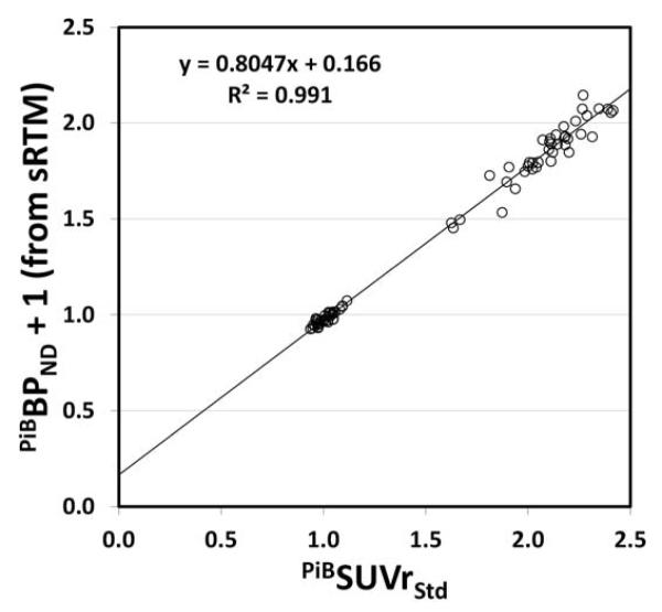
Although amyloid imaging with PiB-PET ([C-11]Pittsburgh Compound-B positron emission tomography), and now with F-18-labeled tracers, has produced remarkably consistent qualitative findings across a large number of centers, there has been considerable variability in the exact numbers reported as quantitative outcome measures of tracer retention. In some cases this is as trivial as the choice of units, in some cases it is scanner dependent, and of course, different tracers yield different numbers. Our working group was formed to standardize quantitative amyloid imaging measures by scaling the outcome of each particular analysis method or tracer to a 0 to 100 scale, anchored by young controls (≤ 45 years) and typical Alzheimer's disease patients. The units of this scale have been named "Centiloids." Basically, we describe a "standard" method of analyzing PiB PET data and then a method for scaling any "nonstandard" method of PiB PET analysis (or any other tracer) to the Centiloid scale.
Keywords: Amyloid imaging; Centiloid scale; Pittsburgh compound B; Positron emission tomography; Standardize.
Copyright © 2015 The Alzheimer's Association. Published by Elsevier Inc. All rights reserved.
Figures
References
-
- Frisoni GB, Jack CR. Harmonization of magnetic resonance-based manual hippocampal segmentation: a mandatory step for wide clinical use. Alzheimers Dement. 2011;7:171–174. - PubMed
-
- Jack CR, Jr., Barkhof F, Bernstein MA, Cantillon M, Cole PE, Decarli C, Dubois B, Duchesne S, Fox NC, Frisoni GB, Hampel H, Hill DL, Johnson K, Mangin JF, Scheltens P, Schwarz AJ, Sperling R, Suhy J, Thompson PM, Weiner M, Foster NL. Steps to standardization and validation of hippocampal volumetry as a biomarker in clinical trials and diagnostic criterion for Alzheimer’s disease. Alzheimers Dement. 2011;7:474–485. e4741. - PMC - PubMed
-
- Boccardi M, Bocchetta M, Ganzola R, Robitaille N, Redolfi A, Duchesne S, Jack CR, Jr, Frisoni GB. Operationalizing protocol differences for EADC-ADNI manual hippocampal segmentation. Alzheimers Dement. 2013 (in press): DOI 10.1016/j.jalz.2013.03.001. - PubMed
-
- Frisoni GB, Bocchetta M, Chetelat G, Rabinovici GD, de Leon MJ, Kaye J, Reiman EM, Scheltens P, Barkhof F, Black SE, Brooks DJ, Carrillo MC, Fox NC, Herholz K, Nordberg A, Jack CR, Jr., Jagust WJ, Johnson KA, Rowe CC, Sperling RA, Thies W, Wahlund LO, Weiner MW, Pasqualetti P, Decarli C. Imaging markers for Alzheimer disease: Which vs how. Neurology. 2013;81:487–500. - PMC - PubMed
-
- Vanderstichele H, Bibl M, Engelborghs S, Le Bastard N, Lewczuk P, Molinuevo JL, Parnetti L, Perret-Liaudet A, Shaw LM, Teunissen C, Wouters D, Blennow K. Standardization of preanalytical aspects of cerebrospinal fluid biomarker testing for Alzheimer’s disease diagnosis: a consensus paper from the Alzheimer’s Biomarkers Standardization Initiative. Alzheimers Dement. 2012;8:65–73. - PubMed
Publication types
MeSH terms
Substances
Grants and funding
LinkOut - more resources
Full Text Sources
Other Literature Sources
Medical
