

Subjective and objective screening tests for hydroxychloroquine toxicity
- PMID: 25444344
- PMCID: PMC8356134
- DOI: 10.1016/j.ophtha.2014.07.056
Subjective and objective screening tests for hydroxychloroquine toxicity
Abstract
Objective: To compare subjective and objective clinical tests used in the screening for hydroxychloroquine retinal toxicity to multifocal electroretinography (mfERG) reference testing.
Design: Prospective, single-center, case control study.
Participants: Fifty-seven patients with a previous or current history of hydroxychloroquine treatment of more than 5 years' duration.
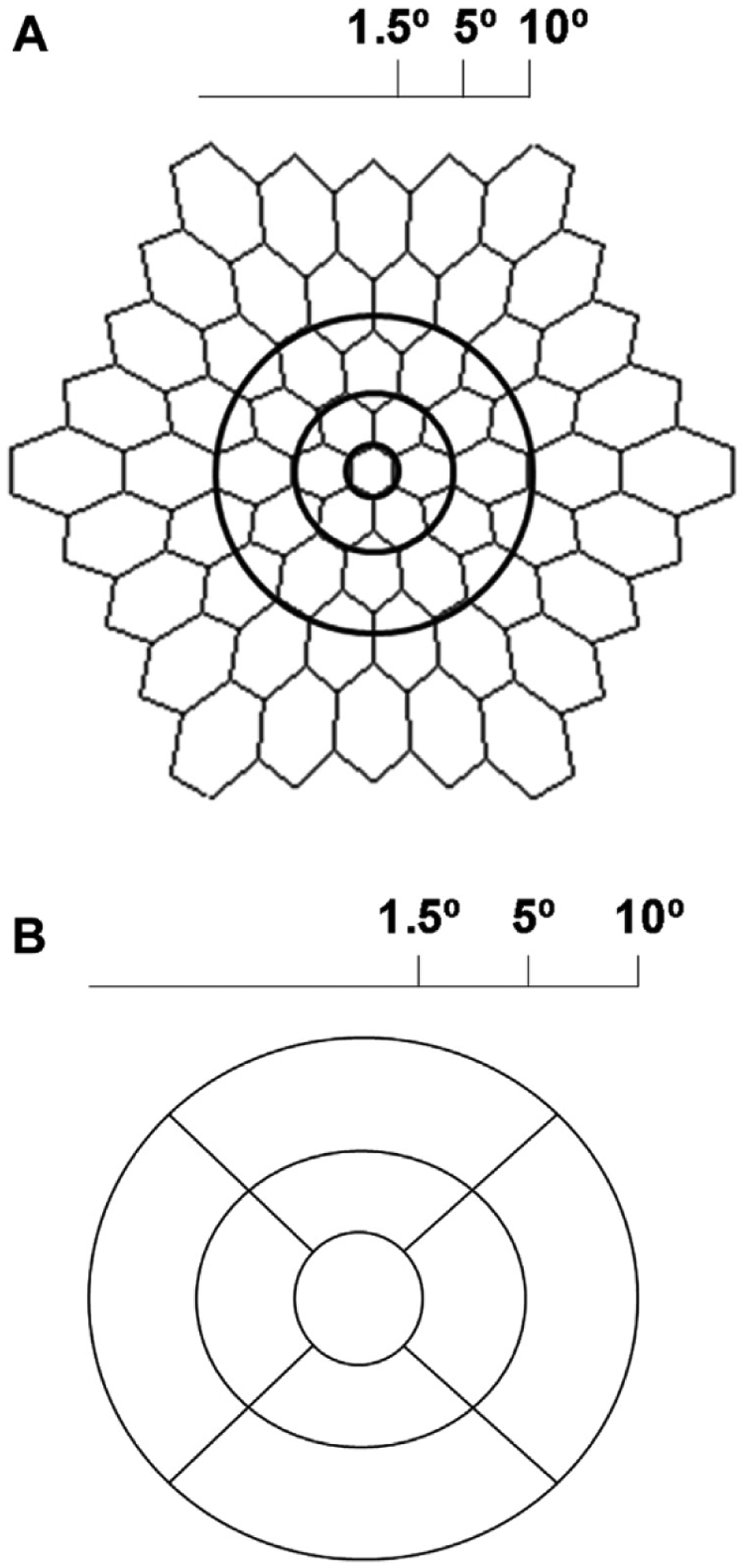
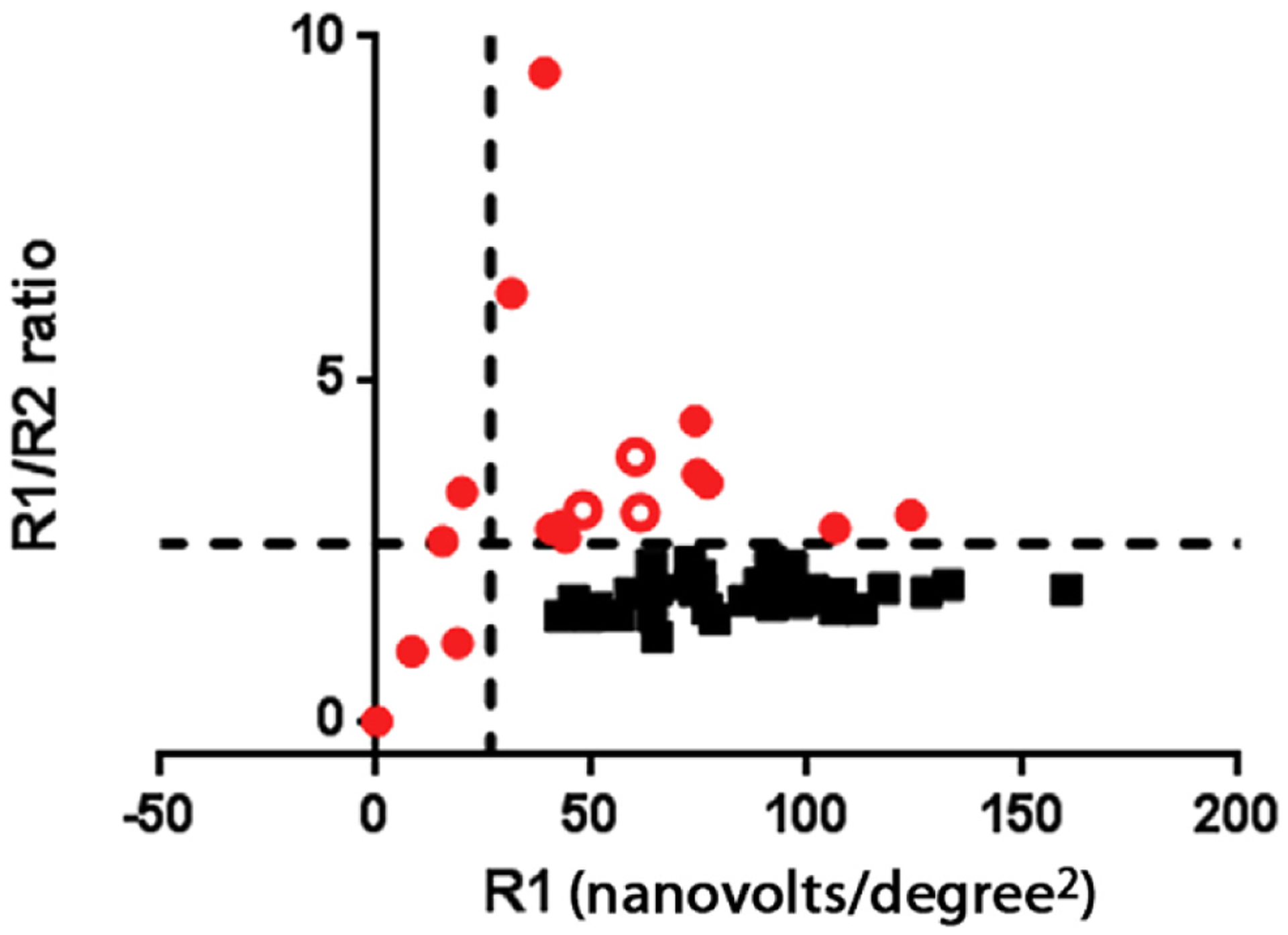
Methods: Participants were evaluated with a detailed medical history, dilated ophthalmologic examination, color fundus photography, fundus autofluorescence (FAF) imaging, spectral-domain (SD) optical coherence tomography (OCT), automated visual field testing (10-2 visual field mean deviation [VFMD]), and mfERG testing. We used mfERG test parameters as a gold standard to divide participants into 2 groups: those affected by hydroxychloroquine-induced retinal toxicity and those unaffected.
Main outcome measures: We assessed the association of various imaging and psychophysical variables in the affected versus the unaffected group.
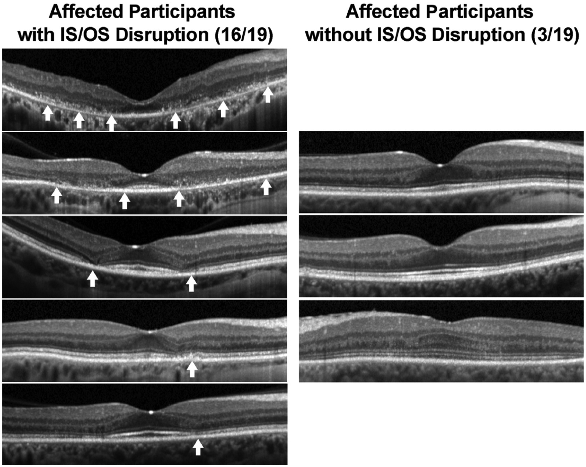
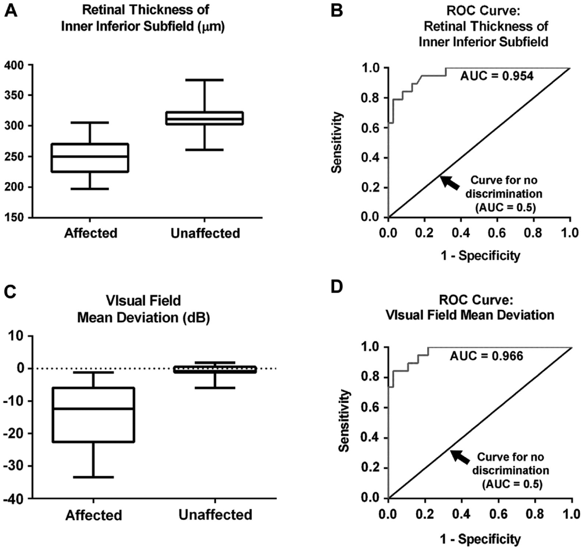
Results: Fifty-seven study participants (91.2% female; mean age, 55.7±10.4 years; mean duration of hydroxychloroquine treatment, 15.0±7.5 years) were divided into affected (n = 19) and unaffected (n = 38) groups based on mfERG criteria. Mean age and duration of hydroxychloroquine treatment did not differ statistically between groups. Mean OCT retinal thickness measurements in all 9 macular subfields were significantly lower (<40 μm) in the affected group (P < 0.01 for all comparisons) compared with those in the unaffected group. Mean VFMD was 11 dB lower in the affected group (P < 0.0001). Clinical features indicative of retinal toxicity were scored for the 2 groups and were detected in 68.4% versus 0.0% using color fundus photographs, 73.3% versus 9.1% using FAF images, and 84.2% versus 0.0% on the scoring for the perifoveal loss of the photoreceptor ellipsoid zone on SD-OCT for affected and unaffected participants, respectively. Using a polynomial modeling approach, OCT inner ring retinal thickness measurements and Humphrey 10-2 VFMD were identified as the variables associated most strongly with the presence of hydroxychloroquine as defined by mfERG testing.
Conclusions: Optical coherence tomography retinal thickness and 10-2 VFMD are objective measures demonstrating clinically useful sensitivity and specificity for the detection of hydroxychloroquine toxicity as identified by mfERG, and thus may be suitable surrogate tests.
Published by Elsevier Inc.
Conflict of interest statement
Financial Disclosure(s):
The author(s) have no proprietary or commercial interest in any materials discussed in this article.
Figures
Similar articles
-
Comparison of screening procedures in hydroxychloroquine toxicity.Arch Ophthalmol. 2012 Apr;130(4):461-9. doi: 10.1001/archophthalmol.2011.371. Epub 2011 Dec 12. Arch Ophthalmol. 2012. PMID: 22159170
-
Optical Coherence Tomography Minimum Intensity as an Objective Measure for the Detection of Hydroxychloroquine Toxicity.Invest Ophthalmol Vis Sci. 2018 Apr 1;59(5):1953-1963. doi: 10.1167/iovs.17-22668. Invest Ophthalmol Vis Sci. 2018. PMID: 29677357 Free PMC article.
-
The Relationship of Hydroxychloroquine Retinopathy Progression to Stage at Cessation of Therapy.Am J Ophthalmol. 2025 Sep;277:335-348. doi: 10.1016/j.ajo.2025.05.030. Epub 2025 May 28. Am J Ophthalmol. 2025. PMID: 40447243
-
Hydroxychloroquine Retinopathy in the Era of Advanced Imaging Modalities.Int Ophthalmol Clin. 2020 Winter;60(1):73-83. doi: 10.1097/IIO.0000000000000294. Int Ophthalmol Clin. 2020. PMID: 31855897 Review. No abstract available.
-
Hydroxychloroquine: A Brief Review on Screening, Toxicity, and Progression.Ophthalmic Surg Lasers Imaging Retina. 2016 Mar;47(3):207-17. doi: 10.3928/23258160-20160229-02. Ophthalmic Surg Lasers Imaging Retina. 2016. PMID: 26985794 Review. No abstract available.
Cited by
-
Machine Learning-Based Automated Detection of Hydroxychloroquine Toxicity and Prediction of Future Toxicity Using Higher-Order OCT Biomarkers.Ophthalmol Retina. 2022 Dec;6(12):1241-1252. doi: 10.1016/j.oret.2022.05.031. Epub 2022 Jun 9. Ophthalmol Retina. 2022. PMID: 35691579 Free PMC article.
-
Early morpho-functional changes in patients treated with hydroxychloroquine: a prospective cohort study.Graefes Arch Clin Exp Ophthalmol. 2018 Nov;256(11):2201-2210. doi: 10.1007/s00417-018-4103-9. Epub 2018 Aug 27. Graefes Arch Clin Exp Ophthalmol. 2018. PMID: 30151601
-
Development of a chiral HPLC method for the separation and quantification of hydroxychloroquine enantiomers.Sci Rep. 2021 Apr 13;11(1):8017. doi: 10.1038/s41598-021-87511-5. Sci Rep. 2021. PMID: 33850241 Free PMC article.
-
Fundus autofluorescence imaging: systematic review of test accuracy for the diagnosis and monitoring of retinal conditions.Eye (Lond). 2017 Jul;31(7):995-1007. doi: 10.1038/eye.2017.19. Epub 2017 Mar 10. Eye (Lond). 2017. PMID: 28282065 Free PMC article.
-
Retinal Diseases that Can Masquerade as Neurological Causes of Vision Loss.Curr Neurol Neurosci Rep. 2020 Sep 15;20(11):51. doi: 10.1007/s11910-020-01071-1. Curr Neurol Neurosci Rep. 2020. PMID: 32930896 Free PMC article. Review.
References
-
- Marmor MF, Kellner U, Lai TY, et al.; American Academy of Ophthalmology. Revised recommendations on screening for chloroquine and hydroxychloroquine retinopathy. Ophthalmology 2011;118:415–22. - PubMed
-
- Kellner S, Kellner U, Weber BH, et al.Lipofuscin- and melanin-related fundus autofluorescence in patients with ABCA4-associated retinal dystrophies. Am J Ophthalmol 2009;147:895–902. - PubMed
-
- Marmor MF. Comparison of screening procedures in hydroxychloroquine toxicity. Arch Ophthalmol 2012;130:461–9. - PubMed
-
- Browning DJ. Impact of the revised American Academy of Ophthalmology guidelines regarding hydroxychloroquine screening on actual practice. Am J Ophthalmol 2013; 155: 418–28. - PubMed
-
- Marmor MF. Efficient and effective screening for hydroxychloroquine toxicity. Am J Ophthalmol 2013; 155: 413–4. - PubMed
Publication types
MeSH terms
Substances
Grants and funding
LinkOut - more resources
Full Text Sources
Medical
Miscellaneous
