Review
. 2015 Feb;39(1):44-54.
doi: 10.1053/j.semperi.2014.10.009. Epub 2014 Nov 28.

Recent advances of genomic testing in perinatal medicine

Affiliations
Review

Recent advances of genomic testing in perinatal medicine

David G Peters et al. Semin Perinatol. 2015 Feb.

Abstract

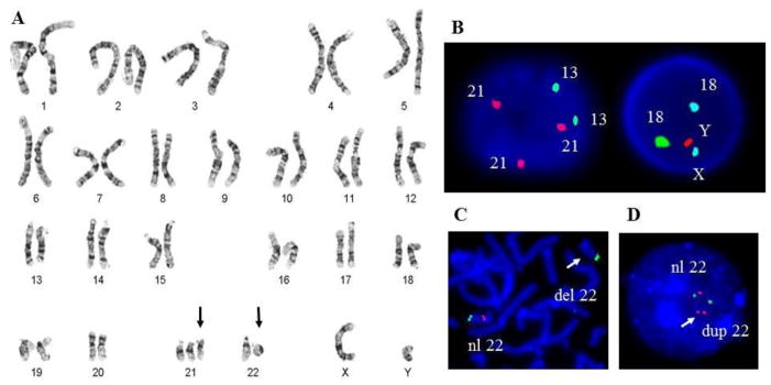
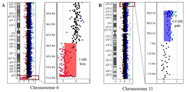
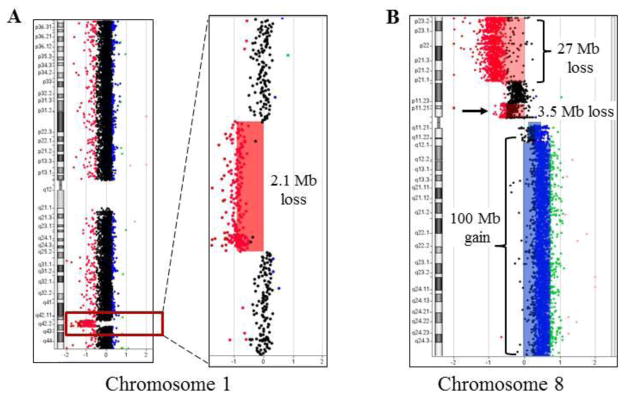
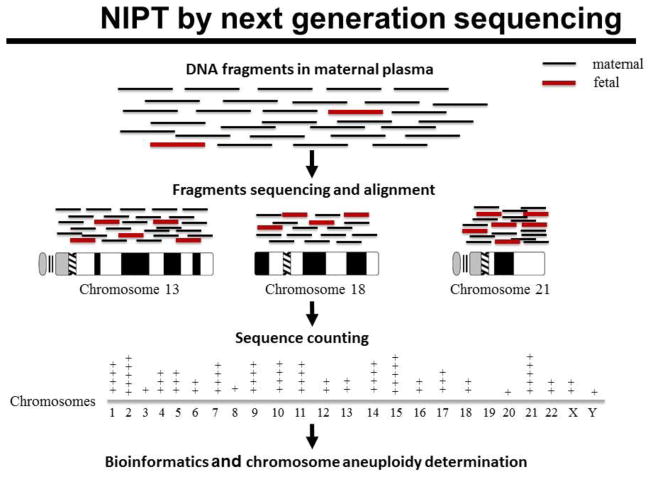
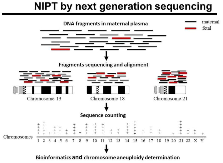
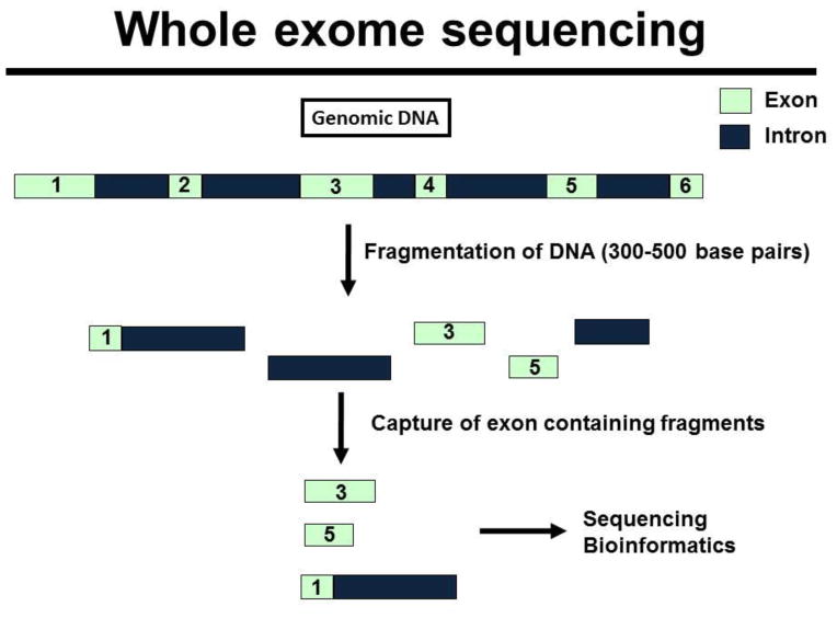
Rapid progress in genomic medicine in recent years has made it possible to diagnose subtle genetic abnormalities in a clinical setting on routine basis. This has allowed for detailed genotype-phenotype correlations and the identification of the genetic basis of many congenital anomalies. In addition to the availability of chromosomal microarray analysis, exome and whole-genome sequencing on pre- and postnatal samples of cell-free DNA has revolutionized the field of prenatal diagnosis. Incorporation of these technologies in perinatal pathology is bound to play a major role in coming years. In this communication, we briefly present the current experience with use of classical chromosome analysis, fluorescence in situ hybridization, and microarray testing, development of whole-genome analysis by next-generation sequencing technology, offer a detailed review of the history and current status of non-invasive prenatal testing using cell-free DNA, and discuss the advents of these new genomic technologies in perinatal medicine.


Figures

Fig 1
Fig 1. Prenatal and postnatal diagnosis of chromosome abnormalities
A. Classical chromosome analysis of product of conception. G-banded karyotype at the 500-band resolution showing a trisomy 21 (black arrow) and an abnormal chromosome 22 (black arrow) with a deletion in the long arm in a male fetus. B. Aneuvysion FISH analysis on the interphase cells showing two signals for chromosome 13 and 18, one signal for each chromosome X and Y, and three signals for chromosome 21- specific probe, indicating a trisomy 21 in a male fetus. C. FISH analysis on a metaphase spread showing a deletion (white arrow) in the DiGeorge/Velocardiofacial critical region on 22q11.2 in a newborn with congenital heart defect. D. Interphase FISH analysis demonstrated duplication (white arrow) in the 22q11.2 region in a stillborn.
Fig 2
Fig 2. Detection of complex submicroscopic chromosome abnormalities using whole genome microarray in a neonate with multiple congenital anomalies and normal karyotype
A. On the left, idiogram and array-CGH plot of chromosome 6, showing a loss (red box) in the subtelomeric region of the long arm (6q27). On the right, a magnified view of chromosome 6 array-CGH plot demonstrates a loss (red shaded area) of ~3,000,000 base pairs (3 Mb). B. On the left, idiogram and array-CGH plot of chromosome 11, showing a gain (red box) in the terminal 11p region. On the right, a magnified view of chromosome 11p with a gain (blue shaded area) of ~900,000 base pairs (0.9 Mb). These results indicate the presence of a derivative chromosome 6 resulted from a translocation of 6q and 11p in the child.
Fig 3
Fig 3
A. Microarray analysis reveals submicroscopic chromosome alteration in a newborn with brain malformation. On the left, idiogram and array-CGH plot of chromosome 1, showing a loss (red box) in the 1q42-q43 chromosome region. On the right, a magnified view of a ~2.1 Mb deletion (red shaded area). B. Microarray analysis from a spontaneous abortion. Karyotype analysis was not available as the cells failed to grow in culture. Array CGH analysis showed complex alterations of chromosome 8, including a loss of ~27 Mb in short arm, a loss of ~3.5 Mb in the pericentromeric region of the 8p, and a gain of entire long arm of chromosome 8 (~100 Mb). These results are consistent with an isochromosome 8q in the fetus.
Fig 4
Fig 4. Regions of homozygosity detected by CGH+SNP combo array analysis can reveal copy number neutral chromosomal abnormalities
A. Contiguous regions of homozygosity (blue shaded areas) detected in a newborn with severe IUGR consistent with consanguineous parents and an increased risk for autosomal recessive disorder. B. CGH+SNP analysis in a newborn with hypotonia and undescended testes reveal absence of heterozygosity for the entire chromosome 15 (blue shaded area), consistent with uniparental disomy for chromosome 15 (UPD15).
Fig 5
Fig 5. Overview of noninvasive aneuploidy detection using next-generation sequencing of maternal plasma
Maternal plasma contains small fraction of fetal DNA (red fragments), while maternal DNA (black fragments) represent 80–95% of circulating DNA. Both maternal and fetal fragments, obtained from maternal plasma sample, are sequenced and aligned to a specific chromosome positions according to the human reference genome. Using bioinformatics analysis, the total number of sequences mapped to each chromosome is counted and quantified to assess copy number for each chromosome. The number of DNA fragments from an aneuploid chromosome is expected to be higher (in cases with trisomy) and lower (in pregnancies with monosomy) in comparison to a normal diploid fetus.
Fig 6
Fig 6. Schematic illustration of the whole exome sequencing
In whole exome sequencing, genomic DNA is fragmented, and specially designed baits are used to capture fragments of DNA that contain exons. Exons constitute only 1% of genomic DNA. The exon containing fragments are eluted, amplified by PCR and sequenced. Large amounts of data are generated and require specialized programs to align the sequences against the reference genome, to determine nucleotide variants that differ from the reference, and to identify potentially pathogenic mutations.

Similar articles

Cited by

References

    1. MacDorman MF, Kirmeyer S. Fetal and perinatal mortality, United States, 2005. Natl Vital Stat Rep. 2009;57:1–19. - PubMed
    1. Carvalho CM, Zhang F, Lupski JR. Evolution in health and medicine Sackler colloquium: Genomic disorders: a window into human gene and genome evolution. Proc Natl Acad Sci U S A. 2010;107(Suppl 1):1765–1771. - PMC - PubMed
    1. Schwartz S. Clinical utility of single nucleotide polymorphism arrays. Clin Lab Med. 2011;31:581–594. - PubMed
    1. Manning M, Hudgins L Professional Practice and Guidelines Committee. Array-based technology and recommendations for utilization in medical genetics practice for detection of chromosomal abnormalities. Genet Med. 2010;12:742–745. - PMC - PubMed
    1. Miller DT, Adam MP, Aradhya S, et al. Consensus statement: chromosomal microarray is a first-tier clinical diagnostic test for individuals with developmental disabilities or congenital anomalies. Am J Hum Genet. 2010;86:749–764. - PMC - PubMed