

Corneal Regeneration After Photorefractive Keratectomy: A Review
- PMID: 25444646
- PMCID: PMC4502084
- DOI: 10.1016/j.optom.2014.09.001
Corneal Regeneration After Photorefractive Keratectomy: A Review
Abstract
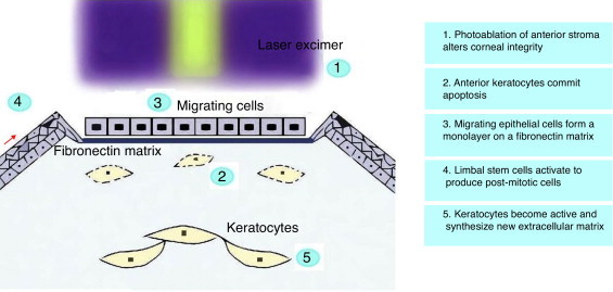
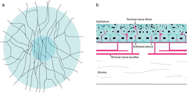
Photorefractive keratectomy (PRK) remodels corneal stroma to compensate refractive errors. The removal of epithelium and the ablation of stroma provoke the disruption of corneal nerves and a release of several peptides from tears, epithelium, stroma and nerves. A myriad of cytokines, growth factors, and matrix metalloproteases participate in the process of corneal wound healing. Their balance will determine if reepithelization and stromal remodeling are appropriate. The final aim is to achieve corneal transparency for restoring corneal function, and a proper visual quality. Therefore, wound-healing response is critical for a successful refractive surgery. Our goal is to provide an overview into how corneal wounding develops following PRK. We will also review the influence of intraoperative application of mitomycin C, bandage contact lenses, anti-inflammatory and other drugs in preventing corneal haze and post-PRK pain.
La queratectomía fotorrefractiva (PRK) remodela el estroma de la córnea para compensar los errores refractivos. La eliminación del epitelio y la ablación del estroma provoca la alteración de los nervios corneales y la liberación de diversos péptidos de la lágrima, epitelio, estroma y nervios. Innumerables citoquinas, factores de crecimiento y metaloproteasas de la matriz participan en el proceso de regeneración y cicatrización corneal. Su equilibrio determinará si la re-epitelización y la remodelación del estroma son adecuados. El objetivo final es el logro de la transparencia corneal para restablecer la función de la córnea, así como la calidad visual adecuada. Por tanto, la respuesta de regeneración y cicatrización corneal es esencial para el éxito de la cirugía refractiva. Nuestro objetivo es aportar una visión general sobre el modo en que se desarrolla dicho proceso tras la PRK. Revisaremos también la influencia de la aplicación intraoperatoria de mitomicina C, lentes de contacto terapéuticas, y otros fármacos para prevenir el haze y el dolor tras la PRK.
Keywords: Contact lenses; Cornea; Curación de heridas; Córnea; Lentes de contacto; Photorefractive keratectomy; Queratectomía fotorrefractiva; Wound healing.
Copyright © 2014 Spanish General Council of Optometry. Published by Elsevier Espana. All rights reserved.
Figures
References
-
- Ghirlando A., Gambato C., Midena E. LASEK and photorefractive keratectomy for myopia: clinical and confocal microscopy comparison. J Refract Surg. 2007;23:694–702. - PubMed
-
- Ginis H., Pentari I., de Brouwere D., Bouzoukis D., Naoumidi I., Pallikaris I. Narrow angle light scatter in rabbit corneas after excimer laser surface ablation. Ophthalmic Physiol Opt. 2009;29:357–362. - PubMed
-
- Erie J.C., Patel S.V., McLaren J.W., Hodge D.O., Bourne W.M. Corneal keratocyte deficits after photorefractive keratectomy and laser in situ keratomileusis. Am J Ophthalmol. 2006;141:799–809. - PubMed
Publication types
MeSH terms
Substances
LinkOut - more resources
Full Text Sources
Other Literature Sources
Medical
