. 2014 Oct 31;453(4):710-21.
doi: 10.1016/j.bbrc.2014.09.144. Epub 2014 Oct 14.

Cdk1, PKCδ and calcineurin-mediated Drp1 pathway contributes to mitochondrial fission-induced cardiomyocyte death

Affiliations

Cdk1, PKCδ and calcineurin-mediated Drp1 pathway contributes to mitochondrial fission-induced cardiomyocyte death

Ivan Zaja et al. Biochem Biophys Res Commun. .

Abstract

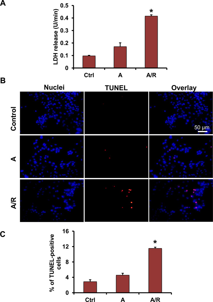
Myocardial ischemia-reperfusion (I/R) injury is one of the leading causes of death and disability worldwide. Mitochondrial fission has been shown to be involved in cardiomyocyte death. However, molecular machinery involved in mitochondrial fission during I/R injury has not yet been completely understood. In this study we aimed to investigate molecular mechanisms of controlling activation of dynamin-related protein 1 (Drp1, a key protein in mitochondrial fission) during anoxia-reoxygenation (A/R) injury of HL1 cardiomyocytes. A/R injury induced cardiomyocyte death accompanied by the increases of mitochondrial fission, reactive oxygen species (ROS) production and activated Drp1 (pSer616 Drp1), and decrease of inactivated Drp1 (pSer637 Drp1) while mitochondrial fusion protein levels were not significantly changed. Blocking Drp1 activity with mitochondrial division inhibitor mdivi1 attenuated cell death, mitochondrial fission, and Drp1 activation after A/R. Trolox, a ROS scavenger, decreased pSer616 Drp1 level and mitochondrial fission after A/R. Immunoprecipitation assay further indicates that cyclin dependent kinase 1 (Cdk1) and protein kinase C isoform delta (PKCδ) bind Drp1, thus increasing mitochondrial fission. Inhibiting Cdk1 and PKCδ attenuated the increases in pSer616 Drp1, mitochondrial fission, and cardiomyocyte death. FK506, a calcineurin inhibitor, blocked the decrease in expression of inactivated pSer637 Drp1 and mitochondrial fission. Our findings reveal the following novel molecular mechanisms controlling mitochondrial fission during A/R injury of cardiomyocytes: (1) ROS are upstream initiators of mitochondrial fission; and (2) the increased mitochondrial fission is resulted from both increased activation and decreased inactivation of Drp1 through Cdk1, PKCδ, and calcineurin-mediated pathways, respectively.

Keywords: Cardiomyocyte; Ischemia–reperfusion injury; Mitochondrial dynamics; Reactive oxygen species.


Figures

Fig. 1
Fig. 1
A/R injury induces cardiomyocyte death and increases mitochondrial fission in cardiomyocytes. (A) A/R significantly induced cardiomyocyte death as evidenced by the increase of lactate-dehydrogenase (LDH) release from cells. (B and C) Confocal images of TUNEL staining (red) was used to identify DNA damage after A/R. Hoechst 33342 stains nuclei (blue). Overlaid images demonstrate that most of TUNEL signals were located in fragmented nuclei in A/R-treated cells. Significant increase in TUNEL-positive cells was observed in A/R-treated cells compared to the non-treated control (* P<0.001 vs. Ctrl, n=3). (D and E) Confocal images of mitochondria stained with TMRE show the elongated, branched, and interconnected mitochondrial networks in non-treated control compared to A/R-treated cells, which mostly displayed round, punctiforme and small mitochondria fragments. Quantification of mitochondria length and branching based on the confocal images of mitochodria showed that significant decreases of aspect ratio and form factor values were observed in A/Rtreated cells (* P<0.001 vs. Ctrl, n=4). (F and G) Western blot assay demonstrates that a decrease in inactivated Drp1 (pSer637 Drp1) was observed in A and in A/R treated cells while an increase in activated Drp1 (pSer616 Drp1) levels was observed only in A/R-treated group (* P<0.05 vs. Ctrl, n=4). (H and I) Western blot analysis showed that there was no significant difference in fusion protein (MFN1, MFN2, and OPA1) levels between non-treated control, A and A/R-treated cells.
Fig. 1
Fig. 1
A/R injury induces cardiomyocyte death and increases mitochondrial fission in cardiomyocytes. (A) A/R significantly induced cardiomyocyte death as evidenced by the increase of lactate-dehydrogenase (LDH) release from cells. (B and C) Confocal images of TUNEL staining (red) was used to identify DNA damage after A/R. Hoechst 33342 stains nuclei (blue). Overlaid images demonstrate that most of TUNEL signals were located in fragmented nuclei in A/R-treated cells. Significant increase in TUNEL-positive cells was observed in A/R-treated cells compared to the non-treated control (* P<0.001 vs. Ctrl, n=3). (D and E) Confocal images of mitochondria stained with TMRE show the elongated, branched, and interconnected mitochondrial networks in non-treated control compared to A/R-treated cells, which mostly displayed round, punctiforme and small mitochondria fragments. Quantification of mitochondria length and branching based on the confocal images of mitochodria showed that significant decreases of aspect ratio and form factor values were observed in A/Rtreated cells (* P<0.001 vs. Ctrl, n=4). (F and G) Western blot assay demonstrates that a decrease in inactivated Drp1 (pSer637 Drp1) was observed in A and in A/R treated cells while an increase in activated Drp1 (pSer616 Drp1) levels was observed only in A/R-treated group (* P<0.05 vs. Ctrl, n=4). (H and I) Western blot analysis showed that there was no significant difference in fusion protein (MFN1, MFN2, and OPA1) levels between non-treated control, A and A/R-treated cells.
Fig. 2
Fig. 2
Inhibiting mitochondrial fission reduces cardiomyocyte death after A/R. (A) Mdivi1, a Drp1 inhibitor, dose-dependently attenuated A/R-induced LDH release. (B and C) A significant increase in TUNEL-positive cells after A/R was attenuated with 50 µM mdivi1 treatment (* P<0.001 vs. Ctrl, # P<0.05 vs. A/R, n=3).
Fig. 3
Fig. 3
An increase in reactive oxygen species (ROS) production during reoxygenation causes activation of Drp1. (A and B) An increase of pSer616 Drp1 after A/R was observed compared to non-treated control analyzed using Western blot. Mdivi1 partially and Trolox completely abolished the increase in the level of activated pSer616 Drp1 after A/R (* P<0.001 vs. Ctrl, # P<0.05 vs. A/R, n=3). (C and D) ROS production was analyzed through confocal images of the cells stained with ROS-sensitive dye CM-H2DCFDA. A significant increase in DCF fluorescence (indicating ROS production) in A/R-treated group was abolished if cells were treated with ROS scavenger Trolox (* P<0.001 vs. Ctrl, # P<0.001 vs. A/R, n=3). (E to H) pSer637 Drp1 expression was decreased after A/R compared to non-treated control. Pretreatment with calcineurin inhibitor FK506 during A/R attenuated the decrease of pSer637 Drp1 after A/R while Trolox did not influence pSer637 Drp1 expression (n=3). (I and J) Confocal images of mitochondria stained with TMRE show that that pretreatment of the cells with mdivi1, Trolox, or FK506 attenuated A/R-induced mitochondrial fission as evidenced by the increased aspect ratio and form factor values compared to A/R alone group (* P<0.05 vs. Ctrl, # P<0.05 vs. A/R, n=4).
Fig. 3
Fig. 3
An increase in reactive oxygen species (ROS) production during reoxygenation causes activation of Drp1. (A and B) An increase of pSer616 Drp1 after A/R was observed compared to non-treated control analyzed using Western blot. Mdivi1 partially and Trolox completely abolished the increase in the level of activated pSer616 Drp1 after A/R (* P<0.001 vs. Ctrl, # P<0.05 vs. A/R, n=3). (C and D) ROS production was analyzed through confocal images of the cells stained with ROS-sensitive dye CM-H2DCFDA. A significant increase in DCF fluorescence (indicating ROS production) in A/R-treated group was abolished if cells were treated with ROS scavenger Trolox (* P<0.001 vs. Ctrl, # P<0.001 vs. A/R, n=3). (E to H) pSer637 Drp1 expression was decreased after A/R compared to non-treated control. Pretreatment with calcineurin inhibitor FK506 during A/R attenuated the decrease of pSer637 Drp1 after A/R while Trolox did not influence pSer637 Drp1 expression (n=3). (I and J) Confocal images of mitochondria stained with TMRE show that that pretreatment of the cells with mdivi1, Trolox, or FK506 attenuated A/R-induced mitochondrial fission as evidenced by the increased aspect ratio and form factor values compared to A/R alone group (* P<0.05 vs. Ctrl, # P<0.05 vs. A/R, n=4).
Fig. 4
Fig. 4
A/R injury induces an increase in the expression of pSer616 Drp1 through PKCδ- and Cdk1-mediated pathway in cardiomyocytes. (A) Input lysates, nonspecific IgG, co-immunoprecipitated PKCδ or Cdk1 were detected by western blotting using an anti-Drp1. Both Cdk1 and PKCδ interacted with Drp1 and these interactions were increased in A/R injury. (B) PKCδ siRNA knockdown efficiency in the cardiomyocytes. (C and D) There was a significant increase of pSer616 Drp1 after A/R compared to non-treated control analyzed using Western blot. When added separately, PKCδ siRNA or roscovitine, partially reduced the increase in expression levels of pSer616 Drp1. Adding both PKCδ siRNA and roscovitine during A/R completely abolished the increase of pSer616 Drp1 (* P<0.001 vs. Ctrl, # P<0.05 vs. A/R, † P<0.05 vs. A/R+PKCδ siRNA and A/R+roscovitine, n=5). (E) Representative confocal images of mitochondria stained with TMRE. (F) Mitochondria fission was evaluated through the analysis of confocal images of mitochondria stained with TMRE. Significant decreases of aspect ratio and form factor values were observed in A/R-treated cells, indicating the increased mitochondrial fission. Treatment of the cells with PKCδ siRNA, roscovitine or both attenuated A/R-induced increases of mitochondria fission and increased aspect ratio and form factor values compared to A/R alone group (*P<0.05 vs. Ctrl, # P<0.05 vs. A/R, n=3). (G) PKCδ siRNA or Cdk1 inhibitor roscovitine significantly attenuated LDH release in cardiomyocytes during A/R (*P<0.001 vs. Ctrl, #P<0.05 vs. A/R, n=3).
Fig. 4
Fig. 4
A/R injury induces an increase in the expression of pSer616 Drp1 through PKCδ- and Cdk1-mediated pathway in cardiomyocytes. (A) Input lysates, nonspecific IgG, co-immunoprecipitated PKCδ or Cdk1 were detected by western blotting using an anti-Drp1. Both Cdk1 and PKCδ interacted with Drp1 and these interactions were increased in A/R injury. (B) PKCδ siRNA knockdown efficiency in the cardiomyocytes. (C and D) There was a significant increase of pSer616 Drp1 after A/R compared to non-treated control analyzed using Western blot. When added separately, PKCδ siRNA or roscovitine, partially reduced the increase in expression levels of pSer616 Drp1. Adding both PKCδ siRNA and roscovitine during A/R completely abolished the increase of pSer616 Drp1 (* P<0.001 vs. Ctrl, # P<0.05 vs. A/R, † P<0.05 vs. A/R+PKCδ siRNA and A/R+roscovitine, n=5). (E) Representative confocal images of mitochondria stained with TMRE. (F) Mitochondria fission was evaluated through the analysis of confocal images of mitochondria stained with TMRE. Significant decreases of aspect ratio and form factor values were observed in A/R-treated cells, indicating the increased mitochondrial fission. Treatment of the cells with PKCδ siRNA, roscovitine or both attenuated A/R-induced increases of mitochondria fission and increased aspect ratio and form factor values compared to A/R alone group (*P<0.05 vs. Ctrl, # P<0.05 vs. A/R, n=3). (G) PKCδ siRNA or Cdk1 inhibitor roscovitine significantly attenuated LDH release in cardiomyocytes during A/R (*P<0.001 vs. Ctrl, #P<0.05 vs. A/R, n=3).

References

    1. Brookes PS, Yoon Y, Robotham JL, Anders MW, Sheu SS. Calcium, ATP, and ROS: a mitochondrial love-hate triangle. Am J Physiol Cell Physiol. 2004;287:C817–C833. - PubMed
    1. Halestrap AP. Calcium, mitochondria and reperfusion injury: a pore way to die. Biochem Soc Trans. 2006;34:232–237. - PubMed
    1. Ong SB, Hall AR, Hausenloy DJ. Mitochondrial dynamics in cardiovascular health and disease. Antioxid Redox Signal. 2013;19:400–414. - PMC - PubMed
    1. Ong SB, Subrayan S, Lim SY, Yellon DM, Davidson SM, Hausenloy DJ. Inhibiting mitochondrial fission protects the heart against ischemia/reperfusion injury. Circulation. 2010;121:2012–2022. - PubMed
    1. Chan DC. Mitochondrial fusion and fission in mammals. Annu Rev Cell Dev Biol. 2006;22:79–99. - PubMed

Publication types