

Spatial regulation of controlled bioactive factor delivery for bone tissue engineering
- PMID: 25445719
- PMCID: PMC4428953
- DOI: 10.1016/j.addr.2014.11.018
Spatial regulation of controlled bioactive factor delivery for bone tissue engineering
Abstract
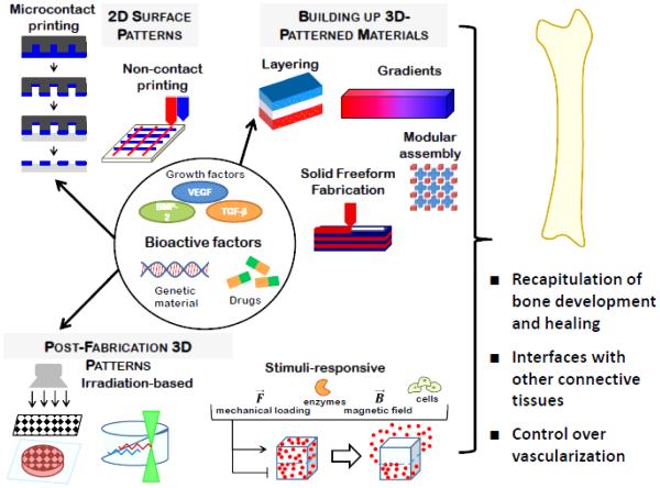
Limitations of current treatment options for critical size bone defects create a significant clinical need for tissue engineered bone strategies. This review describes how control over the spatiotemporal delivery of growth factors, nucleic acids, and drugs and small molecules may aid in recapitulating signals present in bone development and healing, regenerating interfaces of bone with other connective tissues, and enhancing vascularization of tissue engineered bone. State-of-the-art technologies used to create spatially controlled patterns of bioactive factors on the surfaces of materials, to build up 3D materials with patterns of signal presentation within their bulk, and to pattern bioactive factor delivery after scaffold fabrication are presented, highlighting their applications in bone tissue engineering. As these techniques improve in areas such as spatial resolution and speed of patterning, they will continue to grow in value as model systems for understanding cell responses to spatially regulated bioactive factor signal presentation in vitro, and as strategies to investigate the capacity of the defined spatial arrangement of these signals to drive bone regeneration in vivo.
Keywords: Biomaterials patterning; Bone regeneration; Drug delivery; Spatial regulation; Temporal regulation.
Copyright © 2014 Elsevier B.V. All rights reserved.
Figures
References
-
- Giannoudis PV, Dinopoulos H, Tsiridis E. Bone substitutes: an update. Injury. 2005;36(Suppl 3):S20–27. - PubMed
-
- Simon SR. Orthopaedic basic science. Amer Academy of Orthopaedic. 1994
-
- DeCoster TA, Gehlert RJ, Mikola EA, Pirela-Cruz MA. Management of posttraumatic segmental bone defects. The Journal of the American Academy of Orthopaedic Surgeons. 2004;12:28–38. - PubMed
-
- Habal MB, Reddi AH. Bone grafts and bone induction substitutes. Clinics in plastic surgery. 1994;21:525–542. - PubMed
-
- Langer R, Vacanti JP, Vacanti CA, Atala A, Freed LE, Vunjak-Novakovic G. Tissue engineering: biomedical applications. Tissue engineering. 1995;1:151–161. - PubMed
Publication types
MeSH terms
Substances
Grants and funding
LinkOut - more resources
Full Text Sources
Other Literature Sources
Research Materials
