. 2015 Jan 1;397(1):140-9.
doi: 10.1016/j.ydbio.2014.10.020. Epub 2014 Nov 4.

Retinoic acid regulates Kit translation during spermatogonial differentiation in the mouse

Affiliations

Retinoic acid regulates Kit translation during spermatogonial differentiation in the mouse

Jonathan T Busada et al. Dev Biol. .

Abstract

In the testis, a subset of spermatogonia retains stem cell potential, while others differentiate to eventually become spermatozoa. This delicate balance must be maintained, as defects can result in testicular cancer or infertility. Currently, little is known about the gene products and signaling pathways directing these critical cell fate decisions. Retinoic acid (RA) is a requisite driver of spermatogonial differentiation and entry into meiosis, yet the mechanisms activated downstream are undefined. Here, we determined a requirement for RA in the expression of KIT, a receptor tyrosine kinase essential for spermatogonial differentiation. We found that RA signaling utilized the PI3K/AKT/mTOR signaling pathway to induce the efficient translation of mRNAs for Kit, which are present but not translated in undifferentiated spermatogonia. Our findings provide an important molecular link between a morphogen (RA) and the expression of KIT protein, which together direct the differentiation of spermatogonia throughout the male reproductive lifespan.

Keywords: Kit; Retinoic acid; Spermatogenesis; Spermatogonia; Testis; Translation.


Figures

Fig. 1
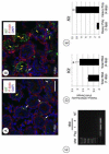
Fig. 1. KIT protein levels increase during neonatal testis development
(A-B) Immunolabeling for KIT on mouse testis sections at 1 dpp (A) or 4 dpp (B). Sections were stained for KIT protein (green), phalloidin-594 was added to visualize F-Actin and outline the testis cords (red), and DAPI was added to visualize nuclei (blue). White arrows indicate KIT+ interstitial cells, and yellow arrows indicate KIT+ germ cells (A-B). (C) Kit amplicons from RT-PCR using total RNA isolated from 2 dpp THY1+ prospermatogonia (Psg), and whole testis lysate of mice aged 1 dpp and 4 dpp. MW = molecular weight ladder, and NT = no template control. (D) Kit mRNA abundance was assessed by qRT-PCR using total testis RNA from 1 and 4 dpp mice. (E) Kit mRNA abundance in heavy polysome fractions. Asterisk indicates statistical significance (P≤0.01). Scale bar (in A) = 40 μm.
Fig. 2
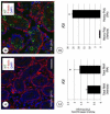
Fig. 2. RA stimulates KIT expression in spermatogonia by increasing mRNA utilization
(A-B) Immunolabeling for KIT on mouse testis sections treated at 1 dpp with DMSO (vehicle (A)) or RA (B) and euthanized 24 hours later. Sections were labeled with anti-KIT (green), phalloidin-594 was added to visualize testis cords (red), and DAPI stained nuclei (blue). (C) Kit mRNA levels were assessed by qRT-PCR from DMSO- and RA-treated mice. (D) Kit mRNA abundance in heavy polysomes was assessed by qRT-PCR using total testis RNA from DMSO and RA dpp mice. Scale bar (in A) = 40 μm. Asterisks indicate statistical significance with P≤0.01.
Fig. 3
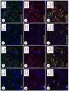
Fig. 3. KIT and STRA8 colocalize in neonatal germ cells
(A-I) Immunolabeling of mouse testis sections from mice aged 1 dpp (A-C) and 4 dpp (D-F) as well as on those from mice treated at 1 dpp with the vehicle (G-I) or RA (J-L) and euthanized 24 hours later. Antibodies were used to detect KIT (green) and STRA8 (red), and F-actin was labeled with phalloidin (blue). Red arrows indicate representative KIT+ germ cells. Green arrows indicate representative STRA8+ germ cells, and yellow arrows indicate double KIT+/STRA8+ germ cells. Scale bar (in A) = 50 μm.
Fig. 4
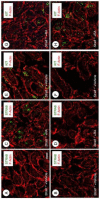
Fig. 4. KIT expression is not dependent upon STRA8
Stra8+/− and Stra8−/− mice were injected with DMSO (A, C, E, G) or RA (B, D, F, H) at 1 dpp and then euthanized 24 hours later. (A-H) Immunolabeling was performed to detect STRA8 (green in A-B and E-F) or KIT (green in C-D and G-H). Phalloidin-594 was used in each section to visualize outline of the testis cords (red). (A-B and E-F) As expected, STRA8 was undetectable in spermatogonia in both Stra8+/− (C) and Stra8−/− (E) testes from DMSO-treated mice. However, RA induced STRA8 in spermatogonia from Stra8+/− (B) but not Stra8−/− (F) mice. (C-D and G-H) KIT protein was undetectable in spermatogonia in both Stra8+/− (C) and Stra8−/− (G) testes from DMSO-treated mice. In contrast, RA induced KIT expression in both Stra8+/− (D) and Stra8−/− (H) testes. Scale bar (in A) = 30 μm.
Fig. 5
Fig. 5. Loss of RA signaling prevents both STRA8 and KIT expression
(A-F) Immunolabeling of mouse testis sections from 4 dpp mice treated with vehicle (A, C, E) or WIN 18,446 (B, D, F). Sections were incubated with antibodies for STRA8 (green in A and B), KIT (green in C and D), or ZBTB16 (green in E and F). Phalloidin-594 was added to visualize testis cords (in red), and nuclei were stained with DAPI (blue). Yellow arrows indicate KIT+ germ cells (C) and white arrows indicate KIT+ interstitial cells (B and D). (G) ZBTB16+ cells quantitated from E-F. (H) Message levels for Stra8 and Kit were determined by qRT-PCR of RNA isolated from whole testis lysate of vehicle- or WIN 18,446-treated mice. Relative mRNA quantities were compared to WIN 18,446-treated samples, which were set at 1. Asterisks indicate statistical significance at P<0.01. Scale bar (in A) = 30 μm.
Fig. 6
Fig. 6. RA signals through PI3K-AKT to stimulate Kit translation
(A) Model of ex vivo testis cultures. Testes are removed from 1 dpp mice cut into pieces and cultured in hanging drops in the presence of the vehicle or RA. (B) Ex vivo testis cultures are pre-treated for 2 hours with inhibitors for either PI3K or AKT followed by treatment with vehicle or RA (+) the inhibitor for 6 hours. (C-F) Immunolabeling was performed on testes cultured with the vehicle or RA for 6 hours. Sections were stained for KIT (Green, C and D) and STRA8 (Red, C-D) or for FOXO1 (Green, E-F). Phalloidin was added to visualize testis cords (Blue in C-D, and Red in E-F). (G-J) Testis cultures were pretreated with inhibitors of PI3K (G,I) or AKT (H,J) for two hours followed by co-culture with the vehicle or RA and inhibitor for 6 hours. Immunolabeling was performed on testis sections stained for FOXO1 (Green G-H) or KIT (Green, I-J) and STRA8 (Red, I-J). Phalloidin was added to visualize testis cords (Blue in G-H, and Red in I-J). Scale bar (in C) = 40 μm.
Fig. 7
Fig. 7. RA may signal spermatogonia differentiation by activating both genomic and non-genomic pathways
(A) Co-immunolabeling was performed for STRA8 (red) and KIT (green) in 4 dpp testes, and phalloidin was added to visualize testis cords (blue). Cords exposed to RA (as indicated by STRA8+/KIT+ spermatogonia) are circled in yellow, and those not exposed are encircled in white. (B) A testis cord, such as the ones shown in A, is depicted in longitudinal form. Regional exposure to RA would result in the differentiation of subpopulations of spermatogonia (yellow bands), while the other areas of the cord are unexposed so that those spermatogonia remain undifferentiated (white band). (C) Diagram of the canonical genomic RA signaling pathway, in which RA regulates transcription by binding to retinoic acid receptors (RAR) at retinoic acid response elements (RARE) in the promoter of target genes such as Stra8. (D) In the proposed alternative non-genomic pathway, RA regulates translation by binding to RARs, which activate the PI3K-AKT-mTOR signaling cascade to stimulate translation of mRNAs such as Kit.

Similar articles

Cited by

References

    1. Al Tanoury Z, Piskunov A, Rochette-Egly C. Vitamin A and retinoid signaling: genomic and nongenomic effects. Journal of lipid research. 2013;54:1761–1775. - PMC - PubMed
    1. Amory JK, Muller CH, Shimshoni JA, Isoherranen N, Paik J, Moreb JS, Amory DW, Sr., Evanoff R, Goldstein AS, Griswold MD. Suppression of spermatogenesis by bisdichloroacetyldiamines is mediated by inhibition of testicular retinoic acid biosynthesis. J Androl. 2011;32:111–119. - PMC - PubMed
    1. Anderson EL, Baltus AE, Roepers-Gajadien HL, Hassold TJ, de Rooij DG, van Pelt AM, Page DC. Stra8 and its inducer, retinoic acid, regulate meiotic initiation in both spermatogenesis and oogenesis in mice. Proc Natl Acad Sci U S A. 2008;105:14976–14980. - PMC - PubMed
    1. Arava Y. Isolation of polysomal RNA for microarray analysis. Methods Mol Biol. 2003;224:79–87. - PubMed
    1. Baltus AE, Menke DB, Hu YC, Goodheart ML, Carpenter AE, de Rooij DG, Page DC. In germ cells of mouse embryonic ovaries, the decision to enter meiosis precedes premeiotic DNA replication. Nat Genet. 2006;38:1430–1434. - PubMed

Publication types

Substances