. 2015 Apr;212(4):479.e1-479.e10.
doi: 10.1016/j.ajog.2014.10.026. Epub 2014 Oct 19.

Metformin inhibits ovarian cancer growth and increases sensitivity to paclitaxel in mouse models

Affiliations

Metformin inhibits ovarian cancer growth and increases sensitivity to paclitaxel in mouse models

Ernst Lengyel et al. Am J Obstet Gynecol. 2015 Apr.

Abstract

Objective: There is increasing preclinical evidence indicating that metformin, a medication commonly used for type 2 diabetes mellitus, may protect against cancer. Motivated by this emerging evidence we asked 2 questions: (1) can metformin prevent ovarian cancer growth by altering metabolism and (2) will metformin increase sensitivity to chemotherapy.

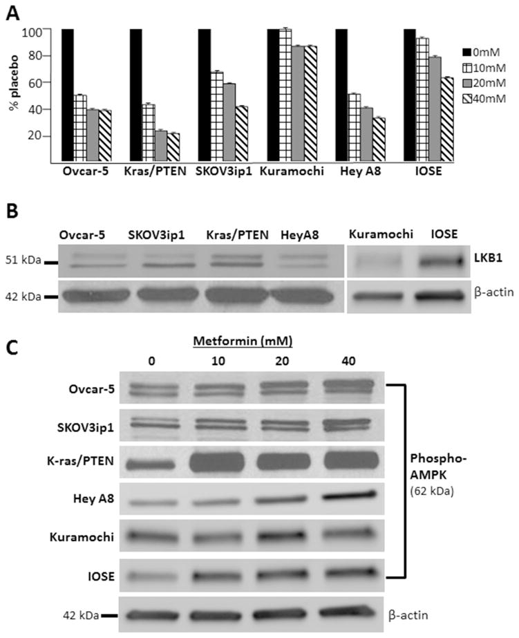
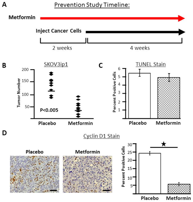
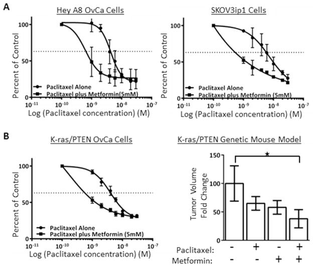
Study design: The effect of metformin in ovarian cancer was tested in vitro and with 2 different mouse models. In vitro, cell lines (n = 6) were treated with metformin (10-40 mmol/L) or phosphate-buffered saline solution and cellular proliferation and metabolic alterations (adenosine monophosphate-activated protein kinase activity, glycolysis, and lipid synthesis) were compared between the 2 groups. In mouse models, a prevention study was performed by treating mice with metformin (250 mg/kg/d intraperitoneally) or placebo for 2 weeks followed by intraperitoneal injection of the SKOV3ip1 human ovarian cancer cell line, and the mean number of tumor implants in each treatment group was compared. In a treatment study, the LSL-K-ras(G12D/+)/PTEN(floxP/floxP) genetic mouse model of ovarian cancer was used. Mice were treated with placebo, paclitaxel (3 mg/kg/wk intraperitoneally for 7 weeks), metformin (100 mg/kg/d in water for 7 weeks), or paclitaxel plus metformin, and tumor volume was compared among treatment groups.

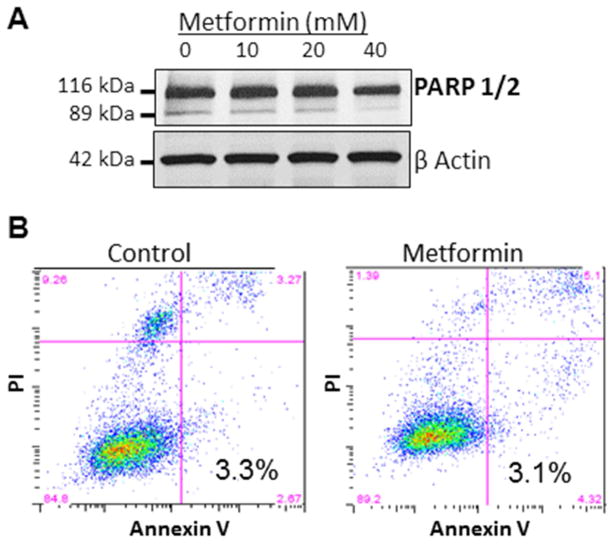
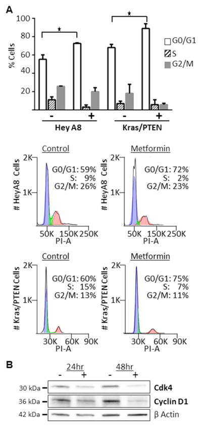
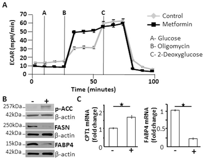
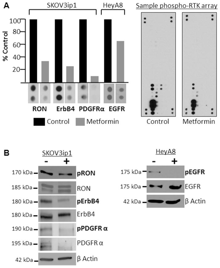
Results: In vitro, metformin decreased proliferation of ovarian cancer cell lines and induced cell cycle arrest, but not apoptosis. Further analysis showed that metformin altered several aspects of metabolism including adenosine monophosphate-activated protein kinase activity, glycolysis, and lipid synthesis. In the prevention mouse model, mice that were pretreated with metformin had 60% fewer tumor implants compared with controls (P < .005). In the treatment study, mice that were treated with paclitaxel plus metformin had a 60% reduction in tumor weight compared with controls (P = .02), which is a level of tumor reduction greater than that resulting from either paclitaxel or metformin alone.

Conclusion: Based on these results, we conclude that metformin alters metabolism in ovarian cancer cells, prevents tumor growth, and increases sensitivity to chemotherapy in vitro and in mouse models. These preclinical findings suggest that metformin warrants further investigation for use as an ovarian cancer therapeutic.

Keywords: cancer; metabolism; metformin; mouse model; ovarian cancer; prevention.


Conflict of interest statement

Disclosure statement: The authors report no conflict of interest.

Figures

Figure 1
Figure 1
Metformin inhibits growth and activates AMPK in ovarian cancer cells. (A) MTT proliferation assays showing that Ovcar-5, Kras/PTEN, SKOV3ip1, Kuramochi, HeyA8 and IOSE cell lines treated with 10, 20 or 40 mM metformin exhibit a dose-dependent reduction in proliferation. Bars represent fold change in proliferation. In 4 out of 6 cell lines the reduction in proliferation is statistically significant (p <0.05) with all metformin concentrations when compared to control, the exceptions are the Kuramochi and IOSE cell lines. (B) Western blots. Protein expression of LKB1 in cell lines. (C) Western blots. Protein expression of phosphorylated (activated) AMPK (Thr 172). B-actin done for each blot, but only shown for the IOSE blot. The indicated cell lines were serum-starved for 24 hours and then treated with 10, 20 or 40 mM metformin or vehicle for 24 hours.
Figure 2
Figure 2
Metformin’s anti-proliferative effect is not a result of apoptosis. (A) Western blot. Protein expression of cleaved and total PARP 1/2. The Ovcar-5 OvCa cell line was treated with 10, 20 or 40 mM metformin or vehicle for 48 hours. (B) Apoptosis analysis. SKOV3ip1 cells were serum-starved for 24 hours before treating with 40 mM metformin for 24 hours. Cells were then stained with propidium iodine (PI) and Annexin V and analyzed using a flow cytometer. The percentage of apoptotic cells is shown.
Figure 3
Figure 3
Cell cycle arrest with metformin treatment. (A) Cell cycle analysis. K-ras/PTEN and HeyA8 cells were serum-starved for 24 hours before treating with 20 mM metformin for 24 hours. Cells were then stained with propidium iodine and analyzed using flow cytometry. Columns, mean percent of cells in each phase of cell cycle; *, P<0.5. Histograms from each cell line are shown. Blue: G0/G1 phase, green: S phase, red: G2/M phase. (B) Western blots. Protein expression of Cdk4 and cyclin D1. The SKOV3ip1 ovarian cancer cell line was treated with 40 mM metformin or vehicle in serum free media for the indicated times.
Figure 4
Figure 4
In ovarian cancer cells, metformin alters glucose and lipid metabolism. (A) Glycolysis levels in cancer cells treated with metformin are increased compared to control. IGROV cells were plated in complete growth medium and treated for 4 hours with 20 mM metformin or vehicle. Glycolytic response of the cell line to pharmacological inhibitors of metabolism was determined by a Seahorse SF96 Extracellular Flux Analyzer. Extracellular acidification rates (ECAR) for the two groups are depicted. (B) Western blots. Protein expression of phosphorylated ACC, fatty acid synthase, and fatty acid binding protein 4. Serum starved Ovcar-5 ovarian cancer cells were treated with 20 mM metformin or vehicle for 48 hours. (C) Quantitative real-time RT-PCR. mRNA levels of the key regulatory enzyme involved in fatty acid oxidation, carnitine palmitoyltransferase, was significantly increased and fatty acid binding protein 4 was significantly decreased with metformin treatment. Ovcar-5 ovarian cancer cells were treated with 20 mM metformin or vehicle for 48 hours, RNA was extracted, and quantitative real-time PCR was performed to determine the level of mRNA. ECAR: extracellular acidification rates, p-ACC: phosphorylated acetyl-coA carboxylase, FASN: fatty acid synthase, FABP4: fatty acid binding protein 4, CPT-1: carnitine palmitoyltransferase. *P<0.05
Figure 5
Figure 5
Metformin inhibits activity of several receptor tyrosine kinases. (A) Receptor tyrosine kinases array. SKOV3ip1 or HeyA8 cells were serum starved for 24 hours and then treated with control or 40 mM metformin for 48 hours prior to performing the arrays. Columns, protein expression level quantified by image J and expressed as percent of control. Image of control and metformin treated arrays. (B) Western blots. Protein expression of phosphorylated and total: RON (Y1238/Y1239), ErbB4 (Tyr1284), PDGFR (Tyr849), and EGFR (Tyr1045). B-actin done for each blot, but only shown for the ErbB4 and EGFR blots. Serum starved SKOV3ip1 and HeyA8 cells were treated with control or 40 mM metformin for 48 hours.
Figure 6
Figure 6
In a preventive study design, metformin inhibits ovarian cancer cell growth. (A) Study timeline. Female athymic nude mice were pretreated with metformin (250 mg/kg/day, n=9) or placebo (100 μl sterile PBS, n=9) by i.p. injection for 2 weeks followed by i.p. injection of SKOV3ip1 ovarian cancer cells. Treatment with metformin or placebo continued until completion of the study. (B) At the end of the experiment tumors are counted to determine tumor number. Each diamond indicates tumor number for an individual mouse and the line indicates mean tumor number for the group. (C) TUNEL staining of tumors from placebo- and metformin- treated mice. (D) Immunohistochemistry for cycling D1 in tumors from control and metformin treated mice. Representative staining from each group. Original magnification, x200. Scale bar, 50μm. Columns, mean percent positive for group; *, P<0.5.
Figure 7
Figure 7
Metformin increases sensitivity to paclitaxel. (A) IC50 curves for human ovarian cancer cell lines. HeyA8 and SKOV3ip1 human ovarian cancer cells were plated in complete medium and treated with paclitaxel concentrations ranging from 1 to 30 nM alone or with 5 mM metformin. MTT assay was performed to determine the level proliferation. (B) Genetic mouse model of ovarian cancer. A cancer cell line was established from the LSL-K-rasG12D/+PtenloxP/loxP genetic mouse model and proliferation was measured after treatment with paclitaxel alone or plus metformin. In a in vivo treatment study design, cancer was initiated in virginal LSL-K-rasG12D/+PtenloxP/loxP mice and two weeks later mice were treated with placebo (100 μl sterile PBS, n=21), paclitaxel alone (3 mg/kg/week injected i.p., n=25), metformin alone administered in drinking water (100 mg/kg/day, n=18) or paclitaxel plus metformin (n=29). Columns, fold change in tumor volume, normalized to control group; *, P=0.02. Dotted line, indicates 50% reduction in percent of proliferating cells.

References

    1. Adams CP, Brantner VV. Estimating the cost of new drug development: Is it really 802 million dollars? Health Aff (Millwood) 2006;25(2):420–8. - PubMed
    1. Chong CR, Sullivan DJ. New uses for old drugs. Nature. 2007;448:645–6. - PubMed
    1. Bowker SL, Majumdar SR, Veugelers P, Johnson JA. Increased cancer-related mortality for patients with type 2 diabetes who use sulfonylureas or insulin. Diabetes Care. 2006;29(2):254–8. - PubMed
    1. Evans JMM, Donnelly LA, Emslie-Smith AM, Alessi DR, Morris AD. Metformin and reduced risk of cancer in diabetic patients. Br Med J. 2005;530:1304–5. - PMC - PubMed
    1. Libby G, Donnelly LA, Donnan PT, Alessi DR, Morris AD, Evans JMM. New users of metformin are at low risk of incident cancer: A cohort study among people with type 2 diabetes. Diabetes Care. 2009;32(9):1620–5. - PMC - PubMed

Publication types

MeSH terms