. 2014 Nov 10;26(5):695-706.
doi: 10.1016/j.ccell.2014.09.009. Epub 2014 Oct 30.

Cells of origin in the embryonic nerve roots for NF1-associated plexiform neurofibroma

Affiliations

Cells of origin in the embryonic nerve roots for NF1-associated plexiform neurofibroma

Zhiguo Chen et al. Cancer Cell. .

Abstract

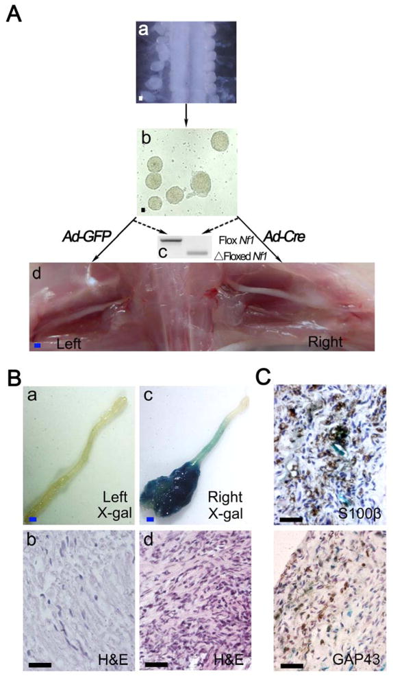
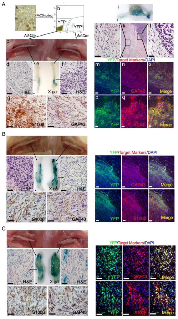
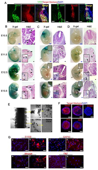
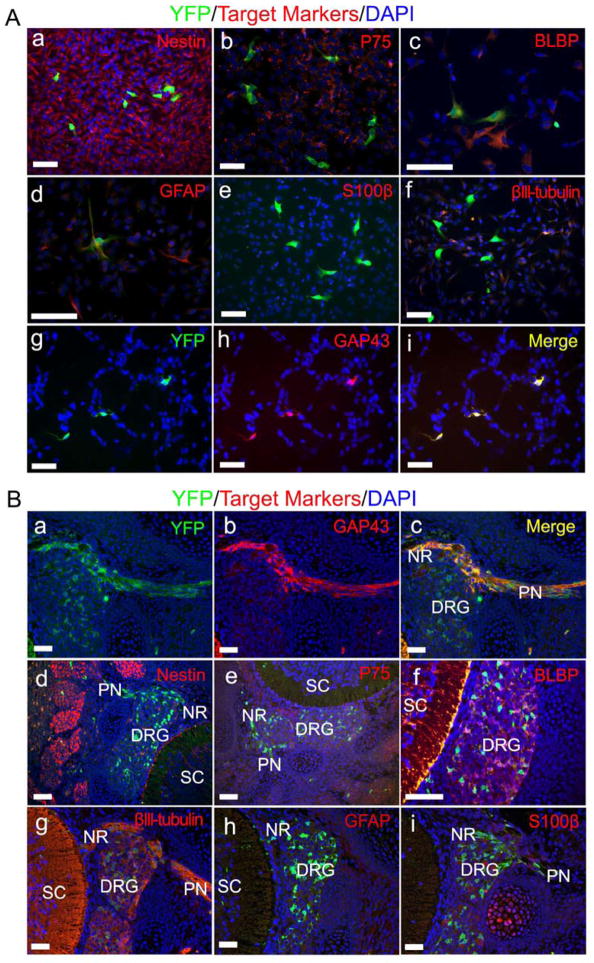
Neurofibromatosis type 1 is a tumor-predisposing genetic disorder. Plexiform neurofibromas are common NF1 tumors carrying a risk of malignant transformation, which is typically fatal. Little is known about mechanisms mediating initiation and identity of specific cell type that gives rise to neurofibromas. Using cell-lineage tracing, we identify a population of GAP43(+) PLP(+) precursors in embryonic nerve roots as the cells of origin for these tumors and report a non-germline neurofibroma model for preclinical drug screening to identify effective therapies. The identity of the tumor cell of origin and facility for isolation and expansion provides fertile ground for continued analysis to define factors critical for neurofibromagenesis. It also provides unique approaches to develop therapies to prevent neurofibroma formation in NF1 patients.


Figures

Figure 1
Figure 1. Loss of Nf1 in embryonic DNSCs gives rise to classic plexiform neurofibroma in vivo
(A) Diagram of experimental design: Isolation of DRGs/nerve roots from E13.5 Nf1flox/flox;R26R-LacZ embryos (a) and DNSCs culture (b); DNSCs were infected with Ad-Cre to ablate Nf1; Ad-GFP was used as a control (c); Nf1−/−;LacZ+ and Nf1flox/flox;LacZ DNSCs were implanted to right and left sciatic nerves of nude mice, respectively (d). (B) X-gal and H&E staining were perform on left (a and b) and right (c and d) sciatic nerve. (C)Tumor on right sciatic nerve was stained with antibody against S100β and GAP43. Scale bars: 500 μm for blue scale bars, 50 μm for white and black scale bars. See also Figure S1.
Figure 2
Figure 2. The cells of origin for para-spinal plexiform neurofibroma are inside the embryonic PLP+ DNSCs
(A) FACS was performed to obtain the YFP+ and YFP DNSCs from E13.5 PlpCre-ERT;Nf1flox/flox;R26R-LacZ-YFP embryo, whose mother was administered with tamoxifen at E11.5 (a and b); Both YFP+ and YFP DNSCs were infected with Ad-Cre to ablate Nf1, then injected to right and left sciatic nerves of nude mice respectively (c); X-gal and H&E staining were perform on left and right sciatic nerve (d – f); Sections of right sciatic nerve were stained with antibody against S100β (g) and GAP43 (h); A fraction of these sciatic plexiform neurofibromas spontaneously transformed into MPNST, exhibiting both benign and malignant histologic characters (i – l); Immunofluorescence staining of tumor on right sciatic nerve shows expression of YFP as well as GAP43 and S100β Schwann cell markers (m – r). (B and C) A similar strategy to that in panel A was used for YFP+ and YFP DNSCs derived from E13.5 embryos with genotype Krox20-Cre;Nf1flox/flox;R26R-LacZ-YFP (B) and Dhh-Cre;Nf1flox/flox;R26R-LacZ-YFP (C). Scale bars: 500 μm for blue scale bars, 50 μm for white and black scale bars.
Figure 3
Figure 3. Cells of origin for para-spinal plexiform neurofibromas are inside the embryonic PLP+ nerve root cells
(A) Representative frozen sections from E13.5 PlpCre-ERT;Nf1flox/flox;R26R-LacZ-YFP embryo, whose mother was administered with tamoxifen at E11.5, were stained for YFP, Krox20, DHH and DAPI. NR=nerve root, DRG=dorsal root ganglia. (B, C, and D) X-gal and H&E staining were performed on embryos with genotype PlpCre-ERT;R26R-LacZ (B, mother mouse was administered with 4-hydroxytamoxifen at E9.5), Krox20-Cre;R26R-LacZ (C) and Dhh-Cre;R26R-LacZ (D) from E10.5 to E13.5. Arrow = area of enlarged view, arrow head = nerve roots, star mark = DRG. (E) DRG and dorsal nerve root were separated from E13.5 embryos and cultured in neurosphere culture conditions. (F) Nerve root derived neurospheres immunostained for Nestin, GFAP, S100β, P75, Ki67 and DAPI. (G) Immunostaining of nerve root derived neurosphere cells cultured in neurosphere medium (a, c, e, and g) or differentiation medium (b, d, f, and h) with antibodies against S100β, GAP43, βIII-tubulin and Olig2. Scale bars: 500 μm for blue scale bars, 50 μm for white and black scale bars. See also Figure S2.
Figure 4
Figure 4. Phenotypic analysis identifies the PLP+ cells of origin as embryonic Schwann cell precursors
(A) DNSCs obtained from E13.5 PlpCre-ERT;Nf1flox/flox;R26R-YFP embryos, whose mother was administered with tamoxifen at E11.5, were stained for YFP, DAPI and lineage markers: Nestin (a), P75 (b), BLBP (c), GFAP(d), S100β (e), βIII-tubulin (f) and GAP43 (g – i). (B) Formalin fixed paraffin embedded sections from E13.5 PlpCre-ERT;Nf1flox/flox;R26R-YFP embryos, whose mother was administered with tamoxifen at E11.5, were stained for YFP, DAPI and lineage markers: GAP43 (a– c), Nestin (d), P75 (e), BLBP (f), βIII-tubulin (g), GFAP (h) and S100β (i). DRG=dorsal root ganglia, NR= nerve root, PN=peripheral nerve, SC=spinal cord. Scale bars: 50 μm for all. See also Figure S3.
Figure 5
Figure 5. Cell lineage tracing reveals PLP+ precursors in the embryonic nerve roots as the cells of origin for plexiform neurofibromas
(A) X-gal staining of whole spinal cord, DRGs and partial peripheral nerves from PlpCre-ERT;Nf1flox/−;R26R-LacZ mouse, whose mother was administered with tamoxifen at E9.5. There is strong X-gal staining in the enlarged DRGs (star mark), enlarged intercostal nerve (arrow head) and nerve root (arrow). (B) H&E and immunohistochemical staining for GAP43 and S100β in sections from these enlarged LacZ+ DRGs (star mark in panel A). (C) H&E staining of the DRG and its associated enlarged intercostal nerve (EIN) in panel A (arrow head). Enlarged intercostal nerve was also stained for GAP43 and S100β. (D) Representative H&E and immunohistochemical analyses of early neurofibroma in dorsal root and associated normal DRG (arrow in panel A). DRG=dorsal root ganglia, DR=dorsal root, SC=spinal cord, VR=ventral root. Scale bars: 500 μm for blue scale bars, 50 μm for the rest.
Figure 6
Figure 6. Therapeutic effect of MAPK signaling inhibitor on DNSC-derived plexiform neurofibroma
(A) Diagram of experimental design for testing therapeutic effect of PD0325901 on plexiform neurofibroma. (B) Immunostaining of ERK1/2 (total) and p-ERK1/2 (phosphorylated ERK) in E13.5 Nf1−/− DNSCs derived sciatic plexiform neurofibroma (a). Fold change of luminescence count measured during PD0325901 or vehicle treatment (b). After 4 weeks of treatment, both PD0325901 and vehicle treatment group were divided into two subgroups: discontinued PD0325901 or continued the PD0325901 and discontinued vehicle treatment or continued vehicle treatment subgroups for an additional 4 weeks. Tumors were then harvested for X-gal staining (c) and immunostaining for ERK1/2 and p-ERK1/2 (d). Statistics were represented as the mean ± SEM (* p < 0.05). Scale bars: 500 μm for blue scale bars, 50 μm for the rest. See also Figure S4.

Comment in

References

    1. Alcantara Llaguno S, Chen J, Kwon CH, Jackson EL, Li Y, Burns DK, Alvarez-Buylla A, Parada LF. Malignant astrocytomas originate from neural stem/progenitor cells in a somatic tumor suppressor mouse model. Cancer Cell. 2009;15:45–56. - PMC - PubMed
    1. Ballester R, Marchuk D, Boguski M, Saulino A, Letcher R, Wigler M, Collins F. The NF1 locus encodes a protein functionally related to mammalian GAP and yeast IRA proteins. Cell. 1990;63:851–859. - PubMed
    1. Barker N, Ridgway RA, van Es JH, van de Wetering M, Begthel H, van den Born M, Danenberg E, Clarke AR, Sansom OJ, Clevers H. Crypt stem cells as the cells-of-origin of intestinal cancer. Nature. 2009;457:608–611. - PubMed
    1. Bonnet D, Dick JE. Human acute myeloid leukemia is organized as a hierarchy that originates from a primitive hematopoietic cell. Nat Med. 1997;3:730–737. - PubMed
    1. Chang T, Krisman K, Theobald EH, Xu J, Akutagawa J, Lauchle JO, Kogan S, Braun BS, Shannon K. Sustained MEK inhibition abrogates myeloproliferative disease in Nf1 mutant mice. The Journal of clinical investigation. 2013;123:335–339. - PMC - PubMed

Publication types

MeSH terms