

Chronic continuous exenatide infusion does not cause pancreatic inflammation and ductal hyperplasia in non-human primates
- PMID: 25447052
- PMCID: PMC4278248
- DOI: 10.1016/j.ajpath.2014.09.009
Chronic continuous exenatide infusion does not cause pancreatic inflammation and ductal hyperplasia in non-human primates
Abstract
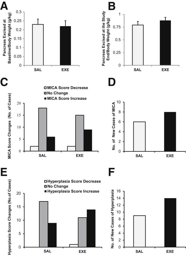
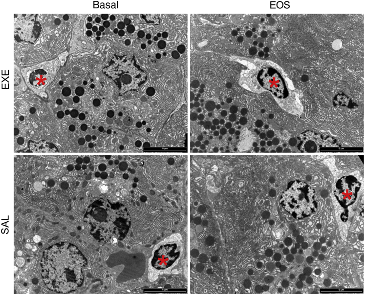
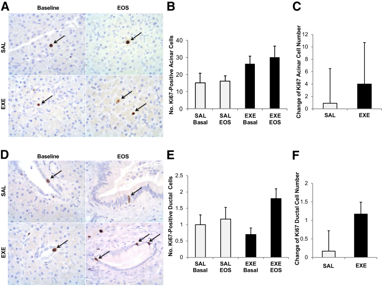
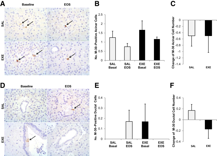
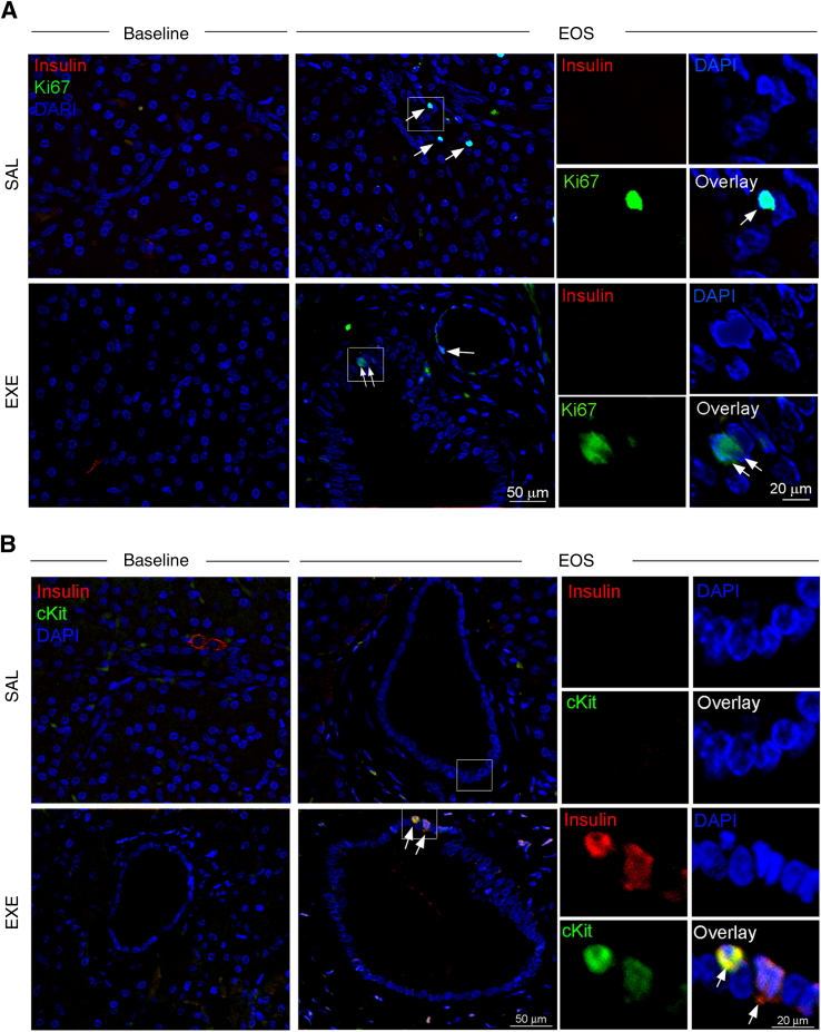
In this study, we aimed to evaluate the effects of exenatide (EXE) treatment on exocrine pancreas of nonhuman primates. To this end, 52 baboons (Papio hamadryas) underwent partial pancreatectomy, followed by continuous infusion of EXE or saline (SAL) for 14 weeks. Histological analysis, immunohistochemistry, Computer Assisted Stereology Toolbox morphometry, and immunofluorescence staining were performed at baseline and after treatment. The EXE treatment did not induce pancreatitis, parenchymal or periductal inflammatory cell accumulation, ductal hyperplasia, or dysplastic lesions/pancreatic intraepithelial neoplasia. At study end, Ki-67-positive (proliferating) acinar cell number did not change, compared with baseline, in either group. Ki-67-positive ductal cells increased after EXE treatment (P = 0.04). However, the change in Ki-67-positive ductal cell number did not differ significantly between the EXE and SAL groups (P = 0.13). M-30-positive (apoptotic) acinar and ductal cell number did not change after SAL or EXE treatment. No changes in ductal density and volume were observed after EXE or SAL. Interestingly, by triple-immunofluorescence staining, we detected c-kit (a marker of cell transdifferentiation) positive ductal cells co-expressing insulin in ducts only in the EXE group at study end, suggesting that EXE may promote the differentiation of ductal cells toward a β-cell phenotype. In conclusion, 14 weeks of EXE treatment did not exert any negative effect on exocrine pancreas, by inducing either pancreatic inflammation or hyperplasia/dysplasia in nonhuman primates.
Copyright © 2015 American Society for Investigative Pathology. Published by Elsevier Inc. All rights reserved.
Figures
References
-
- Xu G., Stoffers D.A., Habener J.F., Bonner-Weir S. Exendin-4 stimulates both beta-cell replication and neogenesis, resulting in increased beta-cell mass and improved glucose tolerance in diabetic rats. Diabetes. 1999;48:2270–2276. - PubMed
-
- Farilla L., Hui H., Bertolotto C., Kang E., Bulotta A., Di Mario U., Perfetti R. Glucagon-like peptide-1 promotes islet cell growth and inhibits apoptosis in Zucker diabetic rats. Endocrinology. 2002;143:4397–4408. - PubMed
-
- Hui H., Wright C., Perfetti R. Glucagon-like peptide 1 induces differentiation of islet duodenal homeobox-1-positive pancreatic ductal cells into insulin-secreting cells. Diabetes. 2001;50:785–796. - PubMed
-
- Fineman M.S., Bicsak T.A., Shen L.Z., Taylor K., Gaines E., Varns A., Kim D., Baron A.D. Effect on glycemic control of exenatide (synthetic exendin-4) additive to existing metformin and/or sulfonylurea treatment in patients with type 2 diabetes. Diabetes Care. 2003;26:2370–2377. - PubMed
-
- Campbell J.E., Drucker D.J. Pharmacology, physiology, and mechanisms of incretin hormone action. Cell Metab. 2013;17:819–837. - PubMed
Publication types
MeSH terms
Substances
Grants and funding
LinkOut - more resources
Full Text Sources
Other Literature Sources
Medical
