. 2014 Dec 20;588(24):4701-7.
doi: 10.1016/j.febslet.2014.10.043. Epub 2014 Nov 11.

Kinetics of the early events of GPCR signalling

Affiliations

Kinetics of the early events of GPCR signalling

Roslin J Adamson et al. FEBS Lett. .

Abstract

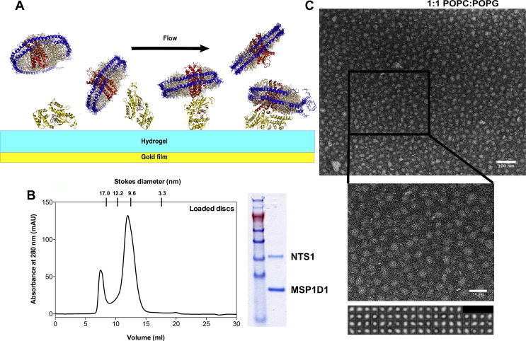
Neurotensin receptor type 1 (NTS1) is a G protein-coupled receptor (GPCR) that affects cellular responses by initiating a cascade of interactions through G proteins. The kinetic details for these interactions are not well-known. Here, NTS1-nanodisc-Gαs and Gαi1 interactions were studied. The binding affinities of Gαi1 and Gαs to NTS1 were directly measured by surface plasmon resonance (SPR) and determined to be 15±6 nM and 31±18 nM, respectively. This SPR configuration permits the kinetics of early events in signalling pathways to be explored and can be used to initiate descriptions of the GPCR interactome.

Keywords: Electron microscopy; G protein; G protein-coupled receptor; Nanodisc; Surface plasmon resonance.


Figures

Fig. 1
Fig. 1
Nanodisc preparation and NTS1-G protein coupling determination. (A) Schematic representation of the experimental setup. G proteins (gold) were amine-coupled to a CM5 Biacore chip (GE Healthcare). FLAG-NTS1-loaded nanodiscs were injected over the sample and reference flow cells. The reference flow cell was activated and blocked or ovalbumin was amine-coupled to it. Empty discs were injected over the sample and reference flow cells as a reference. Single cycle kinetics using serial concentrations of FLAG-NTS1-nanodiscs was performed. Data were double-referenced. (B) Representative SEC profile for nanodisc purification. A peak composed of large aggregates and vesicles elutes in the void volume at approximately 7.5 ml, followed by the nanodisc peak at ∼12.5 ml, corresponding to a calibrated size of approximately 10 nm (left). SDS–PAGE of anti-FLAG enriched nanodiscs, showing approximately twice the amount of MSP1D1 compared to NTS1, which understains with Coomassie Brilliant Blue (right). (C) Negative stain EM images showing nanodisc sample. Nanodiscs prepared with a 1:1 ratio of POPC:POPG form homogeneous populations. Stain was 2% uranyl acetate. Reference-free class averages of 10–12 nm PC:PG discs (prepared using EMAN2 [54]) are shown. Box size is 18.5 nm. Scale bars are 100 nm (upper) and 50 nm (lower).
Fig. 2
Fig. 2
SPR traces of GPCR G coupling. (A) and (B) FLAG-NTS1-PC:PG nanodiscs coupling to His6-Gs or His6-Gi1 immobilised on a CM5 chip. Approximately 7000 RU Gs was amine-coupled to a CM5 Biacore chip (GE Healthcare) in 10 mM sodium acetate, pH 5.5 (A). Approximately 13 000 RU Gi1 was amine-coupled to a CM5 Biacore chip (GE Healthcare) in 10 mM sodium acetate, pH 5.0 (B). The reference flow cell was activated and blocked. Serial concentrations of 41.25–660 nM (A) and 25–400 nM (B) nanodisc-reconstituted and ligand-bound FLAG-NTS1 were injected over the chip surface. Empty nanodiscs at the same concentrations were injected as a reference, and data were double-referenced. The affinity of Gs (A) for FLAG-NTS1 in nanodiscs was 9 nM in this instance, and 9 nM for Gi1 (B). (C) His6-Gs coupling to FLAG-NTS1-PC:PG nanodiscs captured on an L1 chip. FLAG-NTS1-PC:PG nanodiscs (2500 RU) and empty PC:PG nanodiscs (2000 RU) were captured in FC 4 and 3, respectively, of an L1 chip by an 800-s injection at 5 μl/min. The chip was thoroughly washed in running buffer at 50 μl/min for 30–60 min. Serial dilutions of 1000 nM (62.5, 125, 250, 500, 1000 nM) His6-Gs were injected across the flow cells for 150 s per concentration at 50 μl/min. The data (solid lines) were fitted with a 1:1 Langmuir binding model as well as a heterogeneous ligand binding model (dashed lines), giving KD values of 65 nM for the 1:1 fit and 0.5 and 80 nM for KD1 and KD2 respectively. The χ2 values for the fits were 4.4 and 3.3, respectively.
Fig. 3
Fig. 3
Kinetics of the GPCR interactome. Some of the initial steps in the GPCR signalling pathway. The GPCR activates a heterotrimeric G protein (a, b) via the GTPase domain of the Gα subunit, which is regulated by regulators of G protein signalling (RGS), causing hydrolysis of GTP to GDP (c). The heterotrimer dissociates into α and βγ subunits. The hydrolysis of ATP to cyclic AMP (cAMP) (d) is catalysed by interactions of Gα with adenylate cyclase, which regulates Ca2+ channels via activation of protein kinase A (PKA) by cAMP. The Gβγ subunits activate phosphatidylinositide 3-kinase and phospholipase C (PLC). PLC cleaves phosphatidylinositol 4,5-bisphosphate (PIP2) into inositol 1,4,5-trisphosphate (IP3) and diacyl glycerol (DAG) which activate the release of Ca2+ from the endoplasmic reticulum and the activation of protein kinase C (PKC). PKA and G protein-coupled receptor kinases (GRKs) phosphorylate the GPCR, leading to coupling of the receptor to arrestin and subsequent down-regulation of the receptor by internalisation for recycling or degradation in lysosomes. (a, b) Gαi1 and Gαs affinities for NTS1 of 15 ± 6 nM and 31 ± 18 nM (SE), respectively (this study). (c) BODIPY-GTP hydrolysis Km value was 120 ± 60 nM ; GTPγS binding kapp = 0.027 min−1. (d) Basal activity ∼20–65 pmol cAMP/min/mg , kobs of ∼1 × 10−3 − 6 × 10−4 s−1. (e) Spontaneous diffusion-interaction on the ms timescale , with dissociation rates of 1.3 s−1, and a KD of 2–20 nM .

References

    1. Downes G.B., Gautam N. The G protein subunit gene families. Genomics. 1999;62:544–552. - PubMed
    1. Vincent J.P., Mazella J., Kitabgi P. Neurotensin and neurotensin receptors. Trends Pharmacol. Sci. 1999;20:302–309. - PubMed
    1. Harding P.J., Attrill H., Ross S., Koeppe J.R., Kapanidis A.N., Watts A. Neurotensin receptor type 1: Escherichia coli expression, purification, characterization and biophysical study. Biochem. Soc. Trans. 2007;35:760–763. - PubMed
    1. Pelaprat D. Interactions between neurotensin receptors and G proteins. Peptides. 2006;27:2476–2487. - PubMed
    1. Dobner P.R. Neurotensin and pain modulation. Peptides. 2006;27:2405–2414. - PubMed

Publication types

MeSH terms

Substances