. 2015 Jan;1853(1):222-32.
doi: 10.1016/j.bbamcr.2014.10.019. Epub 2014 Oct 30.

Nucleocytoplasmic shuttling of valosin-containing protein (VCP/p97) regulated by its N domain and C-terminal region

Affiliations

Nucleocytoplasmic shuttling of valosin-containing protein (VCP/p97) regulated by its N domain and C-terminal region

Changcheng Song et al. Biochim Biophys Acta. 2015 Jan.

Abstract

Valosin-containing protein (VCP or p97), a member of the AAA family (ATPases associated with diverse cellular activities), plays a key role in many important cellular activities. A genetic deficiency of VCP can cause inclusion body myopathy associated with Paget's disease of bone and frontotemporal dementia (IBMPFD). Previous studies showed that the VCP N domain is essential for the regulation of nuclear entry of VCP. Here we report that IBMPFD mutations, which are mainly located in the N domain, suppress the nuclear entry of VCP. Moreover, the peptide sequence G780AGPSQ in the C-terminal region regulates the retention of VCP in the nucleus. A mutant lacking this sequence can increase the nuclear distribution of IBMPFD VCP, suggesting that this sequence is a potential molecular target for correcting the deficient nucleocytoplasmic shuttling of IBMPFD VCP proteins.

Keywords: Inclusion body myopathy associated with Paget's disease of bone and frontotemporal dementia (IBMPFD); Nuclear export signal; Nucleocytoplasmic shuttling; Valosin containing protein.


Figures

Fig. 1
Fig. 1
VCP-GFP retains biological properties of VCP. (A) HEK293 cells stably expressing VCP-GFP and GFP showed similar morphology in the bright field and green fluorescence images. (B) Cells expressing VCP-GFP and GFP exhibit similar cell cycle. Cells stained with propidium iodide were analyzed using flow cytometry. Cell cycle is presented as the means and standard errors (n=8, P > 0.05). (C) VCP-GFP forms a hexameric structure. VCP-GFP co-immunoprecipited with endogenous VCP (left panel). VCP-GFP protein complexes were immunoprecipitated using anti-GFP antibody followed by Western blotting with anti-VCP antibody. VCP-GFP and VCP were shown as a 124 kDa band (similar to the calculated molecular mass of VCP-GFP) and a 97 kDa band (the molecular mass of monomeric VCP). GAPDH was probed as a loading control. The native gel fluorescence imaging of VCP-GFP showed that VCP-GFP is in a hexameric structure (right panel). The lysates of HEK293 cells expressing VCP-GFP or GFP were resolved using 3–16% native gel. The gel image was taken using a Fluoro Image Analyzer. The 650 to 750 kDa and 27 kDa bands were shown as VCP-GFP and GFP, respectively. (D) VCP-GFP retains the ATPase activity. Protein complexes were immunoprecipitated using anti-GFP or anti-VCP antibody from the lysates of HEK293 cells without transfection (Ctrl) or transfected with GFP or VCP-GFP. The ATPase activities of the immunoprecipited protein complexes were analyzed using a colorimetric ATPase assay. The ATPase activity was shown as the percentage of the ATPase activity of immunoprecipitated protein complexes in the control sample. Data are presented as mean and standard error of four experiments.
Fig. 2
Fig. 2
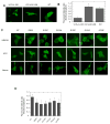
IBMPFD mutations decrease the nuclear distribution of VCP. HEK293 cells were transiently transfected with VCP-Δ1-206, VCP-Δ201-806 and WT VCP and imaged with confocal microscopy (A) and then images were quantitatively analyzed using image J software. The statistical analysis was showed that VCP-Δ1-206 is significantly lower than WT (P<0.01, n=28). (C) IBMPFD mutants were transfected to HEK293, HOS cells and primary neurons, and then imaged with an inverted fluorescence microscope. (D) Quantitative analysis of the fluorescence intensity ratio between the nucleus and the cytoplasm (FIRNC) in HEK293 cells expressing IBMPFD mutants was performed using Olympus CellSens Dimension software. The result shows that the FIRNC of all the IBMPFD mutants is WT is significantly lower than WT (P<0.001, n=50).
Fig. 3
Fig. 3
FRAP analysis of the nuclear entry of VCP–GFP in HEK293 cells. (A) The nucleus of the cells expressing VCP–GFP or GFP were photobleached for 4 s (area indicated by red boxes), and the fluorescence images were obtained at 4 s intervals over 400 s by confocal microscopy. (B) Quantitative analysis from results in (A) for the fluorescence recovery in the nucleus using single exponential FRAP of MIPAV software, where differences in fluorescence recovery rate were observed between VCP-GFP and GFP.
Fig. 4
Fig. 4
The mutation of ATP binding site in D1 or D2 domain of VCP-GFP decreases the nuclear retention. (A) HEK293 cells transfected with VCP-GFP mutant A1 or A2 were imaged using confocal microscopy. (B) Quantitative analysis of the nuclear retention. Data are presented as the fluorescence intensity ratio between the nucleus and the cytoplasm calculated using MIPAV software (n=36, * P<0.05).
Fig. 5
Fig. 5
The sequence 780-785 in the C-terminal region regulates VCP-GFP retention in the nucleus and nucleoli. HEK293 cells were transiently transfected with C-terminal region deletion mutant VCP-Δ780-806, VCP-Δ786-806 and VCP-Δ797-806 for 16 hr. The images were taken under an inverted fluorescence microscope. (A) The images showed that VCP-Δ780-806, but not VCP-Δ786-806 and VCP-Δ797-806, accumulates in the nucleus and forms foci as indicated by red arrows. (B) Quantitative analysis of the results presented in (A). Data are presented as the fluorescence intensity ratio between the nucleus and the cytoplasm calculated using MIPAV software (n=48, * p<0.01). (C) The VCP-Δ780-806 transfected cells were stained using Hoechst 33342 and imaged with fluorescence microscope and the images of the green fluorescence of VCP-Δ780-806 and the nucleus were merged (scale bar = 5 μM). (D) HEK293 cells expressing VCP-Δ780-806 and VCP-Δ786-806 mutants were fixed with cold methanol and stained with anti-nucleolin antibody and TRITC labeled secondary antibody. Images were taken using confocal microscopy (scale bar=5 μM).
Fig. 6
Fig. 6
VCP-Δ780-806 proteins exhibit a higher nuclear import rate than WT VCP-GFP. HEK293 cells were transiently transfected with VCP-Δ780-806 or WT for 16 hr. (A) Large ROIs indicated as red regions in the nucleus or cytoplasm were photobleached. Images were then consecutively taken at 1 min intervals. (A) Representative images show increased fluorescence intensity in the nucleus in cells expressing VCP-Δ780-806, but not WT, noticeable after 5 and 9 min recovery. (B) Fluorescence photobleaching and recovery curves are plotted using single exponential of MIPAV software. Photobleaching in the nucleus or cytoplasm indicated as WT-N and VCP-Δ780-806-N or WT-C and VCP-Δ780-806-C.
Fig. 7
Fig. 7
Effect of VCP-Δ780-806 on the nuclear distribution of VCP-GFP bearing R155H mutation. HEK293 cells were co-transfected with R155H and control vector, VCP-Δ1-206, VCP-Δ780-806 or WT for 16 hr. (A) The cells were imaged with an inverted fluorescence microscope. (B) Fluorescence intensity ratio between nucleus and cytoplasm (FIRNC) were calculated using Olympus CellSens Dimension software. The result showed that the FIRNC of R155H and VCP-Δ780-806 is significant higher than other cotransfectants (p<0.05, n=50). (C) Distribution of the coexpressed VCP variants in the nuclear and cytoplasmic fractions. HEK293 cells were transiently transfected with VCP-GFP variants as indicated and then fractionated into the nuclear fraction (N) and cytoplasmic fraction (C). VCP-GFP variants were detected by anti-GFP antibody. GAPDH and lamin A/C were used as cytoplasmic and nuclear markers, respectively.
Fig. 8
Fig. 8
in cis and in trans effect of the C-terminal sequence 780-806 on VCP-Δ1-206. Cells were transfected with VCP-Δ780-806 with control vector or VCP-Δ1-206, and VCP-Δ1-206 with control vector, or VCP-Δ1-206-Δ780-806 for 16 hr. (A) The cells were imaged with an inverted fluorescence microscope. (B) Fluorescence intensity ratio between the nucleus and cytoplasm (FIRNC) were calculated using Olympus CellSens Dimension software. The comparison of FIRNC between VCP-Δ780-806+VCP-Δ1-206 and VCP-Δ1-206 using ANOVA showed a significant difference (p<0.05, n=50). (C) Distribution of VCP deletion mutants in the nuclear and cytoplasmic fractions. HEK293 cells transiently transfected with VCP-GFP deletion mutants as indicated, and then fractionated into the nuclear fraction (N) and cytoplasmic fraction (C). VCP-GFP variants were detected by anti-GFP antibody. GAPDH and lamin A/C were used as cytoplasmic and nuclear markers, respectively.
Fig. 9
Fig. 9
Schematic representation of VCP-GFP variants and summary of the nuclear retention. The N domain or C-terminal deletion mutants are depicted. The nuclear retention (from Fig. 2, Fig. 4 and Fig. 5) are summarized as +/−, +, ++ and +++, which indicate low to high fluorescence intensities.

Similar articles

Cited by

References

    1. Wang Q, Song C, Li CC. Molecular perspectives on p97-VCP: progress in understanding its structure and diverse biological functions. J Struct Biol. 2004;146:44–57. - PubMed
    1. Meyer H, Bug M, Bremer S. Emerging functions of the VCP/p97 AAA-ATPase in the ubiquitin system. Nat Cell Biol. 2012;14:117–123. - PubMed
    1. Yamanaka K, Sasagawa Y, Ogura T. Recent advances in p97/VCP/Cdc48 cellular functions. Biochim Biophys Acta. 2012;1823:130–137. - PubMed
    1. Partridge JJ, Lopreiato JO, Jr, Latterich M, Indig FE. DNA Damage Modulates Nucleolar Interaction of the Werner Protein with the AAA ATPase p97/VCP. Mol Biol Cell. 2003;14:4221–4229. - PMC - PubMed
    1. Madeo F, Schlauer J, Zischka H, Mecke D, Frohlich KU. Tyrosine phosphorylation regulates cell cycle-dependent nuclear localization of Cdc48p. Mol Biol Cell. 1998;9:131–141. - PMC - PubMed

Publication types

MeSH terms

Supplementary concepts

LinkOut - more resources