. 2015 Feb;43(2):89-99.
doi: 10.1016/j.exphem.2014.10.004. Epub 2014 Oct 22.

Dual targeting of the thioredoxin and glutathione antioxidant systems in malignant B cells: a novel synergistic therapeutic approach

Affiliations

Dual targeting of the thioredoxin and glutathione antioxidant systems in malignant B cells: a novel synergistic therapeutic approach

Michelle Kiebala et al. Exp Hematol. 2015 Feb.

Abstract

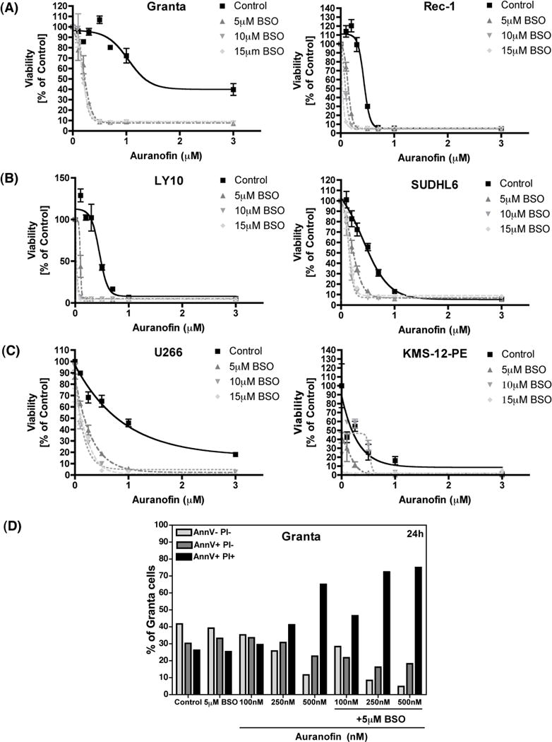
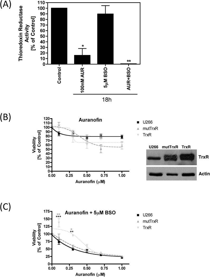
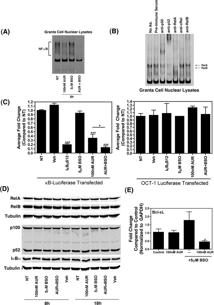
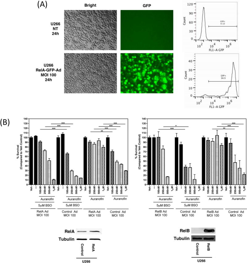
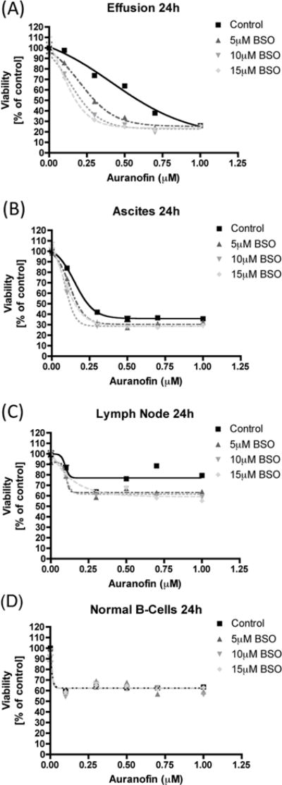
B-cell malignancies are a common type of cancer. One approach to cancer therapy is to either increase oxidative stress or inhibit the stress response systems on which cancer cells rely. In this study, we combined nontoxic concentrations of Auranofin (AUR), an inhibitor of the thioredoxin system, with nontoxic concentrations of buthionine-sulfoximine (BSO), a compound that reduces intracellular glutathione levels, and investigated the effect of this drug combination on multiple pathways critical for malignant B-cell survival. Auranofin interacted synergistically with BSO at low concentrations to trigger death in multiple malignant B-cell lines and primary mantle-cell lymphoma cells. Additionally, there was less toxicity toward normal B cells. Low AUR concentrations inhibited thioredoxin reductase (TrxR) activity, an effect significantly increased by BSO cotreatment. Overexpression of TrxR partially reversed AUR+BSO toxicity. Interestingly, the combination of AUR+BSO inhibited nuclear factor κB (NF-κB) signaling. Moreover, synergistic cell death induced by this regimen was attenuated in cells overexpressing NF-κB proteins, arguing for a functional role for NF-κB inhibition in AUR+BSO-mediated cell death. Together, these findings suggest that AUR+BSO synergistically induces malignant B-cell death, a process mediated by dual inhibition of TrxR and NF-κB, and such an approach warrants further investigation in B-cell malignancies.


Conflict of interest statement

Competing Interests

Authors have no competing interests.

Figures

Figure 1
Figure 1. Toxicity of Auranofin and BSO in malignant B-cell lines
(A) Granta cells and Rec-1 cells (human mantle cell lymphoma cell lines) were treated with Auranofin or BSO, alone or together, at the indicated concentrations for 24 h. Cell viability was measured using the MTT assay. Percent survival was calculated as compared to DMSO-treated control cells. (B) LY10 and SUDHL6 (human diffuse large B-cell lymphoma cell lines) were treated as indicated for 24 h and cell viability was measured as described above. (C) U266 and KMS-12-PE cells (human multiple myeloma cell lines) were treated as indicated for 24 h and cell viability was measured as described above. (D) Granta cells were treated as indicated for 24 h and cell death was analyzed using Annexin V/PI staining followed by flow cytometry. AnnexinV− PI− indicates viable cells, AnnexinV+ PI− indicates early apoptotic cells, and AnnexinV+ PI+ indicates late apoptotic/dead cells.
Figure 2
Figure 2. Effect of Auranofin and BSO on thioredoxin reductase activity
(A) Granta cells were treated with Auranofin or BSO, alone or together, at the indicated concentrations for 18 h. Thioredoxin reducatase activity was measured using the Sigma Thioredoxin Reductase assay kit. Results are shown as percent activity compared to DMSO-treated control cells. (B, C) U266 cells either untransfected (control), or stably over-expressing mutant thioredoxin reductase (mutTrxR) or wild-type thioredoxin reductase (TrxR) were treated with the indicated concentrations of Auranofin either alone (B) or together with 5 μM BSO (C) for 24 h. Cell viability was measured using the MTT assay. Percent survival was calculated as compared to DMSO-treated control cells. Immunoblot analysis was performed on whole cell lysates from U266 cells either untransfected (control), or stably over-expressing mutTR or wild-type TrxR. Over-expression of TrxR was confirmed using an anti-TrxR antibody. Actin was used as a loading control. Statistical significance is indicated, as compared to untransfected control cells (***p<0.001, **p<0.01) or as compared to mutTrxR expressing cells (**p<0.01, *p<0.05).
Figure 3
Figure 3. Effect of Auranofin and BSO on NF-κB activity
(A) Granta cells (1×10⁶) were treated with the indicated concentrations of Auranofin and BSO, either alone or together, for 6 h. Following these treatments, nuclear extracts were prepared and EMSA was performed by incubating nuclear extracts with IR-Dye-700 conjugated, double-stranded DNA probe at room temperature for 10 minutes, followed by resolution of the protein-DNA complexes on native 4 % polyacrylamide gels. The bands were then visualized on an Odyssey infrared imager (LI-COR Biosciences). (B) Nuclear extracts were prepared from untreated Granta cells and super-shift assays were performed by incubating nuclear extracts with IR-Dye-700 conjugated, double-stranded DNA probe at room temperature for 10 minutes, followed by incubation with the indicated antibodies for 5 minutes. The protein-DNA-antibody complexes were resolved on native 4 % polyacrylamide gels. The bands were then visualized as described above. (C) Luciferase reporter plasmids containing NF-κB or OCT-1 responsive elements upstream of a firefly luciferase gene was transfected into Granta cells (5×10⁶), together with either a vector expressing IκBαS1/2 or an empty vector, using Nucleofector (Amaxa/Lonza). The total amount of plasmid DNA was kept constant at 5 μg for each transfection. Thirty minutes after the transfection, cells were plated into single wells and were either left untreated or incubated for 6 h with the indicated doses of Auranofin or BSO, alone or together. Treatment with DMSO (Veh) was used as a control. Cell lysates were prepared using reporter lysis buffer and luciferase activity was measured with a SpectraMax M3 plate reader (Molecular Devices). Data are presented as fold change compared to non-treated cells and are shown as mean ± SEM of values derived from three replicates from each of four combined experiments for NF-κB or from two combined experiments for OCT-1. ***, p<0.001, **, p<0.01 as compared to cells transfected with the luciferase reporter and left untreated. (D) Whole cell lysates from Granta cells treated as indicated were subjected to immunoblot analysis with antibodies against RelA (first panel), RelB (second panel), p100/p52 (fourth panel), IκBα (fifth panel), or α-Tubulin (third and sixth panels). (E) Bcl-xL mRNA levels were determined via qRT-PCR in Granta cells treated as indicated for 24 h. mRNA levels were compared using the ΔΔCt method. *, p<0.05 as compared to DMSO-treated cells (control).
Figure 4
Figure 4. Over-expression of NF-κB molecules rescues U266 cells from Auranofin + BSO-induced toxicity
(A) U266 cells were either left untransduced, or were transduced with an adenoviral vector expressing RelA and GFP at a multiplicity of infection (MOI) of 100 for the indicated time. GFP expression was captured by fluorescence microscopy. Cells were then fixed with 4 % paraformaldehyde and were subjected to flow cytometric analysis. The percentage of GFP-positive cells is indicated. (B) U266 cells were transduced with a control adenoviral vector or with an adenoviral vector expressing RelB (first panel) or RelA (second panel) for 24 h. Cells were then treated with the indicated concentrations of Auranofin and/or BSO for an additional 24 h. Cell viability was measured using the MTT assay. Percent survival was calculated as compared to DMSO-treated (Veh) control cells. Data are presented as mean ± SEM of values derived from a single experiment that was performed in triplicate. ***, p<0.001, **, p<0.01, *, p<0.05 indicates statistical significance.
Figure 5
Figure 5. Toxicity of Auranofin and BSO in primary cells
(A, B, C) Unseparated, single cell suspensions derived from the indicated sources were treated with Auranofin or BSO, alone or together, at the indicated concentrations for 24 h. Cell viability was measured using the alamarBlue® assay. Percent survival was calculated as compared to DMSO-treated control cells. (D) Normal B-cells isolated from a healthy individual were treated as indicated for 24 h and cell viability was measured via the alamarBlue® assay. Percent survival was calculated as compared to DMSO-treated control cells.

Similar articles

Cited by

References

    1. Sharma SV, Settleman J. Exploiting the balance between life and death: targeted cancer therapy and “oncogenic shock”. Biochem Pharmacol. 2010;80:666–673. - PubMed
    1. Luo J, Solimini NL, Elledge SJ. Principles of cancer therapy: oncogene and non-oncogene addiction. Cell. 2009;136:823–837. - PMC - PubMed
    1. Champion GD, Cairns DR, Bieri D, et al. Dose response studies and longterm evaluation of auranofin in rheumatoid arthritis. J Rheumatol. 1988;15:28–34. - PubMed
    1. Williams HJ, Ward JR, Egger MJ, et al. Auranofin, gold sodium thiomalate, and placebo in the treatment of rheumatoid arthritis. Cooperative systematic studies of rheumatic diseases. Clin Rheumatol. 1984;3(Suppl 1):39–50. - PubMed
    1. Furst DE. Mechanism of action, pharmacology, clinical efficacy and side effects of auranofin. An orally administered organic gold compound for the treatment of rheumatoid arthritis. Pharmacotherapy. 1983;3:284–298. - PubMed

Publication types

MeSH terms