Review
. 2014 Dec 2;20(6):939-52.
doi: 10.1016/j.cmet.2014.09.013. Epub 2014 Oct 30.

AMPK--sensing energy while talking to other signaling pathways

Affiliations
Review

AMPK--sensing energy while talking to other signaling pathways

D Grahame Hardie. Cell Metab. .

Abstract

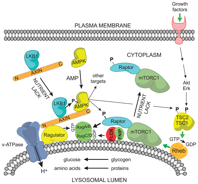
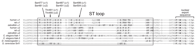
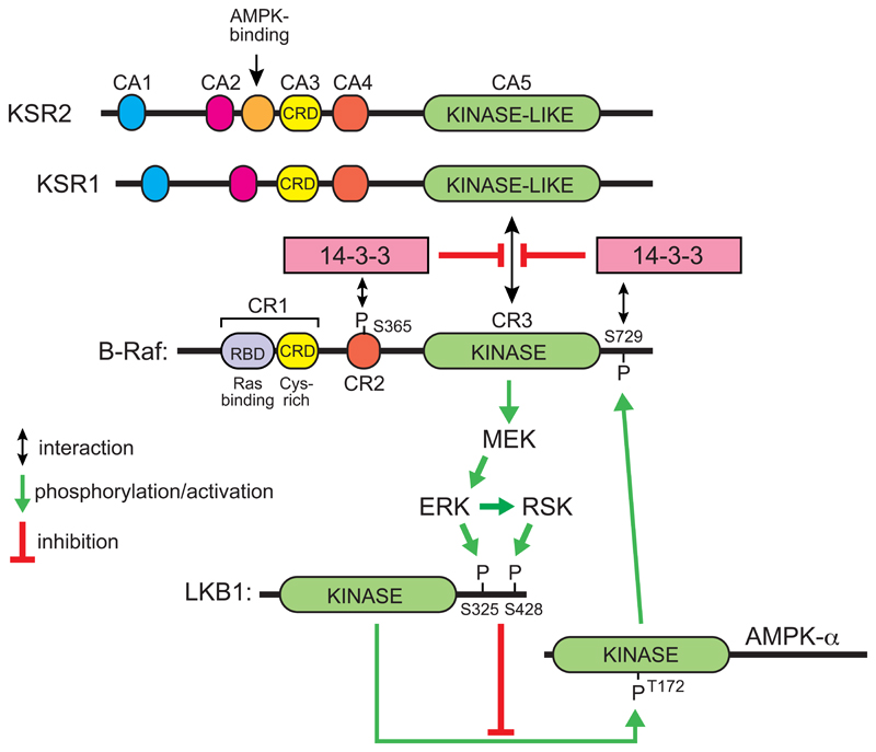
The AMP-activated protein kinase (AMPK) is a sensor of cellular energy and nutrient status, expressed almost universally in eukaryotes as heterotrimeric complexes comprising catalytic (α) and regulatory (β and γ) subunits. Along with the mechanistic target of rapamycin complex-1 (mTORC1), AMPK may have been one of the earliest signaling pathways to have arisen during eukaryotic evolution. Recent crystal structures have provided insights into the mechanisms by which AMPK is regulated by phosphorylation and allosteric activators. Another recent development has been the realization that activation of AMPK by the upstream kinase LKB1 may primarily occur not in the cytoplasm, but at the surface of the lysosome, where AMPK and mTORC1 are regulated in a reciprocal manner by the availability of nutrients. It is also becoming clear that there is a substantial amount of crosstalk between the AMPK pathway and other signaling pathways that promote cell growth and proliferation, and this will be discussed.


Figures

Figure 1
Figure 1. Structures of a selection of three different classes of AMPK-activating compounds.
Figure 2
Figure 2. Structure of complete α2β1γ1 heterotrimer of AMPK.
The model was created with MacPyMol using PDB file 4CFE. The α, β and γ subunits are shown in “cartoon view” with different domains color coded, while the activator 991, the kinase inhibitor staurosporine, the three bound molecules of AMP, and phospho-Thr172 are shown in “sphere view” with C atoms green, O red, N blue and P orange. Various features labeled are discussed in the text.
Figure 3
Figure 3. Comparison of structure of the inactive KD:AID fragment from S. pombe, and the active KD:AID fragment from human α2, showing the dramatic change in the position of the AID.
The models were created with McPymol using PDB entries 3H4J and 4CFE (although the latter was a complete heterotrimer, only the KD and AID are shown). The kinase domains are viewed from approximately the same angle, so that the change in the orientation of the AID is more obvious. The N- and C-lobes of the kinase domain are in yellow and cyan respectively, except that the activation segments within the C-lobe (which have markedly different conformations in the two structures) are in red. The AIDs are in orange, with their three α-helices labeled AID-α1 to -α3 (AID-α2 was poorly resolved in the human structure). In the inactive S. pombe structure, AID-α3 interacts with αC (C-helix) from the N-lobe and αE (E-helix) from the C-lobe, whereas in the active human structure it interacts mainly with the γ subunit (not shown) instead.
Figure 4
Figure 4. Proposed mechanism for reciprocal regulation of mTORC1 and AMPK at the cytoplasmic surface of the lysosome.
The model is based on (Bar-Peled and Sabatini, 2014) and (Zhang et al., 2014). (1) Activation of mTORC1: nutrient availability (amino acids and/or glucose) within the lysosome are sensed by a mechanism requiring the v-ATPase proton pump, and this effect is transmitted to the Ragulator, which converts RagA/BGDP to RagA/BGTP, while the folliculin:FNIP complex converts RagC/DGTP to RagC/DGDP. The RagAGTP:RagCGDP complex binds the Raptor component of mTORC1, whose activation also requires Rheb:GTP. Formation of the latter complex is promoted by growth factors via phosphorylation and dissociation from the lysosome of TSC1:TSC2, a Rheb-GAP (Menon et al., 2014). (2) Activation of AMPK: nutrient lack causes the Ragulator to recruit the axin:LKB1 complex, and elevated AMP also recruits AMPK to this complex. LKB1 phosphorylates and activates AMPK, which then presumably dissociates from the complex to phosphorylate downstream targets. Two of these targets are Raptor and the TSC1:TSC2 complex. Phosphorylation of Raptor by AMPK, coupled with conversion of RagAGTP:RagCGDP to RagAGDP:RagCGTP, may promote the dissociation of mTORC1 from the lysosome. Phosphorylation of the TSC1:TSC2 complex appears to increase its Rheb-GAP activity, thus converting Rheb:GTP to the inactive Rheb:GDP form, although the exact mechanism remains unclear.
Figure 5
Figure 5. Alignment of sequences at the C-terminal end of the α subunits, showing the location of the ST loop.
Sequences were aligned using CLC Main Workbench 6 using a “gap open cost” of 10 and a “gap extension cost” of 1. Serine and threonine residues within the ST loop are highlighted in bold type, with residues where there is evidence for phosphorylation shown with arrows (human residue numbering). Also shown are the nuclear export sequences at the extreme C-termini, with critical hydrophobic residues highlighted in bold.
Figure 6
Figure 6. Proposed reciprocal feedback loops operating between the Raf-MEK-ERK-RSK and LKB1-AMPK signaling pathways.
In the inactive state of B-Raf, phosphorylation of Ser365 and Ser729 [the latter by AMPK (Shen et al., 2013)] causes binding of 14-3-3 proteins that prevent association of B-Raf with the plasma membrane and with the Raf-like scaffold proteins KSR1 or KSR2. When Ras is activated it binds to the Ras-binding region of the B-Raf CR1 domain, relieving the inhibitory effect of the latter, and if 14-3-3 proteins are not bound KSR1 or KSR2 then forms a complex with B-Raf and MEK, causing phosphorylation and activation of the latter by B-Raf. This in turn triggers a cascade of phosphorylation and activation of the downstream kinases, ERK and RSK. The latter phosphorylate LKB1 at Ser325 and Ser428 respectively, reducing its ability to activate AMPK via phosphorylation of Thr172 (Zheng et al., 2009). Activated AMPK then phosphorylates B-Raf at Ser729, causing its interaction with 14-3-3 proteins and down-regulation once again. For more details of the conserved domains on B-Raf, KSR1 and KSR2, see Udell et al (2011).

References

    1. Amodeo GA, Rudolph MJ, Tong L. Crystal structure of the heterotrimer core of Saccharomyces cerevisiae AMPK homologue SNF1. Nature. 2007;449:492–495. - PubMed
    1. Baba M, Hong SB, Sharma N, Warren MB, Nickerson ML, Iwamatsu A, Esposito D, Gillette WK, Hopkins RF, 3rd, Hartley JL, et al. Folliculin encoded by the BHD gene interacts with a binding protein, FNIP1, and AMPK, and is involved in AMPK and mTOR signaling. Proc Natl Acad Sci U S A. 2006;103:15552–15557. - PMC - PubMed
    1. Bar-Peled L, Sabatini DM. Regulation of mTORC1 by amino acids. Trends Cell Biol. 2014 in press. - PMC - PubMed
    1. Bateman A. The structure of a domain common to archaebacteria and the homocystinuria disease protein. Trends Biochem Sci. 1997;22:12–13. - PubMed
    1. Bultot L, Guigas B, Von Wilamowitz-Moellendorff A, Maisin L, Vertommen D, Hussain N, Beullens M, Guinovart JJ, Foretz M, Viollet B, et al. AMP-activated protein kinase phosphorylates and inactivates liver glycogen synthase. Biochem J. 2012;443:193–203. - PubMed

Publication types

MeSH terms

Substances