. 2015 Feb;148(2):379-391.e4.
doi: 10.1053/j.gastro.2014.10.008. Epub 2014 Oct 13.

Liver-specific deletion of augmenter of liver regeneration accelerates development of steatohepatitis and hepatocellular carcinoma in mice

Affiliations

Liver-specific deletion of augmenter of liver regeneration accelerates development of steatohepatitis and hepatocellular carcinoma in mice

Chandrashekhar R Gandhi et al. Gastroenterology. 2015 Feb.

Abstract

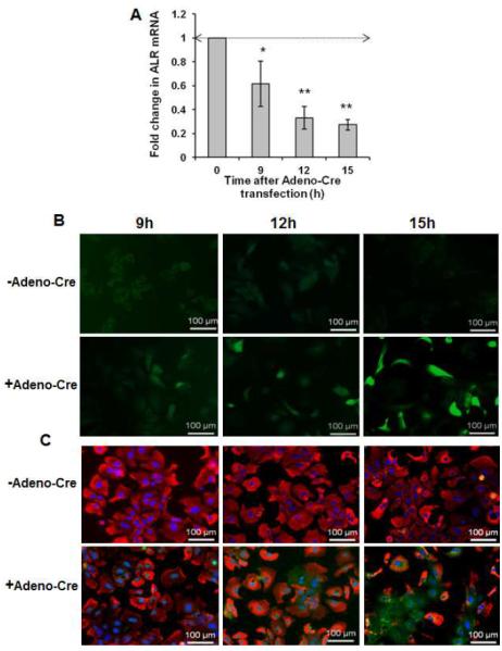
Background & aims: Augmenter of liver regeneration (ALR, encoded by GFER) is a widely distributed pleiotropic protein originally identified as a hepatic growth factor. However, little is known about its roles in hepatic physiology and pathology. We created mice with liver-specific deletion of ALR to study its function.

Methods: We developed mice with liver-specific deletion of ALR (ALR-L-KO) using the albumin-Cre/LoxP system. Liver tissues were collected from ALR-L-KO mice and ALR(floxed/floxed) mice (controls) and analyzed by histology, reverse-transcription polymerase chain reaction, immunohistochemistry, electron microscopy, and techniques to measure fibrosis and lipids. Liver tissues from patients with and without advanced liver disease were determined by immunoblot analysis.

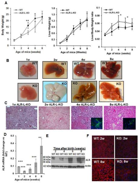
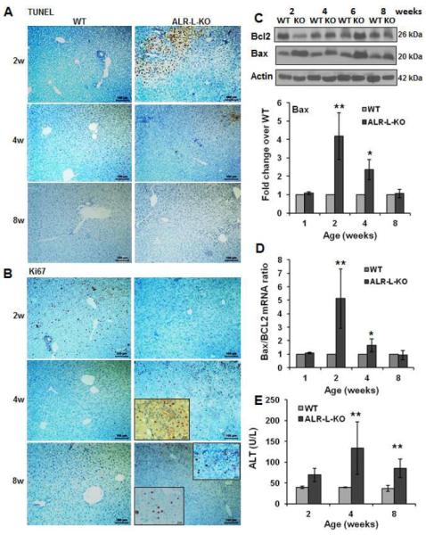
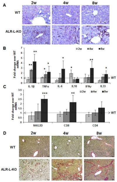
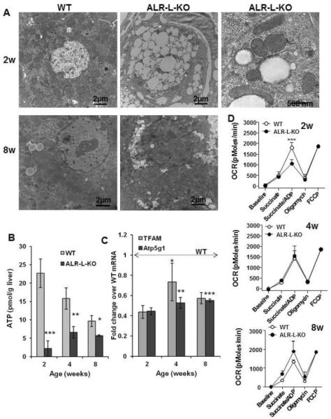
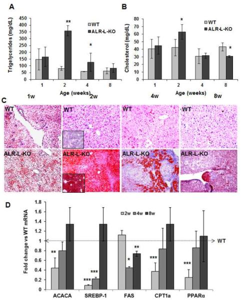
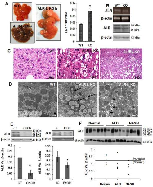
Results: Two weeks after birth, livers of ALR-L-KO mice contained low levels of ALR and adenosine triphosphate (ATP); they had reduced mitochondrial respiratory function and increased oxidative stress, compared with livers from control mice, and had excessive steatosis, and hepatocyte apoptosis. Levels of carbamyl-palmitoyl transferase 1a and ATP synthase subunit ATP5G1 were reduced in livers of ALR-L-KO mice, indicating defects in mitochondrial fatty acid transport and ATP synthesis. Electron microscopy showed mitochondrial swelling with abnormalities in shapes and numbers of cristae. From weeks 2-4 after birth, levels of steatosis and apoptosis decreased in ALR-L-KO mice, and numbers of ALR-expressing cells increased, along with ATP levels. However, at weeks 4-8 after birth, livers became inflamed, with hepatocellular necrosis, ductular proliferation, and fibrosis; hepatocellular carcinoma developed by 1 year after birth in nearly 60% of the mice. Hepatic levels of ALR were also low in ob/ob mice and alcohol-fed mice with liver steatosis, compared with controls. Levels of ALR were lower in liver tissues from patients with advanced alcoholic liver disease and nonalcoholic steatohepatitis than in control liver tissues.

Conclusions: We developed mice with liver-specific deletion of ALR, and showed that it is required for mitochondrial function and lipid homeostasis in the liver. ALR-L-KO mice provide a useful model for investigating the pathogenesis of steatohepatitis and its complications.

Keywords: ALD; Augmenter of Liver Regeneration; Mouse Model; NASH.


Figures

Figure 1
Figure 1. General characteristics of ALR-L-KO mouse
(A) Liver and body weight of the ALR-L-KO mice and their WT littermates. (B) The ALR-L-KO liver turned almost white at 2 weeks, regained normal color at 4 weeks, but developed granularity that increased further at 8 weeks. (C) H/E-stained sections of ALR-L-KO liver show development of steatosis at one week that becames excessive at 2 weeks. At 4 weeks, strong inflammation and ductular proliferation are apparent. Inset shows A6-immunostained oval/biliary epithelial cells. Persistence of inflammation, ductular proliferation with oval/biliary cell expansion (inset) continued at 8 weeks; several necrotic foci are also seen. Hepatic ALR mRNA (D) and protein (E) in ALR-L-KO mice at indicated ages. (F) Immunolabeling show almost no ALR in ALR-L-KO liver at 2 weeks and lower patchy expression at 8 weeks, while all of the hepatocytes in WT liver show strong expression. Fold change of the specific mRNA/GAPDH mRNA ratio in relation to control or WT mRNA/GAPDH ratio normalized to 1 is shown in all figures. (A,D) P*<.05, ** <.01 and *** <0.001 vs WT.
Figure 2
Figure 2. Apoptosis and proliferation of hepatocytes in ALR-L-KO mice
(A) TUNEL staining at 2 weeks shows profound apoptosis of hepatocytes in ALR-L-KO mice that reduces at 4 and 8 weeks. (B) Ki67 labeling of the same sections show no proliferation of hepatocytes at 2 weeks in ALR-L-KO liver in contrast to strong proliferation in the WT liver. At 4 weeks, ALR-L-KO liver shows significant Ki67 labeling in the parenchyma as well as portal area (inset), which reduces significantly at 8 weeks. (C) Western blot (representative of least 4 independent samples) shows increased expression of Bax and reduced expression of Bcl2 at 2 weeks in ALR-L-KO liver. Bax expression decreased and Bcl2 expression increased subsequently in the ALR-L-KO liver and was similar to WT levels at 8 weeks. (D) Bax/Bcl2 mRNA ratio increased strongly in ALR-L-KO liver as compared to WT liver at 2 weeks. (E) Serum ALT in ALR-L-KO mice. Bar graphs show averages of 4 independent values ± S.D. *P<.05 and **P<.01 vs corresponding values in WT liver.
Figure 3
Figure 3. Hepatic inflammation and fibrosis in ALR-L-KO mice
(A) CD45 staining indicates progressively increased inflammatory cells in the ALR-L-KO liver. (B) Hepatic mRNA expression of inflammatory cytokines (IL1β, TNFα, IL6, IFNγ and IL33) increased strongly at 8 weeks in ALR-L-KO relative to WT mice. Anti-inflammatory IL10 increased at 2 weeks and declines to the WT level at 4 and 8 weeks. (C) Hepatic mRNA expression of NKG2D and CD8, but not CD4, increased at 8 weeks. *P<.05, **P<.01 and ***P<.001 vs expression in WT liver. (D) Sirius Red staining shows mostly pericellular fibrosis at 2 weeks and periportal fibrosis at 4 weeks, whereas at 8 weeks there is bridging fibrosis.
Figure 4
Figure 4. Mitochondrial degeneration and reduced respiration in ALR-L-KO liver
(A) TEM showing relatively few mitochondria at 2 weeks in ALR-L-KO hepatocytes, most of which are abnormally shaped with loss or abnormal spacing of cristae. Profound lipid accumulation is also apparent. Bar=500 nm. At 8 weeks, ALR-L-KO liver regains mitochondria, majority of which demonstrating normal structure. (B) ATP was strongly reduced in ALR-L-KO liver at 2 weeks and despite recovery of mitochondria, was still significantly lower in ALR-L-KO liver than in WT liver at 4 and 8 weeks. (C) mRNA expression of Atp5g1 and TFAM was significantly lower (p<0.05) in ALR-L-KO liver than WT values. *p=0.009; **p=0.006; ***p=0.002 Vs. 2 weeks ALR-L-KO. (D) ALR deficiency significantly reduces mitochondrial respiration at 2 week of age. Mitochondrial respiration, as a measure of oxygen consumption rate (OCR), was quantified in both WT and ALR-L-KO mice using Seahorse technology. *P<.05, **P<.01 and ***P<.001 vs WT values.
Figure 5
Figure 5. ALR deficiency causes increased oxidative stress, mitochondrial injury and DNA damage
(A) Infection of ALRfloxed/floxed hepatocytes with Adeno-Cre causes time-dependent decrease in ALR mRNA expression. *P<.05 and **P<0.01 vs expression at “0” time. (B) DCFDA fluorescence assay shows increasing oxidative stress in Adeno-Cre infected ALRfloxed/floxed hepatocytes. (C) Analysis of mitochondrial and DNA damage at indicated times after Adeno-Cre infection in ALRfloxed/floxed hepatocytes using mitotracker (red) and 8 Oxoguanine (green).
Figure 6
Figure 6. Neutral lipid accumulation and changes in genes associated with lipid metabolism in ALR-L-KO mice
(A,B) Triglyceride accumulation increased strongly at 2 weeks in ALR-L-KO mice and declined to the basal value by 8 weeks; increase in cholesterol was modest at 2 weeks compared to WT liver. *P<.05 and **P<.001 vs WT. (C) Intense lipid (Oil Red-O) staining, seen at 2 weeks in ALR-L-KO liver, decreased at 4 and 8 weeks. (D) mRNA expressions of ACACA, SREBP1, CPT1a and PPARα all decreased at 2 weeks and recovered to the WT values by 8 weeks. FAS expression was similar to WT level at 2 weeks but reduced significantly at 4 weeks, and recovered by 8 weeks. *P<.05, **P<0.01 and ***P<.001 vs WT.
Figure 7
Figure 7. Liver tumor development in ALR-L-KO mice and ALR in murine and human fatty liver disease
(A) Macroscopically, there were multiple tumors in ALR-L-KO livers at 1 year. Bar graph shows liver/body weight ratio in ALR-L-KO and WT mice (*P<.01). (B) ALR mRNA and protein expressions as determined by semi-quantitative RT-PCR and Western blot analysis, respectively, were similar in WT and ALR-L-KO livers. (C) The tumor (HCC) and non-tumor regions of the ALR-L-KO liver are shown. HCC is characterized by the loss of liver structure with mitotic figures and anaplastic nuclei. (D) TEM shows lipid accumulation and autophagosomes (arrows) as well as ER dilation (arrowheads) in ALR-L-KO liver at 1 year. Bar=500nm. (E) Western analysis shows hepatic ALR expression in Ob/Ob mice (8 weeks of age), mice fed Lieber-De Carlie isocaloric (IC) or alcoholic (EtOH) diet for 5 weeks that resulted in fatty liver, and (F) humans with advanced ALD or NASH. Bar graphs in (E) show ratio of ALR vs β-actin expression (n=5 each) ± SD. In the graph for (F), ALR vs β-actin ratio was plotted individually.

Comment in

References

    1. Francavilla A, Hagiya M, Porter KA, et al. Augmenter of liver regeneration: Its place in the universe of hepatic growth factors. Hepatology. 1994;20:747–757. - PubMed
    1. Gandhi CR. Augmenter of liver regeneration. Fibrogenesis Tissue Repair. 2012;5:10. - PMC - PubMed
    1. Hagiya M, Francavilla A, Polimeno L, et al. Cloning and sequence analysis of the rat augmenter of liver regeneration (ALR) gene: Expression of biologically active recombinant ALR and demonstration of tissue distribution. Proc Natl Acad Sci. 1994;91:8142–8146. - PMC - PubMed
    1. Giorda R, Hagiya M, Seki T, et al. Analysis of the structure and expression of the augmenter of liver regeneration (ALR) gene. Molecular Medicine. 1996;2:97–108. - PMC - PubMed
    1. Gandhi CR, Kuddus R, Subbotin VM, et al. A fresh look at augmenter of liver regeneration in rats. Hepatology. 1999;29:1435–1445. - PMC - PubMed

Substances