. 2014 Dec:57:123-34.
doi: 10.1016/j.biocel.2014.10.019. Epub 2014 Oct 23.

Cbl-family ubiquitin ligases and their recruitment of CIN85 are largely dispensable for epidermal growth factor receptor endocytosis

Affiliations

Cbl-family ubiquitin ligases and their recruitment of CIN85 are largely dispensable for epidermal growth factor receptor endocytosis

Gulzar Ahmad et al. Int J Biochem Cell Biol. 2014 Dec.

Abstract

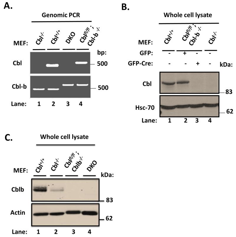
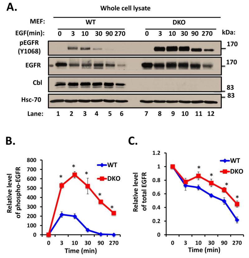
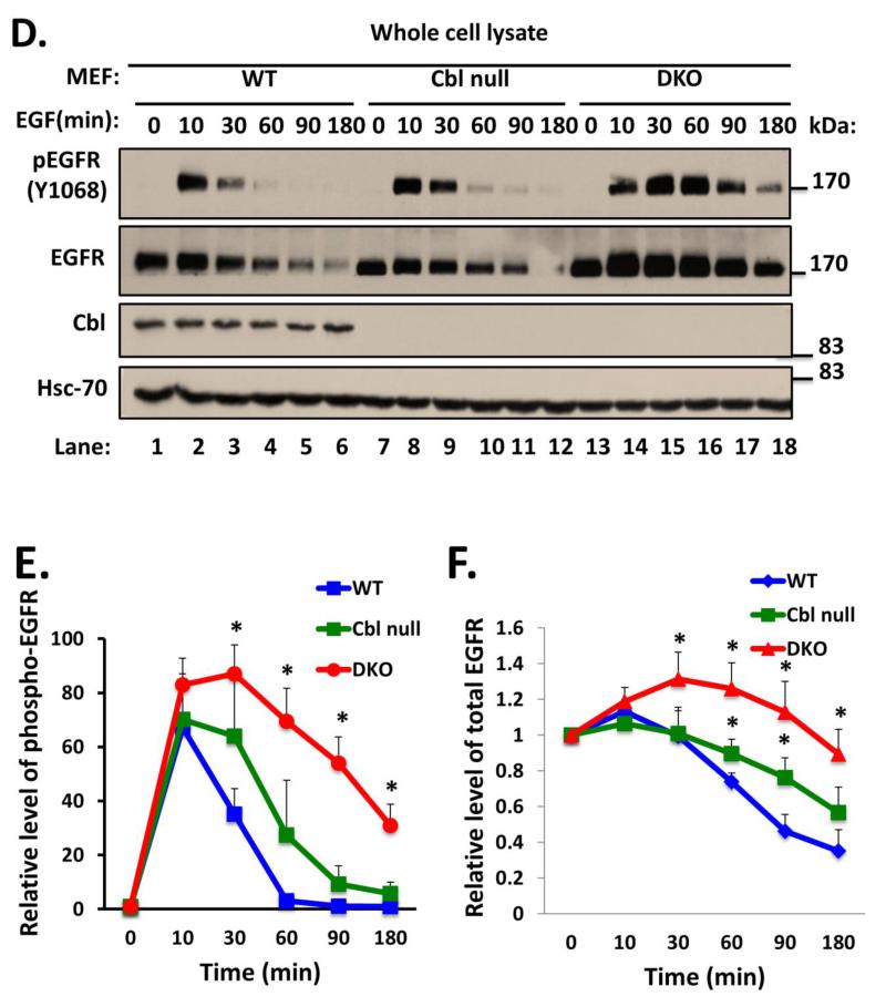
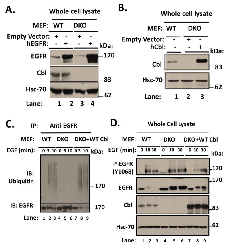
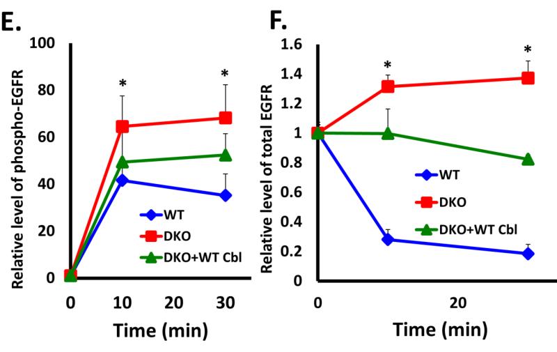
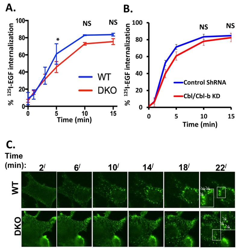
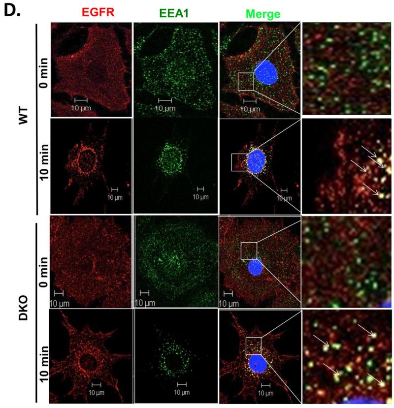
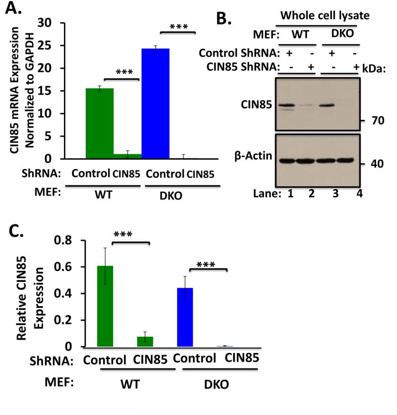
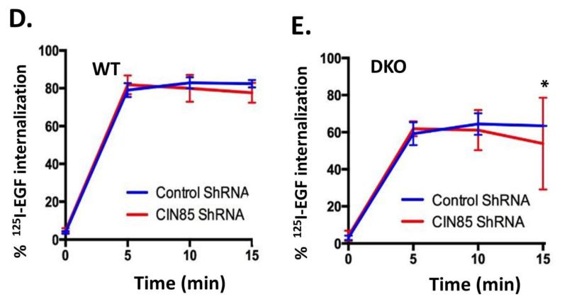
Members of the casitas B-lineage lymphoma (Cbl) family (Cbl, Cbl-b and Cbl-c) of ubiquitin ligases serve as negative regulators of receptor tyrosine kinases (RTKs). An essential role of Cbl-family protein-dependent ubiquitination for efficient ligand-induced lysosomal targeting and degradation is now well-accepted. However, a more proximal role of Cbl and Cbl-b as adapters for CIN85-endophilin recruitment to mediate ligand-induced initial internalization of RTKs is supported by some studies but refuted by others. Overexpression and/or incomplete depletion of Cbl proteins in these studies is likely to have contributed to this dichotomy. To address the role of endogenous Cbl and Cbl-b in the internalization step of RTK endocytic traffic, we established Cbl/Cbl-b double-knockout (DKO) mouse embryonic fibroblasts (MEFs) and demonstrated that these cells lack the expression of both Cbl-family members as well as endophilin A, while they express CIN85. We show that ligand-induced ubiquitination of EGFR, as a prototype RTK, was abolished in DKO MEFs, and EGFR degradation was delayed. These traits were reversed by ectopic human Cbl expression. EGFR endocytosis, assessed using the internalization of (125)I-labeled or fluorescent EGF, or of EGFR itself, was largely retained in Cbl/Cbl-b DKO compared to wild type MEFs. EGFR internalization was also largely intact in Cbl/Cbl-b depleted MCF-10A human mammary epithelial cell line. Inducible shRNA-mediated knockdown of CIN85 in wild type or Cbl/Cbl-b DKO MEFs had no impact on EGFR internalization. Our findings, establish that, at physiological expression levels, Cbl, Cbl-b and CIN85 are largely dispensable for EGFR internalization. Our results support the model that Cbl-CIN85-endophilin complex is not required for efficient internalization of EGFR, a prototype RTK.

Keywords: CIN85; Cbl; Epidermal growth factor receptor; Internalization; Mouse embryonic fibroblast.


Figures

FIGURE 1
FIGURE 1. Characterization of Cbl/Cblb DKO MEFs
A) Cblflox/flox; Cbl-b−/− MEFs were infected with Adeno-GFP-Cre to derive DKO MEFs. Genomic DNA isolated from parental Cblflox/flox; Cbl-b−/− MEFs, their Adeno-GFP-Cre infected DKO derivative, as well as control Cbl−/− and wildtype (WT) MEFs was subjected to PCR using specific primer pairs indicated in Supplementary Table S1. Absence of the floxed Cbl band in DKO MEFs confirmed the deletion. B) 100 μg aliquots of Triton X-100 lysate protein from WT, Cblflox/flox; Cbl-b−/−, adeno-Cre infected Cblflox/flox; Cbl-b−/− (DKO) and Cbl−/− MEFs were subjected to Western blotting with anti-Cbl and anti-HSC-70 (loading control) antibodies. Molecular weight markers in kilodaltons (kDa) are indicated. C) 100 μg aliquots of Triton X-100 lysate protein from WT, Cblflox/flox; Cbl-b−/−, adeno-Cre infected Cblflox/flox; Cbl-b−/− (DKO) and Cbl−/− MEFs were subjected to Western blotting with anti-Cbl-b and anti-β-actin (loading control) antibodies. Molecular weight markers in kilodaltons (kDa) are indicated.
FIGURE 2
FIGURE 2. Delayed ligand-induced downregulation of EGFR in Cbl/Cbl-b DKO MEFs
A) Wildtype (WT) and Cbl/Cbl-b DKO (DKO) MEFs were serum-starved for 48h, and then left unstimulated or were stimulated with 100 ng/ml EGF for the indicated time points in minutes (min). 100 μg aliquots of Triton X-100 lysate protein were subjected to Western blotting with anti-phospho-EGFR (Y1068) antibody to visualize phospho-EGFR (top panel), anti-EGFR antibody to visualize total EGFR (middle panel), anti-Cbl antibody to confirm lack of Cbl expression in DKO MEFs, and with anti-HSC-70 antibody as a loading control. The bands corresponding to phospho-EGFR (B) and total EGFR (C) were quantified using Image J software, and normalized relative to band intensities for the corresponding HSC-70 loading controls. The bars represent mean ± SEM of data from three independent experiments. D). WT, Cbl-null (Cbl-/-) and Cbl/Cbl-b DKO (DKO) MEFs were serum-starved for 48h, and then left unstimulated or were stimulated with 100 ng/ml EGF for the indicated time points. 100 μg aliquots of Triton X-100 lysate protein were subjected to Western blotting with anti-phospho EGFR (Y1068) antibody to visualize phospho-EGFR (top panel), anti-EGFR antibody to visualize total EGFR (middle panel), anti-Cbl antibody to confirm lack of Cbl expression in Cbl-null (Cbl-/-) and DKO MEFs and with anti-HSC-70 antibody as a loading control. The bands corresponding to phospho-EGFR (E) and total EGFR (F) were quantified using Image J software, and normalized relative to band intensities for the corresponding HSC-70 loading controls. The bars represent mean ± SEM of data from three independent experiments.
FIGURE 3
FIGURE 3. Restoration of defective ligand-induced EGFR turnover in Cbl/Cbl-b DKO MEFs by ectopically-expressed human Cbl
A) Wildtype (WT) or Cbl/Cbl-b DKO (DKO) MEF lines retrovirally transduced with human EGFR or the corresponding vector control were derived. 100 μg aliquots of Triton X-100 lysate protein from these lines were subjected to Western blotting with an anti-EGFR antibody to assess the increased EGFR levels in hEGFR-expressing lines (upper panel), anti-Cbl antibody to confirm lack of Cbl expression in DKO MEFs and anti-HSC-70 antibody (loading control). B) The human EGFR-expressing Cbl/Cbl-b DKO MEF lines in (A) were further transduced with a retrovirus encoding human Cbl and GFP (separated by IRES), and successfully transduced cells were FACS sorted to obtain stably transduced lines. 100 μg aliquots of Triton X-100 lysate protein from these lines and WT were subjected to Western blotting with antibodies against Cbl (top panel) and HSC-70 (loading control). C) WT, Cbl/Cbl-b DKO (DKO) and hCbl-reconstituted DKO MEFs were serum-starved for 48 hours and then stimulated with 100 ng/ml EGF for the indicated time points. Anti-EGFR immunoprecipitations from 1 mg aliquots of RIPA lysate protein were subjected to Western blotting with anti-ubiquitin (upper panel) and anti-EGFR antibodies. D) WT, Cbl/Cbl-b DKO (DKO) and human Cbl-reconstituted DKO MEFs were serum-starved and stimulated as in (C) for the indicated time points. 100 μg aliquots of Triton X-100 lysate protein from these lines were subjected to Western blotting with antibodies against phospho EGFR (Y1068) to visualize phospho-EGFR (top panel), anti-EGFR to visualize total EGFR (second panel), Cbl (third panel) and HSC-70 (bottom panel; loading control). The bands corresponding to phospho-EGFR (E) and total EGFR (F) were quantified using Image J software, and normalized relative to band intensities for the corresponding HSC-70 loading controls. The bars represent mean ± SEM of data from three independent experiments.
FIGURE 4
FIGURE 4. Complete lack of Cbl-family protein expression does not impair ligand-induced EGFR internalization
A) Triplicate wells of 6-well plates seeded with wildtype (WT) and Cbl/Cbl-b DKO (DKO) MEFs, and serum-starved for 24h, were incubated with 1 ng/ml 125I-EGF (about 200,000 cpm) at 4 °C for 120 minutes to allow EGF binding. Following washes, the cells were incubated at 37°C for the indicated times and the % of internalized relative to total 125I-EGF was plotted against time. The data points are mean ± SEM of data from three independent experiments. B) Control and Cbl/Cbl-b double knockdown MCF-10 human mammary epithelial cells described previously (Duan et al. 2011) and confirmed via anti-Cbl immunoblotting (Supplementary Fig. S4) were used for 125I-EGF internalization as in (A). The data points are mean ± SEM of three independent experiments. C) WT (upper panel) and Cbl/Cbl-b DKO (DKO; lower panel) MEFs expressing hEGFR (Fig. 3A) were serum-starved for 24h and Alexa Fluor® 488-conjugated EGF was allowed to bind to the surface of live cells at 4°C for 1h. The cells were then switched to 37°C and confocal images captured at 2 minutes intervals to assess the transfer of surface fluorescence into intracellular endocytic vesicles. D) WT (upper two panels) and Cbl/Cbl-b DKO (DKO; lower two panels) MEFs expressing human EGFR (Fig. 3A) were serum-starved for 24h, and either left unstimulated or stimulated with EGF (10 ng/ml) for 10 minutes. Cells were fixed and stained for human EGFR and EEA1. Co-localization was assessed in merged images using ZEN 2010 software. The panels on right are magnified images of the insets in third column, as shown. The arrows indicate the EGFR co-localization with the early endosome marker EEA1 (Scale bars: 10 μm).
FIGURE 5
FIGURE 5. CIN85 knockdown does not impair EGFR internalization
(A) Wildtype (WT) and Cbl/Cbl-b DKO (DKO) MEFs stably expressing the doxycycline-inducible control or CIN85 shRNA were induced with doxycycline for 4 days and real-time qPCR analysis was performed using the primers indicated in Supplementary Table S1 to confirm the CIN85 knockdown. GAPDH was used as a normalization control. B) WT and Cbl/Cbl-b DKO MEFs were induced with doxycycline as in (A) and 100 μg aliquots of Triton X-100 lysate protein from these cell lines were subjected to Western blotting with antibodies against CIN85 and β-actin (loading control). C) The bands corresponding to CIN85 in (B) were quantified using Image J software, and normalized relative to band intensities for the corresponding β-actin loading controls. The bars represent mean ± SEM of data from three independent experiments. D and E) WT (D) and Cbl/Cbl-b DKO (E) MEFs were induced with doxycycline as in (A), and used for 125I-EGF internalization as in Fig. 4A. The data points are mean ± SEM of three independent experiments.

References

    1. Bertelsen V, Sak MM, Breen K, Rodland MS, Johannessen LE, Traub LM, Stang E, Madshus IH. A chimeric pre-ubiquitinated EGF receptor is constitutively endocytosed in a clathrin-dependent, but kinase-independent manner. Traffic. 2011;12(4):507–20. - PubMed
    1. Dikic I. Mechanisms controlling EGF receptor endocytosis and degradation. Biochem Soc Trans. 2003;31(Pt 6):1178–81. - PubMed
    1. Dikic I. CIN85/CMS family of adaptor molecules. FEBS Lett. 2002;529(1):110–5. - PubMed
    1. Dou H, Buetow L, Hock A, Sibbet GJ, Vousden KH, Huang DT. Structural basis for autoinhibition and phosphorylation-dependent activation of c-cbl. Nat Struct Mol Biol. 2012;19(2):184–92. - PMC - PubMed
    1. Duan L, Raja SM, Chen G, Virmani S, Williams SH, Clubb RJ, Mukhopadhyay C, Rainey MA, Ying G, Dimri M, et al. Negative regulation of EGFR-Vav2 signaling axis by cbl ubiquitin ligase controls EGF receptor-mediated epithelial cell adherens junction dynamics and cell migration. J Biol Chem. 2011;286(1):620–33. - PMC - PubMed