. 2015 Feb;21(2):172-9.
doi: 10.1261/rna.048272.114. Epub 2014 Dec 1.

Efficient backsplicing produces translatable circular mRNAs

Affiliations

Efficient backsplicing produces translatable circular mRNAs

Yang Wang et al. RNA. 2015 Feb.

Abstract

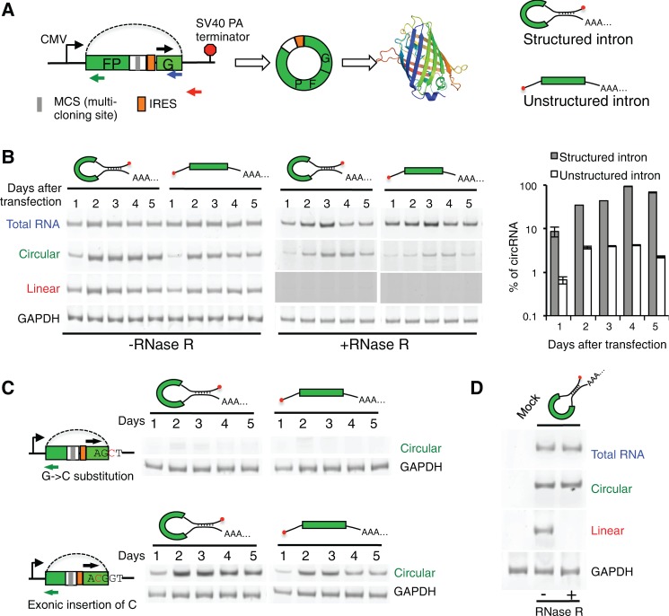
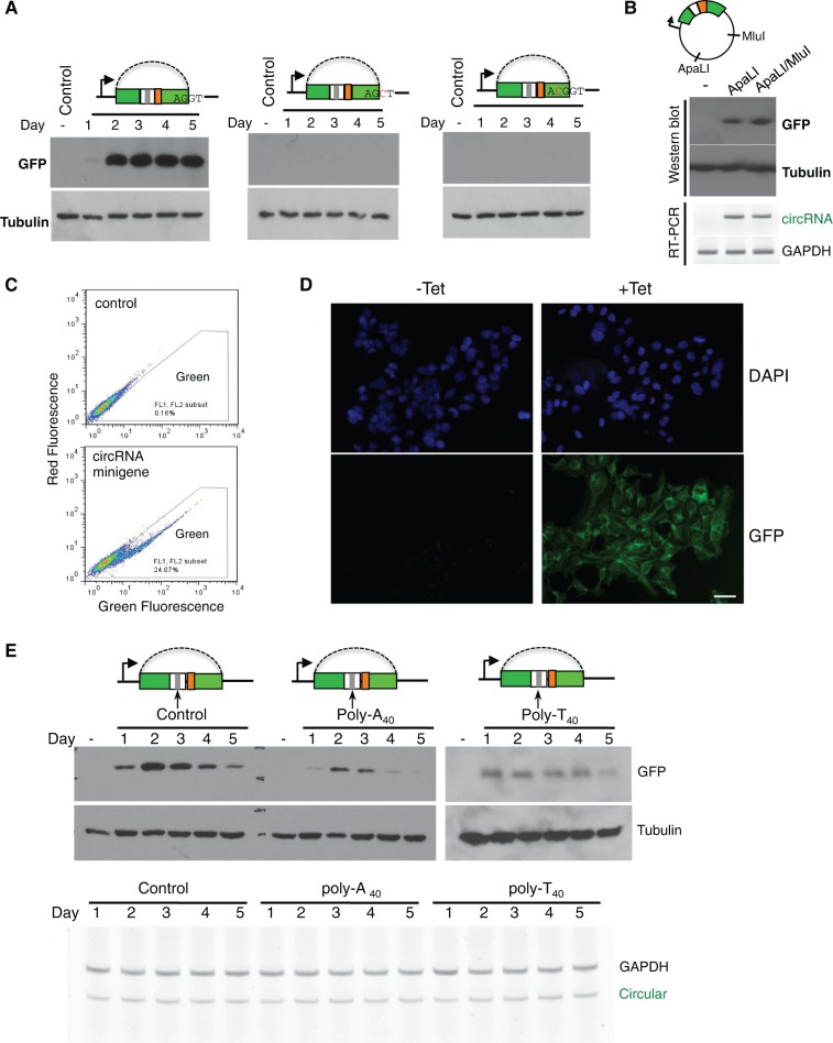
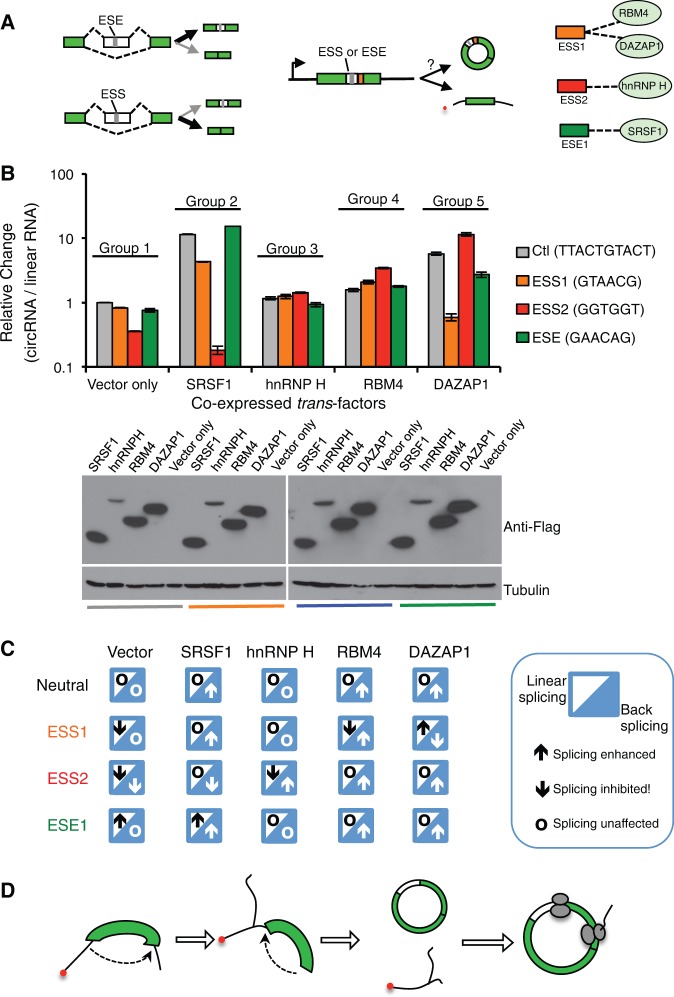
While the human transcriptome contains a large number of circular RNAs (circRNAs), the functions of most circRNAs remain unclear. Sequence annotation suggests that most circRNAs are generated from splicing in reversed orders across exons. However, the mechanisms of this backsplicing are largely unknown. Here we constructed a single exon minigene containing split GFP, and found that the pre-mRNA indeed produces circRNA through efficient backsplicing in human and Drosophila cells. The backsplicing is enhanced by complementary introns that form double-stranded RNA structure to bring splice sites in proximity, but such structure is not required. Moreover, backsplicing is regulated by general splicing factors and cis-elements, but with regulatory rules distinct from canonical splicing. The resulting circRNA can be translated to generate functional proteins. Unlike linear mRNA, poly-adenosine or poly-thymidine in 3' UTR can inhibit circular mRNA translation. This study revealed that backsplicing can occur efficiently in diverse eukaryotes to generate circular mRNAs.

Keywords: alternative splicing; backsplicing; circular RNA; splicing factors; translation.


Figures

FIGURE 1.
FIGURE 1.
circRNA generated from backsplicing. (A) Schematic diagram of minigene with split GFP in a reverse order. The transcription of minigene is driven by a CMV promoter and terminated by SV40 polyadenylation signal. The exon is represented by box, and the backsplicing is indicated with dotted line. Different pairs of primers were used to detect total, linear, or circular RNAs in RT-PCR. Two versions of the minigene, with structured or unstructured introns, were generated. (B) Different RNAs produced from minigenes with structured or unstructured introns. Total RNAs were purified at day 1 to 5 after transfection, treated with or without RNase R, and analyzed by semiquantitative RT-PCRs (22 cycles) using various primer sets. To ensure no linear product is detected in RNase R treated samples, we used a longer exposure time for the corresponding lanes. The amounts of different RNAs were also quantified by real-time RT-PCR, and the percent of circular RNA is plotted in the right panel. (C) Production of circular RNAs in minigenes with splice site mutation or exonic insertion. The circRNAs were detected at different days after transfection. (D) Production of circRNA in S2 cells. The minigene with structured intron was transfected to S2 cells, and different RNAs were detected as described in panel B.
FIGURE 2.
FIGURE 2.
Stability of circRNAs. (A) The RNA levels relative to day 1 of transfection were measured by qRT-PCR using primer sets for linear or circRNAs. The transfections were conducted with minigene containing structured intron, and all experiments are performed in triplicates with the means and SD of RNA levels plotted. In all panels, the RNA level of GAPDH was used to calibrate all other RNA levels. (B) Change of RNA levels after transcription inhibition by actinomycin D or DMSO control. Two minigenes containing structured or unstructured intron were transfected into 293T cells, and the levels of linear or circular RNAs were determined at different times after drug treatment. The level of a typical endogenous mRNA, SRSF1, was also measured as a control.
FIGURE 3.
FIGURE 3.
In vivo translation directed by circular mRNA. (A) Cells were transfected with wild-type or mutated minigenes containing structured introns, and total proteins were purified at different days after transfection to detect production of GFP. One mutated minigenes has a disrupted 5′ splice site (second panel), while the other contains a C residue inserted into exon to disrupt GFP reading frame (third panel). (B) The circRNA minigene vector has single ApaLI and MluI cut sites in the plasmid backbone. We linearized the wild-type minigene with ApaLI or ApaL1/MluI digestion, and transfected the linearized DNA into 293 cells to detect GFP expression at 2 d after transfection. The production of circRNA was also measured by RT-PCR as described in Figure 1. (C) Cells transiently transfected with backsplicing minigene are assayed by flow cytometry at 2 d after transfection. (D) 293 FlpIn T-REX cells were stably transfected with backsplicing minigene containing structured intron, and the production of GFP was induced by addition of tetracycline at a final concentration of 1 μg/mL. The cells were assayed by fluorescence microscopy 48 h after induction. Scale bar = 50 μm. (E) Production of GFPs from minigenes inserted with poly(A40), poly(T40), or nonrepetitive control sequence downstream from the stop codon was detected with Western blot. The tubulin antibody was used as a loading control. The Western blot at the right panel (poly-T40) was overexposed to show protein production. Bottom, the corresponding RNA levels from each transfected sample were determined with semiquantitative RT-PCR.
FIGURE 4.
FIGURE 4.
Regulation of backsplicing by cis-elements and splicing factors. (A) Schematic diagram of splicing regulation by SREs and splicing factors in linear and backsplicing. Linear splicing can be promoted or inhibited by ESEs or ESSs, respectively, which is mediated by their cognate splicing factors (left panel). The same ESSs or ESEs were inserted into the backspliced exons, and the resulting minigenes were coexpressed with various splicing factors to measure levels of circular or linear RNA using quantitative RT-PCR. The interactions between SREs and cognate splicing factors were also shown. (B) Backsplicing minigenes inserted with various ESSs or ESE were coexpressed with four different splicing factors (SRSF1, hnRNP H, RBM4, and DAZAP1). The relative levels of circular RNA were compared with the control vector inserted with a neutral sequence. The expression of splicing factors was confirmed by Western blots. (C) Summary of regulatory rules for linear and backsplicing by various SREs and splicing factors. In each case, the enhancement or inhibition of linear splicing and backsplicing was indicated by an upward or downward arrow. The circles indicate the case where splicing was not affected. The activities of each combination of the SRE and splicing factor are based on data from panel B and previous results (Wang et al. 2012, 2013). (D) A model of backsplicing and translation from circular mRNA.

References

    1. Burd CE, Jeck WR, Liu Y, Sanoff HK, Wang Z, Sharpless NE 2010. Expression of linear and novel circular forms of an INK4/ARF-associated non-coding RNA correlates with atherosclerosis risk. PLoS Genet 6:e1001233. - PMC - PubMed
    1. Chen CY, Sarnow P 1995. Initiation of protein synthesis by the eukaryotic translational apparatus on circular RNAs. Science 268:415–417. - PubMed
    1. Choudhury R, Roy SG, Tsai YS, Tripathy A, Graves LM, Wang Z 2014. The splicing activator DAZAP1 integrates splicing control into MEK/Erk-regulated cell proliferation and migration. Nat Commun 5:3078. - PMC - PubMed
    1. Cocquerelle C, Daubersies P, Majerus MA, Kerckaert JP, Bailleul B 1992. Splicing with inverted order of exons occurs proximal to large introns. EMBO J 11:1095–1098. - PMC - PubMed
    1. De Conti L, Baralle M, Buratti E 2013. Exon and intron definition in pre-mRNA splicing. Wiley Interdiscip Rev RNA 4:49–60. - PubMed

Publication types