

Ferumoxytol administration does not alter infarct volume or the inflammatory response to stroke in mice
- PMID: 25449870
- PMCID: PMC4268374
- DOI: 10.1016/j.neulet.2014.10.041
Ferumoxytol administration does not alter infarct volume or the inflammatory response to stroke in mice
Abstract
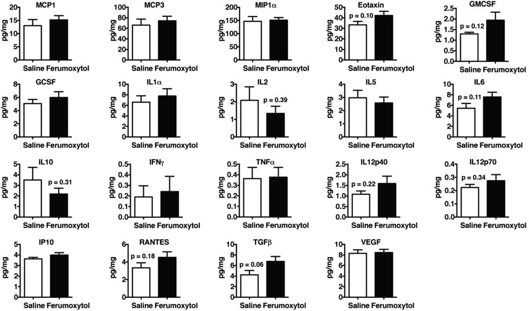
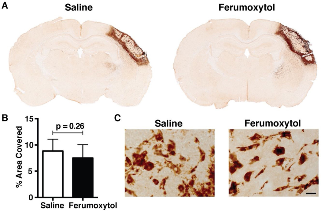
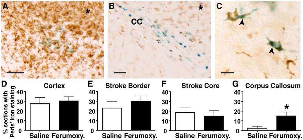
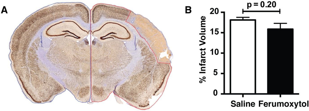
Ferumoxytol is an ultrasmall superparamagnetic iron oxide (USPIO) nanoparticle that is FDA-approved as an intravenous iron replacement therapy for the treatment of iron deficiency anemia in patients with chronic kidney disease. Ferumoxytol has also been used as a contrast agent for cerebral blood volume mapping by magnetic resonance imaging (MRI), which suggests it could be used for imaging hemodynamic abnormalities after stroke. However, circulating macrophages can internalize USPIOs, and recent data indicate that the accumulation of iron in macrophages can lead them to adopt the M1 pro-inflammatory phenotype. Therefore, the uptake of intravenously administered iron particles by circulating macrophages that home to the stroke core could potentially alter the inflammatory response to stroke. To test this possibility in vivo we administered a dose of ferumoxytol previously used to obtain cerebral blood volume maps in healthy humans by steady-state susceptibility contrast (SSC) MRI to BALB/cJ mice 48h after stroke and examined cytokine levels, microglial/macrophage activation, and lesion volume in the brain 5 days later. Treatment with ferumoxytol did not lead to any differences in these parameters. These data indicate that the use of ferumoxytol as a contrast agent for brain imaging after stroke does not alter the inflammatory response to stroke in mice, and is therefore unlikely to do so in human subjects.
Keywords: Ferumoxytol; Inflammation; Magnetic resonance imaging; Stroke.
Copyright © 2014 Elsevier Ireland Ltd. All rights reserved.
Figures
Similar articles
-
Emerging applications for ferumoxytol as a contrast agent in MRI.J Magn Reson Imaging. 2015 Apr;41(4):884-98. doi: 10.1002/jmri.24691. Epub 2014 Jun 30. J Magn Reson Imaging. 2015. PMID: 24974785 Review.
-
Differential uptake of ferumoxtran-10 and ferumoxytol, ultrasmall superparamagnetic iron oxide contrast agents in rabbit: critical determinants of atherosclerotic plaque labeling.J Magn Reson Imaging. 2005 Apr;21(4):432-42. doi: 10.1002/jmri.20283. J Magn Reson Imaging. 2005. PMID: 15779033
-
In vivo analysis of neuroinflammation in the late chronic phase after experimental stroke.Neuroscience. 2015 Apr 30;292:71-80. doi: 10.1016/j.neuroscience.2015.02.024. Epub 2015 Feb 18. Neuroscience. 2015. PMID: 25701708
-
Macrophage Imaging of Cerebral Aneurysms with Ferumoxytol: an Exploratory Study in an Animal Model and in Patients.J Stroke Cerebrovasc Dis. 2017 Oct;26(10):2055-2064. doi: 10.1016/j.jstrokecerebrovasdis.2016.10.026. Epub 2017 Jul 31. J Stroke Cerebrovasc Dis. 2017. PMID: 28774792 Clinical Trial.
-
USPIO-Enhanced MRI Neuroimaging: A Review.J Neuroimaging. 2016 Mar-Apr;26(2):161-8. doi: 10.1111/jon.12318. Epub 2015 Dec 3. J Neuroimaging. 2016. PMID: 26932522 Review.
Cited by
-
Magnetic-Driven Torque-Induced Electrical Stimulation for Millisecond-Scale Wireless Neuromodulation.Adv Healthc Mater. 2025 Aug;14(20):e2500805. doi: 10.1002/adhm.202500805. Epub 2025 Jun 16. Adv Healthc Mater. 2025. PMID: 40522190 Free PMC article.
-
Ferumoxytol-enhanced MRI assessment of venous Thrombus resolution and macrophage content in a murine deep vein thrombosis model.Thromb Res. 2024 Aug;240:109063. doi: 10.1016/j.thromres.2024.109063. Epub 2024 Jun 13. Thromb Res. 2024. PMID: 38878741 Free PMC article.
-
Pharmacokinetic Profiling of Unlabeled Magnetic Nanoparticles Using Magnetic Particle Imaging as a Novel Cold Tracer Assay.Nano Lett. 2024 Dec 11;24(49):15557-15564. doi: 10.1021/acs.nanolett.4c03553. Epub 2024 Nov 26. Nano Lett. 2024. PMID: 39591368 Free PMC article.
-
Advances in Vascular Diagnostics using Magnetic Particle Imaging (MPI) for Blood Circulation Assessment.Adv Healthc Mater. 2024 Sep;13(23):e2400612. doi: 10.1002/adhm.202400612. Epub 2024 Jun 28. Adv Healthc Mater. 2024. PMID: 38879782 Review.
-
Superparamagnetic iron oxide nanoparticles as a tool to track mouse neural stem cells in vivo.Mol Biol Rep. 2019 Feb;46(1):191-198. doi: 10.1007/s11033-018-4460-9. Epub 2018 Nov 12. Mol Biol Rep. 2019. PMID: 30421128
References
-
- Atkinson DJ, Burstein D, Edelman RR. First-pass cardiac perfusion: evaluation with ultrafast MR imaging. Radiology. 1990;174:757–762. - PubMed
-
- Christen T, Ni W, Qiu D, Schmiedeskamp H, Bammer R, Moseley M, Zaharchuk G. High-resolution cerebral blood volume imaging in humans using the blood pool contrast agent ferumoxytol. Magnetic resonance in medicine : official journal of the Society of Magnetic Resonance in Medicine / Society of Magnetic Resonance in Medicine. 2012 - PubMed
Publication types
MeSH terms
Substances
Grants and funding
LinkOut - more resources
Full Text Sources
Other Literature Sources
Medical
