

Neuro-ophthalmic manifestations of prematurity
- PMID: 25449932
- PMCID: PMC4278126
- DOI: 10.4103/0301-4738.145990
Neuro-ophthalmic manifestations of prematurity
Abstract
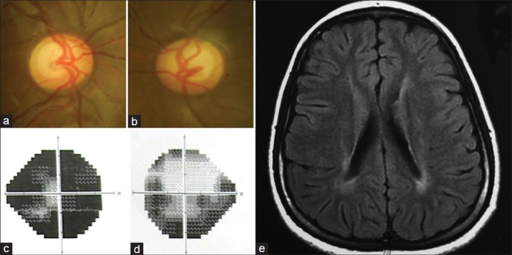
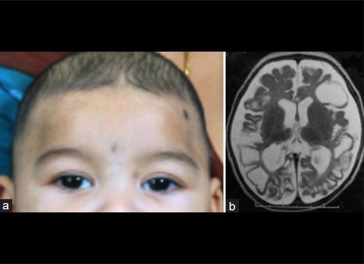
Increasing rates of preterm births coupled with better survival of these infants have resulted in higher prevalence of systemic and ocular complications associated with prematurity. In addition to retinopathy of prematurity, infants who are born preterm may suffer from severe visual impairment as a result of hypoxic ischemic encephalopathy, hypoglycemia, and other metabolic imbalances. The effect of these processes on the anterior visual pathway may result in optic atrophy, optic nerve hypoplasia or optic disc cupping and affection of the posterior visual pathway leads to cortical visual impairment (CVI). Other ocular associations include strabismus, nystagmus, and ocular motor abnormalities such as tonic down gaze and defective saccades and pursuits. Cortical and subcortical involvement also manifests as defects in functional vision and these have not yet been completely understood. Children with CVI may have visual field defects, photophobia, defective visual processing, and deficient color vision. Since most of these children also suffer from additional systemic disabilities, evaluation, and management remains a challenge. However, early diagnosis and initiation of rehabilitation therapy can prove to be of significant benefit in these children.
Conflict of interest statement
Figures

References
-
- Blencowe H, Cousens S, Oestergaard MZ, Chou D, Moller AB, Narwal R, et al. National, regional, and worldwide estimates of preterm birth rates in the year 2010 with time trends since 1990 for selected countries: A systematic analysis and implications. Lancet. 2012;379:2162–72. - PubMed
-
- Robaei D, Kifley A, Gole GA, Mitchell P. The impact of modest prematurity on visual function at age 6 years: Findings from a population-based study. Arch Ophthalmol. 2006;124:871–7. - PubMed
-
- O’Connor AR, Wilson CM, Fielder AR. Ophthalmological problems associated with preterm birth. Eye (Lond) 2007;21:1254–60. - PubMed
Publication types
MeSH terms
LinkOut - more resources
Full Text Sources
Other Literature Sources
Medical
