. 2015 Feb;24(2):253-63.
doi: 10.1002/pro.2612. Epub 2014 Dec 26.

A moonlighting function of Mycobacterium smegmatis Ku in zinc homeostasis?

Affiliations

A moonlighting function of Mycobacterium smegmatis Ku in zinc homeostasis?

Ambuj K Kushwaha et al. Protein Sci. 2015 Feb.

Abstract

Ku protein participates in DNA double-strand break repair via the nonhomologous end-joining pathway. The three-dimensional structure of eukaryotic Ku reveals a central core consisting of a β-barrel domain and pillar and bridge regions that combine to form a ring-like structure that encircles DNA. Homologs of Ku are encoded by a subset of bacterial species, and they are predicted to conserve this core domain. In addition, the bridge region of Ku from some bacteria is predicted from homology modeling and sequence analyses to contain a conventional HxxC and CxxC (where x is any residue) zinc-binding motif. These potential zinc-binding sites have either deteriorated or been entirely lost in Ku from other organisms. Using an in vitro metal binding assay, we show that Mycobacterium smegmatis Ku binds two zinc ions. Zinc binding modestly stabilizes the Ku protein (by ∼3°C) and prevents cysteine oxidation, but it has little effect on DNA binding. In vivo, zinc induces significant upregulation of the gene encoding Ku (∼sixfold) as well as a divergently oriented gene encoding a predicted zinc-dependent MarR family transcription factor. Notably, overexpression of Ku confers zinc tolerance on Escherichia coli. We speculate that zinc-binding sites in Ku proteins from M. smegmatis and other mycobacterial species have been evolutionarily retained to provide protection against zinc toxicity without compromising the function of Ku in DNA double-strand break repair.

Keywords: DNA binding; Ku protein; MarR; PAR assay; thermal stability; zinc binding.


Figures

Figure 1
Figure 1
Model of M. smegmatis Ku illustrating the predicted zinc-binding sites. Each monomer (in purple and light pink) is modeled on template strands 1jeyA and 1jeyB. The residues previously predicted to coordinate zinc are highlighted in different colors: cysteine (red), histidine (orange), and aspartic acid (cyan). The model was created using SwissModel in automatic mode. The image was prepared with PyMOL (www.pymol.org).
Figure 2
Figure 2
Multiple sequence alignment of bacterial Ku homologs. The first and last residue numbers are indicated before and after each sequence in the alignment. Positions corresponding to zinc-binding residues are boxed and marked by asterisks below the alignment. B_meg, Bacillus megaterium; B_ant, Bacillus anthracis; B_pum, Bacillus pumilus; B_sub, Bacillus subtilis; M_smg, Mycobacterium smegmatis; M_JLS, Mycobacterium Sp. JLS; M_KMS, Mycobacterium Sp. KMS; M_gil, Mycobacterium gilvum; M_van, Mycobacterium vanbaalenii; M_tub, Mycobacterium tuberculosis; M_bov, Mycobacterium bovis; M_mar, Mycobacterium marinum; M_ulc, Mycobacterium ulcerans; M_kan, Mycobacterium kansasii; M_avi, Mycobacterium avium; M_int, Mycobacterium intracellulare; S_coe, Streptomyces coelicolor; P_stu, Pseudomonas stutzeri; P_aeu, Pseudomonas aeruginosa; P_put, Pseudomonas putida.
Figure 3
Figure 3
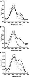
Zinc binding by Ku. Release of zinc from Ku monitored by its complexation with PAR, resulting in a diagnostic absorbance at 500 nm; uncomplexed PAR has an absorbance maximum at 416 nm. (A) Zinc release from native Ku. Absorbance spectrum of zinc-free native Ku (gray line); zinc-bound native Ku (black line); zinc-bound native Ku without His6-tag (dashed line). (B) Zinc release from denatured Ku. Denatured zinc-free Ku (gray line); denatured zinc-bound Ku (protein denatured after incubation with zinc; black line); denatured zinc-bound Ku without His6-tag (dashed line). Denaturation by SDS resulted in an overall increase in the baseline. (C) Zinc release from KuC8891A. Absorbance spectrum of PAR (dashed line); denatured KuC8891A (gray line); denatured zinc-bound KuC8891A (black line).
Figure 4
Figure 4
Affinity of Ku for zinc. Concentration of PAR2Zn, calculated based on the absorbance at 500 nm, as a function of added protein. His6-tagged protein was titrated into a solution of 210 µM PAR and 10 µM ZnCl2. An apparent Kd of 0.8 ± 0.3 nM was estimated. A representative experiment is shown.
Figure 5
Figure 5
Melting temperature determination by differential scanning fluorimetry. Fluorescence of SYPRO Orange bound to exposed hydrophobic protein regions as a function of temperature. (A) Thermal denaturation curve of zinc-free Ku. Tm (with S.D.) is 44.3 ± 0.3°C. (B) Thermal denaturation curve of bipyridyl-treated Ku. Tm (with S.D.) is 44.3 ± 0.2°C. C. Thermal denaturation curve of zinc-bound Ku. Tm (with S.D.) is 47.1 ± 0.1°C.
Figure 6
Figure 6
SDS-PAGE analysis of protein oxidation. (A) The effect of oxidizing and reducing agent on zinc-free native Ku and bipyridyl-treated Ku. Lane 1: molecular weight markers (identified at the left in kDa), lane 2: 5 µg of zinc-free native Ku with DTT (10 mM); lane 3: 5 µg of zinc-free native Ku with H2O2 (10 mM); lane 4: 5 µg of bipyridyl-treated native Ku with DTT (10 mM); lane 5: 5 µg of bipyridyl-treated native Ku with H2O2 (10 mM). Oxidized species with lower electrophoretic mobility identified by an arrow. (B) The effect of oxidizing and reducing agent on zinc-bound native Ku. Lane 1: molecular weight markers (identified at the left in kDa); lane 2: 5 µg of zinc-bound native Ku with H2O2 (10 mM); lane 3: 5 µg of zinc-bound native Ku with DTT (10 mM).
Figure 7
Figure 7
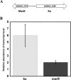
The effect of zinc on the expression of M. smegmatis ku and marR. (A) Genetic locus organization of M. smegmatis ku and marR genes. (B) Relative abundance of transcript levels of ku and marR genes after the addition of 2 mM of zinc. mRNA levels were measured with qRT-PCR and the relative abundance was calculated by the comparative CT method with reference to transcript level of control. The error bars represent the S.D. of the three experiments.
Figure 8
Figure 8
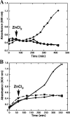
Growth of E. coli Rosetta cells in the presence of 1 mM of ZnCl2. The horizontal axis shows time after the induction of protein expression. (A) Growth curve of cells induced for Ku expression (solid diamond); growth curve of uninduced cells (solid circle); growth curve of E. coli cells harboring plasmid without ku gene (solid square). (B) Growth curve of cells induced for Ku mutant expression in the presence of ZnCl2 (solid circle); growth curve of uninduced cells in the presence of ZnCl2 (solid square); growth curve of E. coli cells in the absence of ZnCl2 (solid triangle). Addition of ZnCl2 is marked with an arrow. Representative experiments are shown.

References

    1. Doherty AJ, Jackson SP, Weller GR. Identification of bacterial homologues of the Ku DNA repair proteins. FEBS Lett. 2001;500:186–188. - PubMed
    1. Featherstone C, Jackson SP. Ku, a DNA repair protein with multiple cellular functions? Mutat Res. 1999;434:3–15. - PubMed
    1. Yoo S, Dynan WS. Geometry of a complex formed by double strand break repair proteins at a single DNA end: recruitment of DNA-PKcs induces inward translocation of Ku protein. Nucleic Acids Res. 1999;27:4679–4686. - PMC - PubMed
    1. Weller GR, Kysela B, Roy R, Tonkin LM, Scanlan E, Della M, Devine SK, Day JP, Wilkinson A, d'Adda di Fagagna F, Devine KM, Bowater RP, Jeggo PA, Jackson SP, Doherty AJ. Identification of a DNA nonhomologous end-joining complex in bacteria. Science. 2002;297:1686–1689. - PubMed
    1. Aravind L, Koonin EV. Prokaryotic homologs of the eukaryotic DNA-end-binding protein Ku, novel domains in the Ku protein and prediction of a prokaryotic double-strand break repair system. Genome Res. 2001;11:1365–1374. - PMC - PubMed

Publication types

LinkOut - more resources