. 2015 Jan:78:179-89.
doi: 10.1016/j.freeradbiomed.2014.10.582. Epub 2014 Nov 5.

Redox regulation of mitophagy in the lung during murine Staphylococcus aureus sepsis

Affiliations

Redox regulation of mitophagy in the lung during murine Staphylococcus aureus sepsis

Alan L Chang et al. Free Radic Biol Med. 2015 Jan.

Abstract

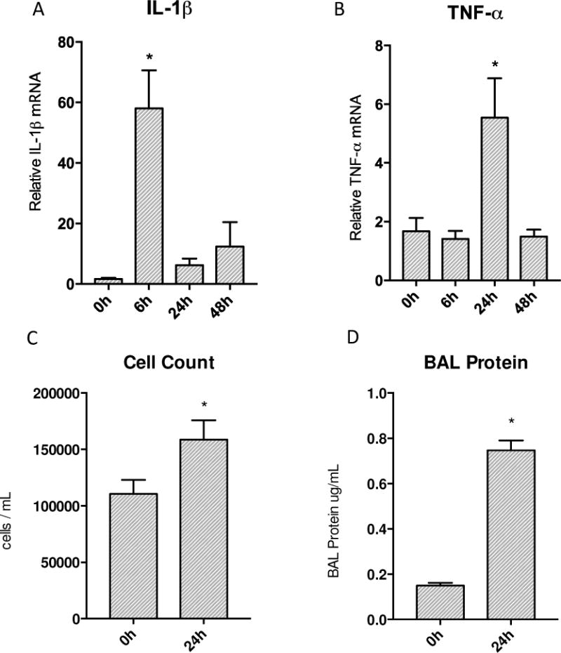
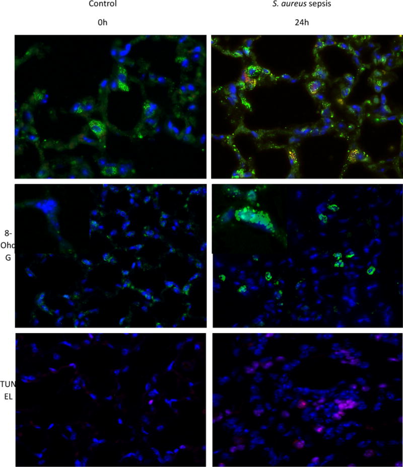
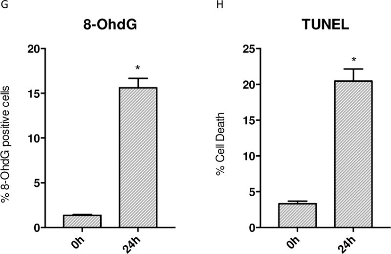
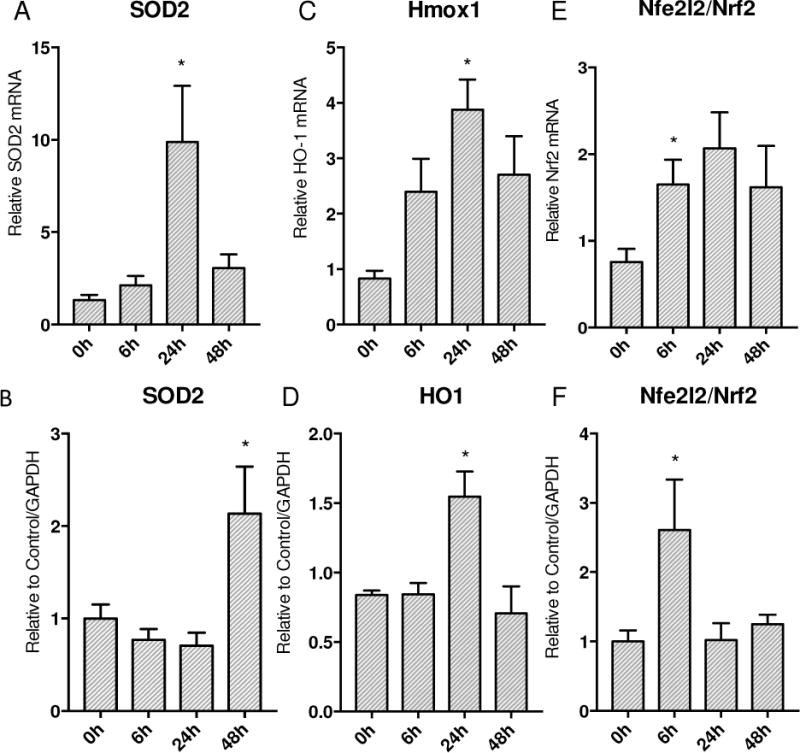
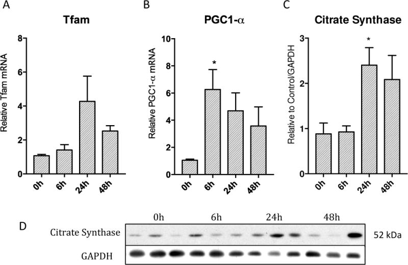
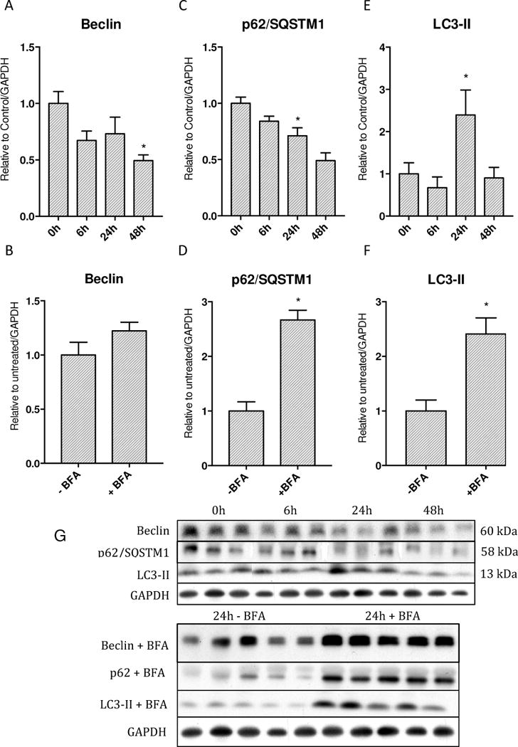
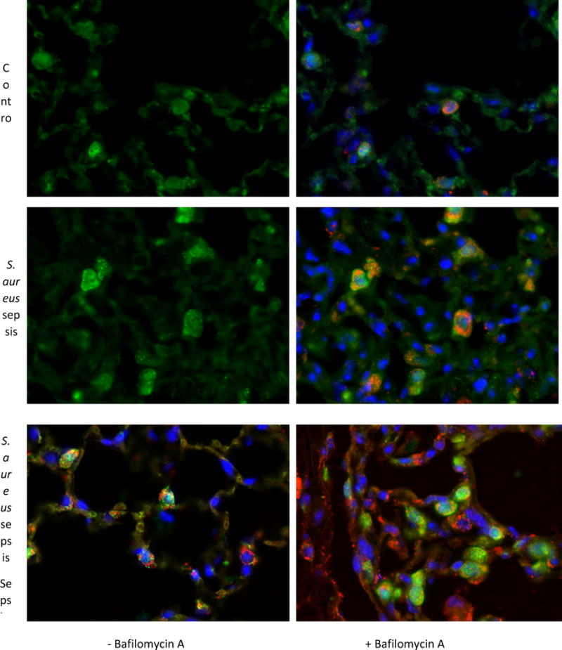
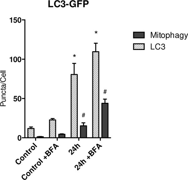
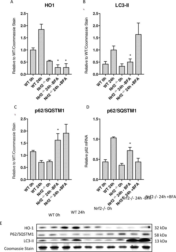
Oxidative mitochondrial damage is closely linked to inflammation and cell death, but low levels of reactive oxygen and nitrogen species serve as signals that involve mitochondrial repair and resolution of inflammation. More specifically, cytoprotection relies on the elimination of damaged mitochondria by selective autophagy (mitophagy) during mitochondrial quality control. This aim of this study was to identify and localize mitophagy in the mouse lung as a potentially upregulatable redox response to Staphylococcus aureus sepsis. Fibrin clots loaded with S. aureus (1×10(7) CFU) were implanted abdominally into anesthetized C57BL/6 and B6.129X1-Nfe2l2tm1Ywk/J (Nrf2(-/-)) mice. At the time of implantation, mice were given vancomycin (6mg/kg) and fluid resuscitation. Mouse lungs were harvested at 0, 6, 24, and 48h for bronchoalveolar lavage (BAL), Western blot analysis, and qRT-PCR. To localize mitochondria with autophagy protein LC3, we used lung immunofluorescence staining in LC3-GFP transgenic mice. In C57BL/6 mice, sepsis-induced pulmonary inflammation was detected by significant increases in mRNA for the inflammatory markers IL-1β and TNF-α at 6 and 24h, respectively. BAL cell count and protein also increased. Sepsis suppressed lung Beclin-1 protein, but not mRNA, suggesting activation of canonical autophagy. Notably sepsis also increased the LC3-II autophagosome marker, as well as the lung׳s noncanonical autophagy pathway as evidenced by loss of p62, a redox-regulated scaffolding protein of the autophagosome. In LC3-GFP mouse lungs, immunofluorescence staining showed colocalization of LC3-II to mitochondria, mainly in type 2 epithelium and alveolar macrophages. In contrast, marked accumulation of p62, as well as attenuation of LC3-II in Nrf2-knockout mice supported an overall decrease in autophagic turnover. The downregulation of canonical autophagy during sepsis may contribute to lung inflammation, whereas the switch to noncanonical autophagy selectively removes damaged mitochondria and accompanies tissue repair and cell survival. Furthermore, mitophagy in the alveolar region appears to depend on activation of Nrf2. Thus, efforts to promote mitophagy may be a useful therapeutic adjunct for acute lung injury in sepsis.

Keywords: Acute lung injury; Autophagy; Inflammation; LC3; Mitochondria; Mitophagy; Nrf2; Oxidative stress; Sepsis; p62.


Figures

Figure 1
Figure 1
Transcription of inflammatory cytokines and end organ inflammation produced by S. aureus peritonitis. A&B) Transcription of IL-1β and TNF-α increased significantly post inoculation in the lung. IL-1β showed a robust increase at 6h then returned to baseline values while TNF-α increased significantly by 24h and then returned to base line afterwards. C&D) Significant increases seen in both cell counts and protein in bronchial alveolar lavage. (* P < 0.05 vs. time 0h)
Figure 2
Figure 2
Immunofluorescence of WT mice given S. aureus 1×10⁷ CFU at 24h stained for ssDNA. A) The control mouse shows low fluorescence of ssDNA (stained red) that is colocalized with SOD2 (green) B) Sepsis increases the amount of ssDNA present. C) Control WT mouse lung stained for 8-OhdG (green) D) WT mouse lung stained for 8-OhdG at 24h after clot implantation. E) Control WT mouse lung with TUNEL staining at 0h. F) WT mouse lung with TUNEL staining after 24h after clot implantation. G) Quantified panels C&D for 8-OhdG positive cells. H) Quantified data from panels E&F for presence of cell death. (* P < 0.05 vs. time 0h)
Figure 2
Figure 2
Immunofluorescence of WT mice given S. aureus 1×10⁷ CFU at 24h stained for ssDNA. A) The control mouse shows low fluorescence of ssDNA (stained red) that is colocalized with SOD2 (green) B) Sepsis increases the amount of ssDNA present. C) Control WT mouse lung stained for 8-OhdG (green) D) WT mouse lung stained for 8-OhdG at 24h after clot implantation. E) Control WT mouse lung with TUNEL staining at 0h. F) WT mouse lung with TUNEL staining after 24h after clot implantation. G) Quantified panels C&D for 8-OhdG positive cells. H) Quantified data from panels E&F for presence of cell death. (* P < 0.05 vs. time 0h)
Figure 3
Figure 3
Response of ARE regulated gene expression to S. aureus 1×10⁷ CFU murine sepsis in the lung. A&B) SOD2 mRNA transcription expression increase by 24h with protein levels following by 48h. C&D) HO-1 mRNA transcription increases significantly by 24h with subsequent protein increase at 24h. E) mRNA transcription of Nrf2 increases significantly at 24h. F) Nrf2 protein shows early up-regulation by 6h. (* P < 0.05 vs. time 0h)
Figure 4
Figure 4
Activation of PGC1-α co-activator, nuclear encoded Tfam, and resulting increase in mitochondrial protein citrate synthase over 48h in lung parenchyma after S. aureus sepsis. A) Tfam mRNA increases in transcription robustly at 24h then starts to decrease by 48h. B) PGC1-α mRNA transcription increases significantly early on at 6h then remains elevated throughout 48h C) Mitochondrial protein citrate synthase is significantly increased by 24h and remains elevated through 48h. D) Western blots of above proteins (* p < 0.05)
Figure 5
Figure 5
Levels of autophagy proteins and assessment of autophagic flux with Bafilomycin A. A&B) Beclin protein levels decrease starting at 6h and continue to decrease through 48h, however Bafilomycin produces nonsignificant increase. C&D) p62 protein levels decrease starting at 6h and reach significance vs. 0h at 48h while inhibition of autophagy with Bafilomycin shows significant accumulation E&F) LC3-II protein levels increase significantly at 24h then return to baseline by 48h. Bafilomycin produces significant accumulation G) Western blots of above proteins. (* p < 0.05).
Figure 6
Figure 6
Immunofluorescence of LC3-GFP reporter mice lungs after S. aureus 1×10⁷ CFU at 24 hours as well as Bafilomycin A. A) The control mouse shows a low number of LC3 puncta B) Citrate synthase is stained red and nuclei are stained blue. C) Sepsis induces more LC3 to form puncta at 24 hours namely in alveolar type 2 as well as macrophages. D) Sepsis induces increased citrate synthase as well as a LC3 puncta expression in alveolar type 2 cells and macrophages. There is more co-localization of LC3 to citrate synthase than in panels A and B. E) There are definite LC3 puncta in alveolar type 2 cells as well as a macrophage that are co-localized with mitochondria as well. F) With Bafilomycin A, there is an increase in LC3 puncta as well as citrate synthase.
Figure 7
Figure 7
Number of autophagic and mitophagic sites (puncta) in LC3-GFP mice during sepsis. Sepsis causes a significant increase in GFP puncta per cell. Colocalization of GFP with mitochondria shows an increase in mitophagic puncta per cell in the setting of sepsis. ( * = p < 0.05 compared to LC3 puncta in WT control; # = p < 0.05 compared to colocalization in WT control)
Figure 8
Figure 8
Decreased autophagy in Nrf2 deficient mice during S. aureus 5×10⁶ peritonitis. A) HO-1 in Nrf2 KO mice show an overall diminished response to sepsis. At 24h Nrf2 KO mice had significantly lower HO-1 levels than WT mice at 24h in the same sepsis model. B) LC3-II is significantly decreased in Nrf2 deficient mice at 24h compared to wild-type C57BL/6 mice. C) p62 levels show accumulation at 24h at the protein level significantly compared to wild-type mice at 24h. D) p62 mRNA shows increased transcription compared to 0h wild-type and Nrf2 deficient mice although decreased copy number compared to wild-type mice at 24h. E) Western blots of above proteins. (* = p < 0.05 compared to WT 24h)

References

    1. Annane D, Aegerter P, Jars-Guincestre MC, Guidet B. Current Epidemiology of Septic Shock. American Thoracic Society; 2012. http://dx.doi.org/10.1164/rccm.2201087. - DOI - PubMed
    1. Hotchkiss RS, Karl IE. The Pathophysiology and Treatment of Sepsis. N Engl J Med. 2003;348:138–150. - PubMed
    1. Galley HF. Oxidative stress and mitochondrial dysfunction in sepsis. British Journal of Anaesthesia. 2011;107:57–64. - PubMed
    1. Cash TP, Pan Y, Simon MC. Reactive oxygen species and cellular oxygen sensing. Free Radical Biology and Medicine. 2007;43:1219–1225. - PMC - PubMed
    1. Singer M. The role of mitochondrial dysfunction in sepsis-induced multi-organ failure. Virulence Landes Bioscience. 2013;5:0–1. - PMC - PubMed

MeSH terms