

Mitochondrial calcium and the regulation of metabolism in the heart
- PMID: 25450609
- PMCID: PMC6534814
- DOI: 10.1016/j.yjmcc.2014.10.019
Mitochondrial calcium and the regulation of metabolism in the heart
Abstract
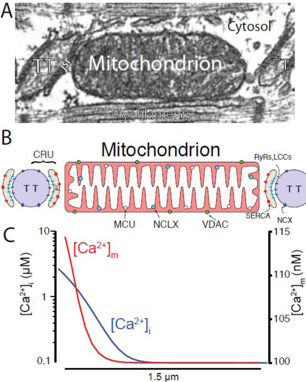
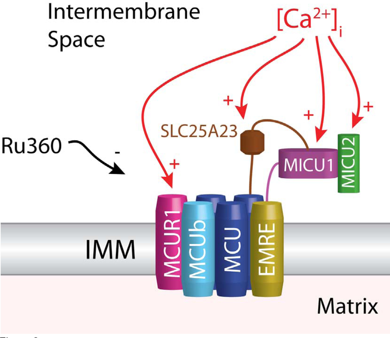
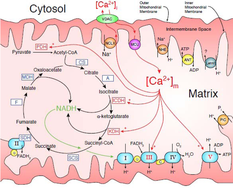
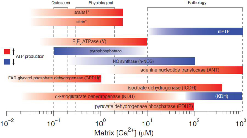
Consumption of adenosine triphosphate (ATP) by the heart can change dramatically as the energetic demands increase from a period of rest to strenuous activity. Mitochondrial ATP production is central to this metabolic response since the heart relies largely on oxidative phosphorylation as its source of intracellular ATP. Significant evidence has been acquired indicating that Ca(2+) plays a critical role in regulating ATP production by the mitochondria. Here the evidence that the Ca(2+) concentration in the mitochondrial matrix ([Ca(2+)]m) plays a pivotal role in regulating ATP production by the mitochondria is critically reviewed and aspects of this process that are under current active investigation are highlighted. Importantly, current quantitative information on the bidirectional Ca(2+) movement across the inner mitochondrial membrane (IMM) is examined in two parts. First, we review how Ca(2+) influx into the mitochondrial matrix depends on the mitochondrial Ca(2+) channel (i.e., the mitochondrial calcium uniporter or MCU). This discussion includes how the MCU open probability (PO) depends on the cytosolic Ca(2+) concentration ([Ca(2+)]i) and on the mitochondrial membrane potential (ΔΨm). Second, we discuss how steady-state [Ca(2+)]m is determined by the dynamic balance between this MCU-based Ca(2+) influx and mitochondrial Na(+)/Ca(2+) exchanger (NCLX) based Ca(2+) efflux. These steady-state [Ca(2+)]m levels are suggested to regulate the metabolic energy supply due to Ca(2+)-dependent regulation of mitochondrial enzymes of the tricarboxylic acid cycle (TCA), the proteins of the electron transport chain (ETC), and the F1F0 ATP synthase itself. We conclude by discussing the roles played by [Ca(2+)]m in influencing mitochondrial responses under pathological conditions. This article is part of a Special Issue entitled "Mitochondria: From BasicMitochondrial Biology to Cardiovascular Disease."
Keywords: ATP; Calcium; Metabolism; Mitochondria; mPTP.
Copyright © 2014 Elsevier Ltd. All rights reserved.
Figures
References
-
- Bers DM. Excitation-Contraction Coupling and Cardiac Contractile Force. Springer; 2001.
-
- Shoshan-Barmatz V, Ben-Hail D. VDAC, a multi-functional mitochondrial protein as a pharmacological target. Mitochondrion 2012;12:24–34. - PubMed
-
- Kirichok Y, Krapivinsky G, Clapham DE. The mitochondrial calcium uniporter is a highly selective ion channel. Nature 2004;427:360–4. - PubMed
Publication types
MeSH terms
Substances
Grants and funding
LinkOut - more resources
Full Text Sources
Other Literature Sources
Miscellaneous
