. 2015 Feb:79:54-68.
doi: 10.1016/j.yjmcc.2014.10.017. Epub 2014 Nov 6.

Redox regulation of cardiomyocyte cell cycling via an ERK1/2 and c-Myc-dependent activation of cyclin D2 transcription

Affiliations

Redox regulation of cardiomyocyte cell cycling via an ERK1/2 and c-Myc-dependent activation of cyclin D2 transcription

Thomas V A Murray et al. J Mol Cell Cardiol. 2015 Feb.

Abstract

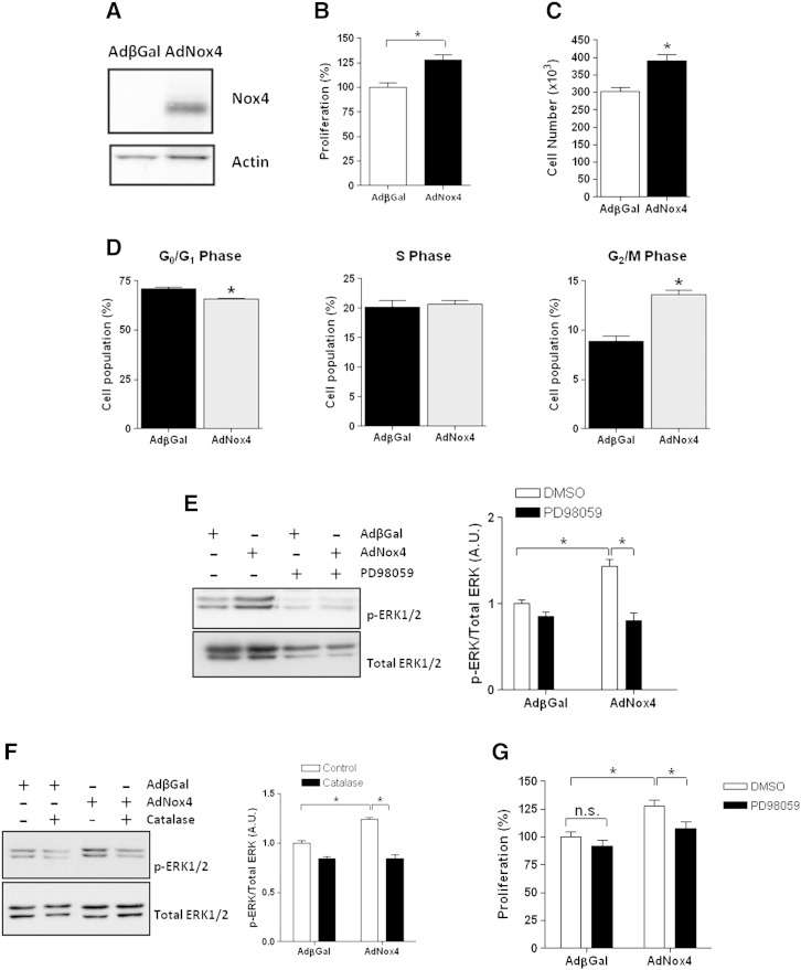
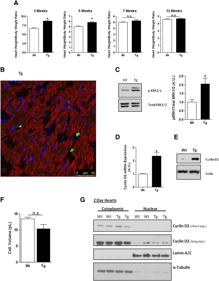
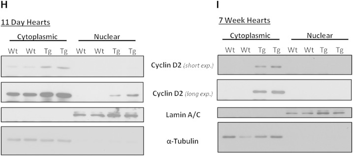
Adult mammalian cardiomyocytes have a very limited capacity to proliferate, and consequently the loss of cells after cardiac stress promotes heart failure. Recent evidence suggests that administration of hydrogen peroxide (H2O2), can regulate redox-dependent signalling pathway(s) to promote cardiomyocyte proliferation in vitro, but the potential relevance of such a pathway in vivo has not been tested. We have generated a transgenic (Tg) mouse model in which the H2O2-generating enzyme, NADPH oxidase 4 (Nox4), is overexpressed within the postnatal cardiomyocytes, and observed that the hearts of 1-3week old Tg mice pups are larger in comparison to wild type (Wt) littermate controls. We demonstrate that the cardiomyocytes of Tg mouse pups have increased cell cycling capacity in vivo as determined by incorporation of 5-bromo-2'-deoxyuridine. Further, microarray analyses of the transcriptome of these Tg mouse hearts suggested that the expression of cyclin D2 is significantly increased. We investigated the molecular mechanisms which underlie this more proliferative phenotype in isolated neonatal rat cardiomyocytes (NRCs) in vitro, and demonstrate that Nox4 overexpression mediates an H2O2-dependent activation of the ERK1/2 signalling pathway, which in turn phosphorylates and activates the transcription factor c-myc. This results in a significant increase in cyclin D2 expression, which we show to be mediated, at least in part, by cis-acting c-myc binding sites within the proximal cyclin D2 promoter. Overexpression of Nox4 in NRCs results in an increase in their proliferative capacity that is ablated by the silencing of cyclin D2. We further demonstrate activation of the ERK1/2 signalling pathway, increased phosphorylation of c-myc and significantly increased expression of cyclin D2 protein in the Nox4 Tg hearts. We suggest that this pathway acts to maintain the proliferative capacity of cardiomyocytes in Nox4 Tg pups in vivo and so delays their exit from the cell cycle after birth.

Keywords: Cardiomyocyte proliferation; Cyclin D2; ERK1/2; Nox4; Redox signalling; c-Myc.


Figures

Fig. 1
Fig. 1
Forced postnatal expression of Nox4 in  cardiomyocytes in vivo results in overt cardiac hypertrophy. (A) Q-PCR analysis of Nox4 mRNA expression in Tg and Wt littermate control mouse hearts at embryonic day 18 and 2 days and 7 weeks post-birth. Expression levels are normalised to those of β-actin and as expressed as arbitrary units relative to endogenous Nox4 expression within the embryo (A.U.). (B) Nox4 protein levels in 2 day postnatal Wt and Tg hearts. (C) Representative picture depicting the proportional increase in the size of hearts isolated from 2 week old Wt and Tg mice. (D) Quantification of heart/body weight ratios of Wt (n = 15) and Tg (n = 12) mice. (E) Echocardiographic measurements of interventricular septum thickness, left ventricular posterior wall thickness and relative wall thickness in 3 week old Wt (n = 5) and Tg (n = 7) mice taken at the end of diastole. (F & G) Representative transverse heart sections for cardiomyocyte area determination from Wt and Tg mice stained with either WGA (F) or an antibody against laminin (G). Mean data are shown in a histogram beneath each stained section. Scale bar, 10 μm. (H) Comparison of isolated cardiomyocyte cell volumes from 2 week old Wt and Tg animals, assessed on a Coulter Counter. All data are presented as mean ± S.E. *P < 0.05, **P < 0.01.
Fig. 2
Fig. 2
Nox4 transgenic hearts demonstrated increased cardiomyocyte cell cycling. (A) ELISA assay of BrdU incorporation from hearts isolated from BrdU injected 2 week old Wt and Tg pups. Pups were injected with BrdU and sacrificed after 16 h. (B) Representative immunofluorescence staining of transverse sections of equivalent paraffin-embedded hearts from BrDU-injected Wt and Tg mouse pups. BrdU-labelled, DNA-synthesising nuclei (green) and DAPI-stained nuclei (blue) are visible. Scale bar, 60 μm. (C) Magnified section from Tg heart section above (white box) depicting BrdU-labelled nuclei. Elongated nuclei consistent with cardiomyocyte morphology are indicated by white arrows. Scale bar, 25 μm. (D) Cardiomyocytes isolated from equivalent BrDU-injected Wt and Tg hearts were co-stained with antibodies against BrdU (green) and cardiac troponin T (red) and the cell nuclei stain DAPI (blue). Representative images are shown at low magnification (10×; scale bar, 110 μm) and at higher magnification (20 ×; scale bar, 54 mm). (D) Quantitation of percentage of BrdU-stained cardiomyocyte nuclei/field of view (n = 13 & 7 fields of view, each comprising approx. 200 cardiomyocyte nuclei). All data are presented as mean ± S.E. *P < 0.05.
Fig. 3
Fig. 3
Nox4 overexpression induces the upregulation of cyclin D2 in vivo. (A) Expression levels of cyclin D2 protein during development from Wt mice at embryonic day 14.5, birth (day 0), and 7 days and 14 days postnatal. (B) Q-PCR analysis of the relative expression of endogenous cyclin D2 mRNA in 2 week old Wt and Tg mouse hearts. Triplicate, independent Tg and Wt littermate controls were analysed, and relative expression was normalised to β-actin in all cases. (C) Protein expression levels of cyclin D2 in Wt and Tg hearts at 2 weeks of age. The histogram depicts the mean of triplicate, independent samples normalised to β  − actin and expressed in arbitrary units (A.U.). (D) Q-PCR analysis of the relative mRNA expression of the indicated cell cycle-related proteins, normalised to β-actin from 2 week old Wt and Tg mice (n = 3 in each case). (E) Representative immunoblots and quantitative histograms of protein expression levels normalised to β-actin of the indicated cell cycle-related proteins in 2 week old Wt and Tg mice (n = 3 in all cases). All data are presented as mean ± S.E. *P < 0.05; ns: not significant.
Fig. 4
Fig. 4
Nox4 expression increased NRC proliferation via an ERK1/2-dependent pathway. NRCs were transduced with either Nox4 (AdNox4) or β-galactosidase (Ad βGal) and incubated for 30 h. (A) Immunoblot of Nox4 expression in transduced NRC cells. (B) MTS-based assessment of NRC proliferation after transduction with either AdNox4 or AdβGal (n = 3). (C) Physical cell count assessment of NRC proliferation after transduction with either AdNox4 or AdβGal (n = 3). (D) Cell cycle analysis of the G0/G1, S and G2/M populations of NRCs after AdβGal or AdNox4 transduction (n = 3). (E & F) Representative immunoblots of phosphorylated and total ERK1/2 levels in NRCs after transduction with AdβGal or AdNox4 in the presence or absence of 20 μM PD98059 (E) or after catalase treatment (400 U/ml overnight; F). Histograms represent the ratio of phosphorylated ERK1/2 to total ERK1/2 protein levels in each case (n = 3). (G) MTS-based cell proliferation assay of NRCs after AdβGal or AdNox4 transduction in the presence or absence of 20 μM PD98059 (n = 3). (H) Cell cycle analysis of NRCs after AdβGal or AdNox4 transduction in the absence (vehicle) or presence of 20 μM PD98059 (n = 3). (I) Representative immunoblot and quantitative histogram showing increased ERK1/2 phosphorylation in 2 week-old Nox4 Tg hearts compared to Wt controls (n = 3). The histogram represents the ratio of phosphorylated ERK1/2 to total ERK1/2 protein levels. All data are presented as mean ± S.E. *P < 0.05.
Fig. 4
Fig. 4
Nox4 expression increased NRC proliferation via an ERK1/2-dependent pathway. NRCs were transduced with either Nox4 (AdNox4) or β-galactosidase (Ad βGal) and incubated for 30 h. (A) Immunoblot of Nox4 expression in transduced NRC cells. (B) MTS-based assessment of NRC proliferation after transduction with either AdNox4 or AdβGal (n = 3). (C) Physical cell count assessment of NRC proliferation after transduction with either AdNox4 or AdβGal (n = 3). (D) Cell cycle analysis of the G0/G1, S and G2/M populations of NRCs after AdβGal or AdNox4 transduction (n = 3). (E & F) Representative immunoblots of phosphorylated and total ERK1/2 levels in NRCs after transduction with AdβGal or AdNox4 in the presence or absence of 20 μM PD98059 (E) or after catalase treatment (400 U/ml overnight; F). Histograms represent the ratio of phosphorylated ERK1/2 to total ERK1/2 protein levels in each case (n = 3). (G) MTS-based cell proliferation assay of NRCs after AdβGal or AdNox4 transduction in the presence or absence of 20 μM PD98059 (n = 3). (H) Cell cycle analysis of NRCs after AdβGal or AdNox4 transduction in the absence (vehicle) or presence of 20 μM PD98059 (n = 3). (I) Representative immunoblot and quantitative histogram showing increased ERK1/2 phosphorylation in 2 week-old Nox4 Tg hearts compared to Wt controls (n = 3). The histogram represents the ratio of phosphorylated ERK1/2 to total ERK1/2 protein levels. All data are presented as mean ± S.E. *P < 0.05.
Fig. 5
Fig. 5
Nox4-induced proliferation in NRCs is mediated by the transcriptional upregulation of cyclin D2. (A) Q-PCR analyses showing relative cyclin D2 mRNA expression, normalised to β-actin (n = 3) and (B) representative immunoblots and quantitative histograms of protein expression levels normalised to those of laminin A/C in NRCs transduced with either Nox4 (AdNox4) or β-galactosidase (AdβGal) as control, for 30 h (n = 3). (C) Q-PCR analyses of cyclin D2 mRNA levels in NRCs transduced with either AdNox4 or AdβGal for 30 h in the presence or absence of the MEK inhibitor, PD98059 (n = 3). (D) Representative immunoblot and quantitative histograms of cyclin D2 levels (relative to those of β-actin) in protein extracted from cells as described in (C) (n = 3). (E) Representative immunoblot and quantitative histogram of cyclin D2 protein in NRCs after either scrambled (siScr) or cyclin D2 (siD2) siRNA treatment (n = 3). (F) MTS-based cell proliferation assay of NRCs transduced with either AdNox4 or AdβGal for 30 h in the presence of scrambled, or cyclin D2 siRNA (n = 3). All data are presented as mean ± S.E. *P < 0.05.
Fig. 6
Fig. 6
Nox4 overexpression activates c-myc resulting in increased cyclin D2 expression. (A) Representative immunoblot showing increased c-myc phosphorylation in 2 week-old Nox4 Tg hearts compared with Wt littermate controls. The histogram represents the ratio of phosphorylated c-myc to total c-myc protein levels (n = 3). (B) Representative immunoblot and quantitative histogram showing phosphorylated and total c-myc levels in NRCs transduced with either AdNox4 or AdβGal for 30 h in the presence or absence of PD98059 as indicated (n = 3). (C) Q-PCR analyses of c-myc mRNA expression, relative to that of β-actin (n = 3), and (D) representative immunoblot indicating c-myc protein expression, in NRCs after 48 h of c-myc siRNA treatment as indicated. β-Actin is shown as a loading control for protein expression. (E) Representative immunoblot and quantitative histogram showing cyclin D2 protein expression, relative to that of β-actin, in NRCs transduced with either AdNox4 or AdβGal for 30 h in the presence of scrambled or c-myc siRNA as indicated (n = 3). All data are presented as mean ± S.E. *P < 0.05.
Fig. 7
Fig. 7
Nox4-generated ROS increases the rate of cyclin D2 transcription via a highly conserved, cis-acting promoter fragment. (A) A DNA sequence comparison showing high conservation between regions of the proximal human and mouse cyclin D2 promoter regions, upstream of exon 1. Functional binding sites for indicated transcription factors are indicated and shaded in grey. Stars indicate nucleotide homology between species. Nucleotides are numbered relative to start of translation of each gene. (B) Relative luciferase activity (RLA) resulting from a promoterless firefly luciferase vector (empty vector) or from a construct comprising a 1766 bp genomic fragment of the proximal mouse cyclin D2 promoter ligated upstream of the luciferase reporter gene (cyclin D2-luc) in NRCs transduced with either AdNox4 or AdβGal for 28 h. (C & D) RLA resulting from the cyclin D2-luc in NRCs transduced with either AdNox4 or AdβGal for 28 h in the presence of scrambled or cmyc siRNA(C), or PD90859 (D), as indicated. In each case, luciferase activities are normalised to that of a co-transfected Renilla luciferase vector and expressed as relative luminescence units (RLUs; n = 3). The RLU resulting from cyclin D2-luc transduced with AdβGal was set to 1.0 in each case. All data are presented as mean ± S.E. *P < 0.05.
Fig. 8
Fig. 8
Forced expression of Nox4 in the postnatal heart prolongs the period of myocyte proliferation. (A) Quantification of heart/body weight ratios of Wt and Nox4 Tg mice at 3 weeks (n = 3/4), 5 weeks (n = 6/6), 7 weeks (n = 13/9) and 12 weeks (n = 13/13) post-birth. *P < 0.05, 2-tailed Student's t-test. Ns: not significant. Heart/body weight ratios of 5 week and 7 week-old Wt and Tg mice additionally analysed by one-way ANOVA with Bonferroni post-hoc test showed significant change at 5 weeks (P < 0.05) are lost at 7 weeks. (B) Representative immunofluorescence staining of transverse frozen heart sections from 7 week old Nox4 Tg mice co-stained with an antibody against the proliferation marker Ki67 (green), cardiac troponin T (red) and the cell nuclei stain DAPI (blue). No Ki67-positive cardiomyocytes are visible at this time point. Scale bar, 50 μm. (C) Representative blot and quantitative histogram showing increased ERK1/2 phosphorylation in 7 week old Nox4 Tg hearts when compared with Wt controls. The histogram represents the ratio of phosphorylated ERK1/2 to total ERK1/2 protein levels (n = 3). (D) Q-PCR analyses of cyclin D2 mRNA levels in 7 week old Wt and Nox4 Tg mice, relative to levels of β-actin mRNA (n = 3). (E) Representative immunoblot showing cyclin D2 protein expression levels in 7 week old Wt and Nox4 Tg mice, compared to those of β-actin. (F) Histogram indicating cell volumes in pico litres (pl) of Wt and Nox4 Tg adult cardiomyocytes isolated from 13 week old male mice (n = 3/3). Cell volumes were assessed by physical measurements of length and cross sectional diameter, and subsequent calculation of volume based on these parameters (see Section 2, Materials and methods). (C, D, F) *P < 0.05, 2-tailed Student's t-test. Ns: not significant. (G, H, I) Representative immunoblots demonstrating cytoplasmic and nuclear localisation of cyclin D2 in Wt and Nox4 Tg mouse hearts at 2 days (G), 11 days (H) and 7 weeks (I) after birth. Staining for lamin A/C and α-tubulin demonstrates successful fractionation of nuclear and cytoplasmic proteins, respectively. Nuclear cyclin D2 is apparent in Wt and Tg mouse hearts at 2 days, and in Tg hearts only at 11 days after birth. No nuclear cyclin D2 is apparent in Wt or Tg hearts of the adult mice.
Fig. 8
Fig. 8
Forced expression of Nox4 in the postnatal heart prolongs the period of myocyte proliferation. (A) Quantification of heart/body weight ratios of Wt and Nox4 Tg mice at 3 weeks (n = 3/4), 5 weeks (n = 6/6), 7 weeks (n = 13/9) and 12 weeks (n = 13/13) post-birth. *P < 0.05, 2-tailed Student's t-test. Ns: not significant. Heart/body weight ratios of 5 week and 7 week-old Wt and Tg mice additionally analysed by one-way ANOVA with Bonferroni post-hoc test showed significant change at 5 weeks (P < 0.05) are lost at 7 weeks. (B) Representative immunofluorescence staining of transverse frozen heart sections from 7 week old Nox4 Tg mice co-stained with an antibody against the proliferation marker Ki67 (green), cardiac troponin T (red) and the cell nuclei stain DAPI (blue). No Ki67-positive cardiomyocytes are visible at this time point. Scale bar, 50 μm. (C) Representative blot and quantitative histogram showing increased ERK1/2 phosphorylation in 7 week old Nox4 Tg hearts when compared with Wt controls. The histogram represents the ratio of phosphorylated ERK1/2 to total ERK1/2 protein levels (n = 3). (D) Q-PCR analyses of cyclin D2 mRNA levels in 7 week old Wt and Nox4 Tg mice, relative to levels of β-actin mRNA (n = 3). (E) Representative immunoblot showing cyclin D2 protein expression levels in 7 week old Wt and Nox4 Tg mice, compared to those of β-actin. (F) Histogram indicating cell volumes in pico litres (pl) of Wt and Nox4 Tg adult cardiomyocytes isolated from 13 week old male mice (n = 3/3). Cell volumes were assessed by physical measurements of length and cross sectional diameter, and subsequent calculation of volume based on these parameters (see Section 2, Materials and methods). (C, D, F) *P < 0.05, 2-tailed Student's t-test. Ns: not significant. (G, H, I) Representative immunoblots demonstrating cytoplasmic and nuclear localisation of cyclin D2 in Wt and Nox4 Tg mouse hearts at 2 days (G), 11 days (H) and 7 weeks (I) after birth. Staining for lamin A/C and α-tubulin demonstrates successful fractionation of nuclear and cytoplasmic proteins, respectively. Nuclear cyclin D2 is apparent in Wt and Tg mouse hearts at 2 days, and in Tg hearts only at 11 days after birth. No nuclear cyclin D2 is apparent in Wt or Tg hearts of the adult mice.

References

    1. Bicknell K.A., Coxon C.H., Brooks G. Can the cardiomyocyte cell cycle be reprogrammed? J Mol Cell Cardiol. 2007;42:706–721. - PubMed
    1. Drenckhahn J.D., Schwarz Q.P., Gray S., Laskowski A., Kiriazis H., Ming Z. Compensatory growth of healthy cardiac cells in the presence of diseased cells restores tissue homeostasis during heart development. Dev Cell. 2008;15:521–533. - PubMed
    1. Porrello E.R., Mahmoud A.I., Simpson E., Hill J.A., Richardson J.A., Olson E.N. Transient regenerative potential of the neonatal mouse heart. Science. 2011;331:1078–1080. - PMC - PubMed
    1. Ahuja P., Sdek P., MacLellan W.R. Cardiac myocyte cell cycle control in development, disease, and regeneration. Physiol Rev. 2007;87:521–544. - PMC - PubMed
    1. Chiu J., Dawes I.W. Redox control of cell proliferation. Trends Cell Biol. 2012;22:592–601. - PubMed

Publication types

MeSH terms

LinkOut - more resources