. 2015 Jan 29:285:215-26.
doi: 10.1016/j.neuroscience.2014.10.035. Epub 2014 Nov 4.

Caveolin expression changes in the neurovascular unit after juvenile traumatic brain injury: signs of blood-brain barrier healing?

Affiliations

Caveolin expression changes in the neurovascular unit after juvenile traumatic brain injury: signs of blood-brain barrier healing?

J Badaut et al. Neuroscience. .

Abstract

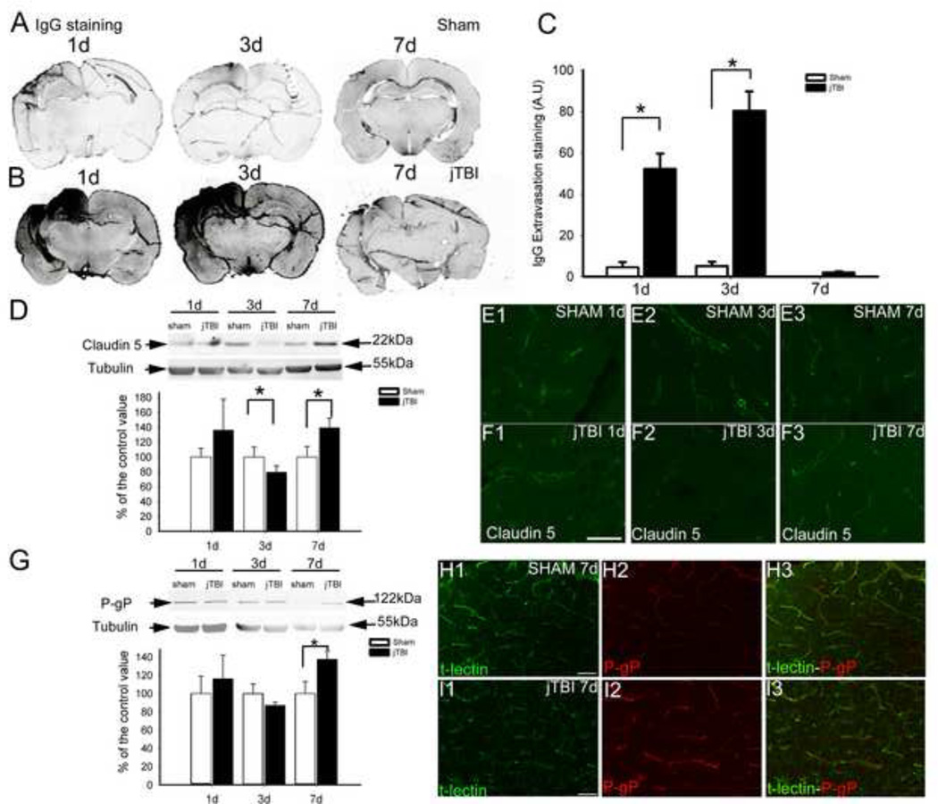
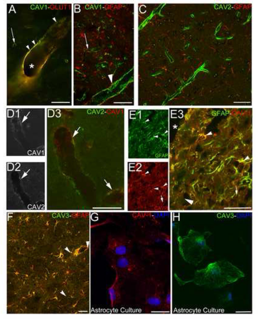
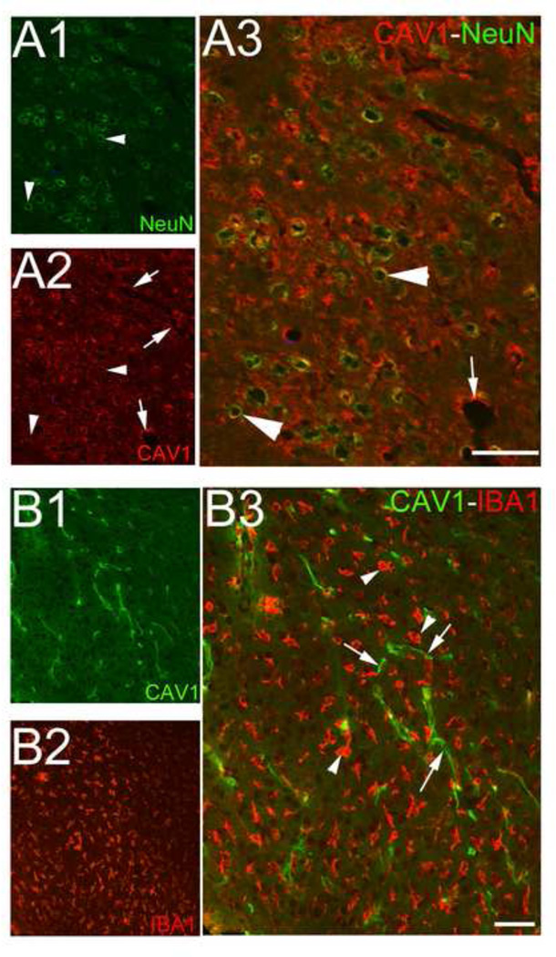
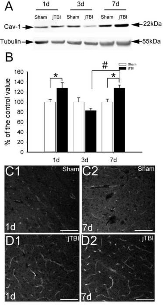
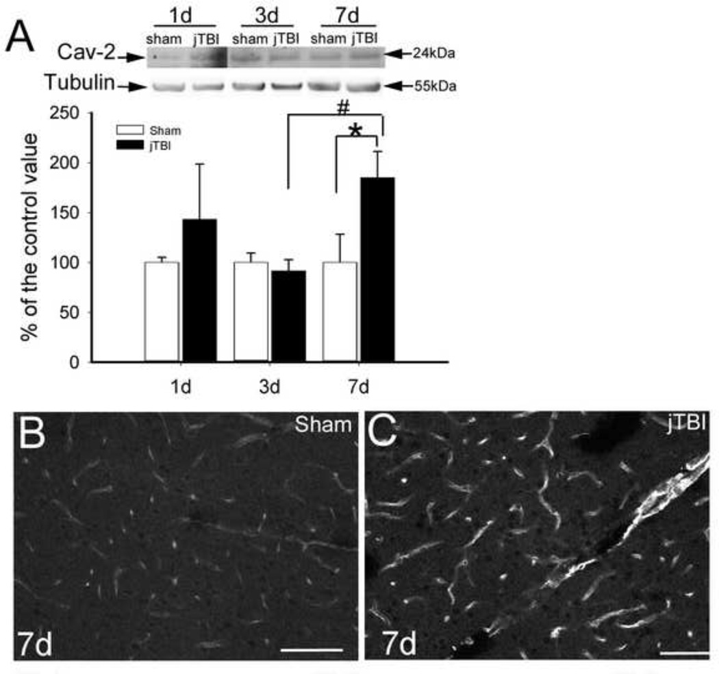
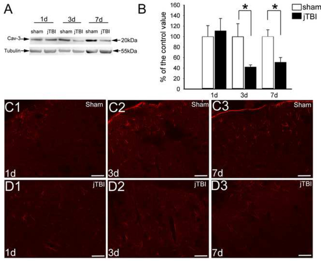
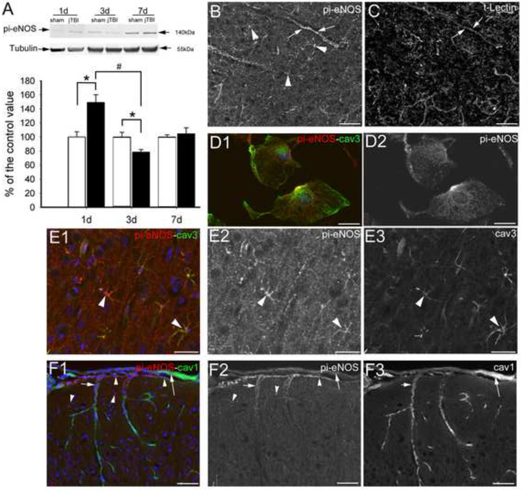
Traumatic brain injury (TBI) is one of the major causes of death and disability in pediatrics, and results in a complex cascade of events including the disruption of the blood-brain barrier (BBB). A controlled-cortical impact on post-natal 17-day-old rats induced BBB disruption by IgG extravasation from 1 to 3 days after injury and returned to normal at day 7. In parallel, we characterized the expression of three caveolin isoforms, caveolin 1 (cav-1), caveolin 2 (cav-2) and caveolin 3 (cav-3). While cav-1 and cav-2 are expressed on endothelial cells, both cav-1 and cav-3 were found to be present on reactive astrocytes, in vivo and in vitro. Following TBI, cav-1 expression was increased in blood vessels at 1 and 7 days in the perilesional cortex. An increase of vascular cav-2 expression was observed 7 days after TBI. In contrast, astrocytic cav-3 expression decreased 3 and 7 days after TBI. Activation of endothelial nitric oxide synthase (eNOS) (via its phosphorylation) was detected 1 day after TBI and phospho-eNOS was detected both in association with blood vessels and with astrocytes. The molecular changes involving caveolins occurring in endothelial cells following juvenile-TBI might participate, independently of eNOS activation, to a mechanism of BBB repair while, they might subserve other undefined roles in astrocytes.

Keywords: astrocyte; blood–brain barrier; caveolin; endothelium; juvenile traumatic brain injury.


Conflict of interest statement

Disclosure/Conflict of Interest

The authors declare no conflict of interest

Figures

Figure 1
Figure 1. BBB disruption after jTBI
(A, B) IgG extravasation staining in sham (A) and jTBI (B) at 1, 3, and 7d post injury. Area of IgG staining (black) is enlarged at 1 and 3d in jTBI animals (B) compared to sham (A), but normalizes by 7d (A, B). (C) Quantification of IgG immunoreactivity shows a significant increase at 1 and 3d, but not at 7d (E; p<0.05). (D) Western blot of claudin-5 showed a band around 22kDa and is significantly decreased in jTBI compared to sham at 3 and increased 7d. The quantification of the intensity of the band for claudin-5 was normalized to the tubulin (55kDa). (E, F) A tight junction protein Claudin-5 staining in the sham (E) and jTBI (F) at 1d (E1, F1), 3d (E2, F2) and 7d (E3, F3). Accordingly to western blot (D), the staining of claudin-5 is lower in jTBI (E2) than sham group (F2) at 1d and 3d. Intensity of claudin-5 staining in jTBI group is increased at 7d after the injury (E3, F3). (G) Western blot of P-gp showed a band around 122kDa and is significantly increased in jTBI compared to sham at 7d. The quantification of the intensity of the band for P-gp was normalized to the tubulin (55kDa). (H, I) Tomato-lectin (t-lectin, green, H1, I1) was co-stained with P-gp immunolabeling (red, H2, I2) in the sham (H) and and jTBI (I) at 7d. Accordingly to western blot (G), the staining of P-gp is increased in intensity (red) in blood vessels (outlined by t-lectin, green) in jTBI animals compared to shams. E, F, H, I bar = 100µm
Figure 2
Figure 2. Caveolin distribution in vascular endothelium and astrocytes
(A) Mouse anti-cav1 (green) exhibit a staining in the cerebral blood vessels (asterisk), from the large blood vessels to capillary bed (arrows). Co-staining cav-1 (green) and anti-GLUT1, marker for the endothelial cells, shows a co-localization in yellow (arrow heads) in large blood vessels as well as in capillaries. (B) The co-staining cav-1 (green) and anti-GFAP (red) did not show overlap in parietal cortex at distance to the injury site, astrocytic cav-1 staining may be contained within reactive astrogliosis around the injury (arrowhead = astrocyte endfeet, arrow = cav1 in endothelium). (C) Caveolin-2 (cav-2, green) and GFAP (red) did not exhibit co-localization, only endothelial staining was observed. (D) Double immunolabeling cav-2 (C1, green C3) and cav-1 (C2, red C3) showed co-localization between both staining in the endothelial cells (arrows). (E) In the perilesional areas, rabbit anti-cav-1 staining (red, C2, C3) was co-localized with GFAP labeling (green, C1, C3) (arrowheads), which is an astrocytic marker and increased in reactive astrogliosis. (F) Cav-3 staining (red) and GFAP labeling (green) showed co-localization (yellow, arrowheads), showing the presence of cav-3 in astrocyte process and cell bodies. (G) Cav-1 labeling (red) and nuclear staining with DAPI (blue) in primary astrocyte culture shows presence of cav-1 in the astrocytes. (H) Cav-3 labeling (green) and nuclear staining DAPI (blue) in primary astrocyte culture show the presence of cav-3 protein in the astrocyte. A = 50µm B, C, D, E, bar = 100µm; F, G, bar=10µm
Figure 3
Figure 3. Caveolin 1 expression in neurons but not microglial cells after jTBI
(A) NeuN labeling (green, A1, A3) and Cav-1 staining (red, A2, A3) co-localized (yellow, arrowheads), suggesting the presence of cav-1 in neurons in the periphery of the lesion. Cav-1 staining (red, A2, A3) is also observed in intracortical microvessels (arrows). (B) Cav-1 labeling (green, B1, B3, arrows) and IBA1 staining (red, B2, B3, arrowhead), a marker of microglial cells, did not show co-localization. A, bar = 500µm; B, bar = 100µm
Figure 4
Figure 4. Caveolin 1 Expression after jTBI
(A) Western blot for cav-1 showed monomer at 22 kDa and tubulin at 55kDa in sham and jTBI ipsilateral cortex. Cav-1 is increased at 1d and 7d in jTBI animal compared to shams. In contrast at 3d, the intensity of the band for cav-1 is lower in jTBI group than sham (C) Quantification of cav-1 from western blots showed significant increases in jTBI animals compared to sham at 1d and 7d (*, p<0.05). The increase is significantly higher at 7d than at 3d in jTBI animals (#, p<0.05). (C, D) Caveolin-1 (cav-1) staining in the sham (C) and jTBI (D) at 1d (C1, D1) and 7d (C2, B2) after the injury. Less endothelial cav-1 staining was observed throughout the time-points studied in sham (A) compared to jTBI (B). C, D, bar = 100µm
Figure 5
Figure 5. Caveolin 2 Expression expression after jTBI
(A) Western blot of cav-2 showed a band around 20kDa with a significant increase in intensity between sham and jTBI group at 7d post-injury (*p<0.05). In jTBI group, cav-2 is significantly increase between 3 and 7d (#, p<0.05) (B, C) Cav-2 staining in the sham (C) and jTBI (D) at 7d exhibits higher intensity of staining in cortical blood vessels of the jTBI group than in sham B, C, bar = 100µm
Figure 6
Figure 6. Caveolin-3 Expression and Distribution after jTBI
(A) Western blot of cav-3 (red) showed a band around 20kDa and is significantly decreased in jTBI compared to sham at 3 and 7d. (B) The quantification of the intensity of the band for cav-3 was normalized to the tubulin (55kDa). (C, D) Cav-3 staining in the sham (C) and jTBI (D) at 1d (C1, D1), 3d (C2, D2) and 7d (C3, D3). In accordance with the western blot, no major changes were observed in cav-3 staining at 1d (C1, D1). However, the pattern of cav-3 staining differs between the jTBI and sham animals at 3d (C2, D2) and 7d (C3, D3). In the glia limitans, cav-3 staining is decreased in jTBI animals (D2, D3) compared to sham (C2, C3). The intensity of cav-3 staining is decreased in jTBI animals (D2, D3) compared to sham (D2, D3) despite an increase of the extend of the surface staining for cav-3 in jTBI group at both time points. C, D, bar = 100µm
Figure 7
Figure 7. pi-eNOS expression and distribution after jTBI
(A) Western blot of pi-eNOS showed a band around 140kDa and is significantly increased in jTBI compared to sham at 1d (*, p< 0.05) and decreased at 3d (*, p<0.05). The quantification of the intensity of the band for claudin-5 was normalized to the tubulin (55kDa). (B, C) The anti pi-eNOS staining (B, arrows) is present in the blood vessels revealed by tomato-lectin staining (t-lectin, C, arrows), but also in the astrocytes (arrowheads) in perilesional cortex of jTBI animals. (D). Double immunolabeling cav-3 (green, D1) and pi-eNOS immunolabeling (red, D1, D2) showed a co-localization in primary astrocyte cultures. (E) Double immunolabeling cav-3 (green, E1, E3) and pi-eNOS immunolabeling (red, E1, E2) showed a co-localization in astrocyte like cells (arrowheads) in the cortex of jTBI rats. (F) Double immunolabeling cav-1 (green, F1, F3) and pi-eNOS immunolabeling (red, F1, F2) showed a co-localization in endothelial cells (arrows) in the cortex and the pial blood vessels in jTBI rats. In the glia limitans, pi-eNOS staining was observed in the astrocytes of the glia limitans (arrowheads). B, C, E, F bar = 50µm; D bar = 25µm

References

    1. Ajao DO, Pop V, Kamper JE, Adami A, Rudobeck E, Huang L, Vlkolinsky R, Hartman RE, Ashwal S, Obenaus A, Badaut J. Traumatic brain injury in young rats leads to progressive behavioral deficits coincident with altered tissue properties in adulthood. J Neurotrauma. 2012;29:2060–2074. - PMC - PubMed
    1. Armstead WM. Cerebral hemodynamics after traumatic brain injury of immature brain. Exp Toxicol Pathol. 1999;51:137–142. - PubMed
    1. Badaut J, Ashwal S, Adami A, Tone B, Recker R, Spagnoli D, Ternon B, Obenaus A. Brain water mobility decreases after astrocytic aquaporin-4 inhibition using RNA interference. J Cereb Blood Flow Metab. 2011;31:819–831. - PMC - PubMed
    1. Bernatchez PN, Bauer PM, Yu J, Prendergast JS, He P, Sessa WC. Dissecting the molecular control of endothelial NO synthase by caveolin-1 using cell-permeable peptides. Proc Natl Acad Sci U S A. 2005;102:761–766. - PMC - PubMed
    1. Bucci M, Gratton JP, Rudic RD, Acevedo L, Roviezzo F, Cirino G, Sessa WC. In vivo delivery of the caveolin-1 scaffolding domain inhibits nitric oxide synthesis and reduces inflammation. Nat Med. 2000;6:1362–1367. - PubMed

Publication types

MeSH terms

LinkOut - more resources