. 2015 Jan 22:284:872-887.
doi: 10.1016/j.neuroscience.2014.10.043. Epub 2014 Nov 1.

Spinally projecting preproglucagon axons preferentially innervate sympathetic preganglionic neurons

Affiliations

Spinally projecting preproglucagon axons preferentially innervate sympathetic preganglionic neurons

I J Llewellyn-Smith et al. Neuroscience. .

Abstract

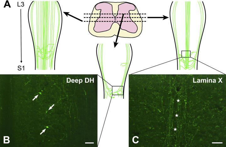
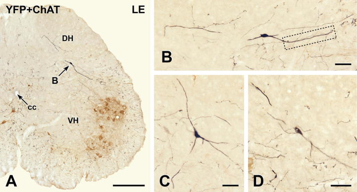
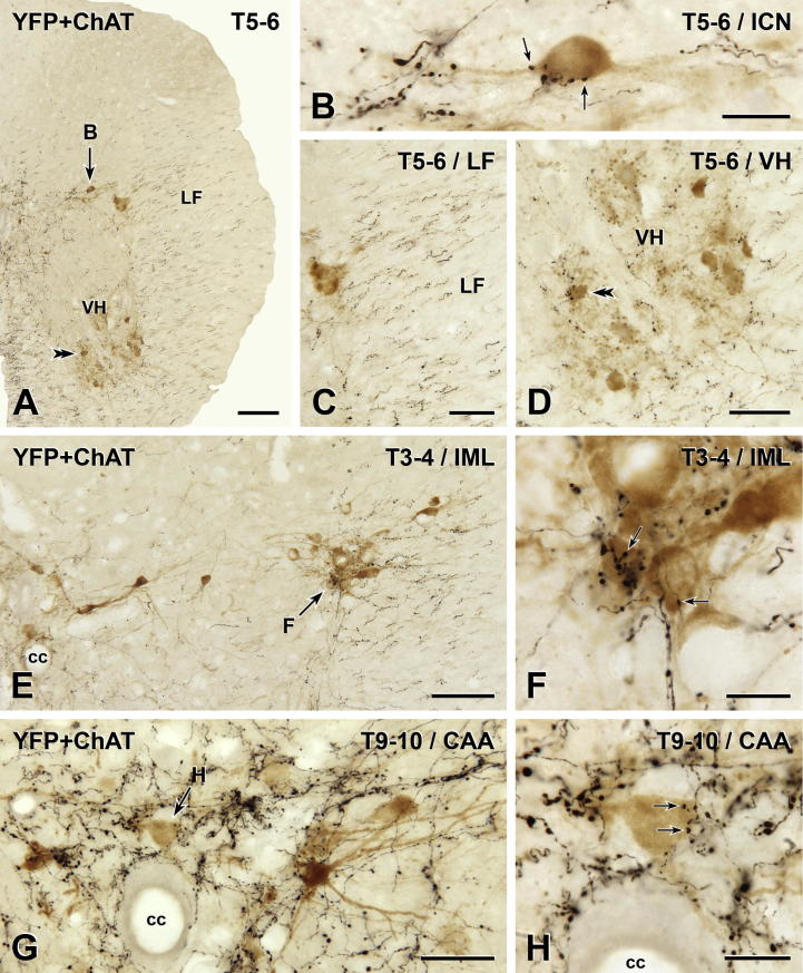
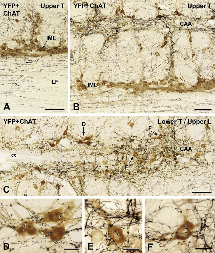
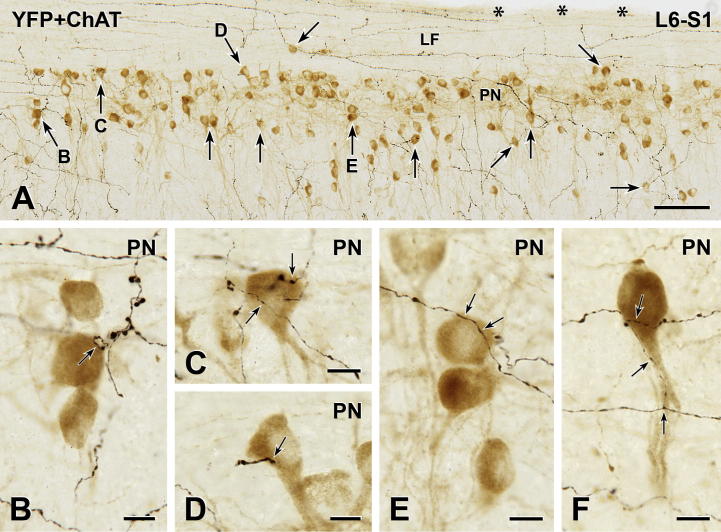
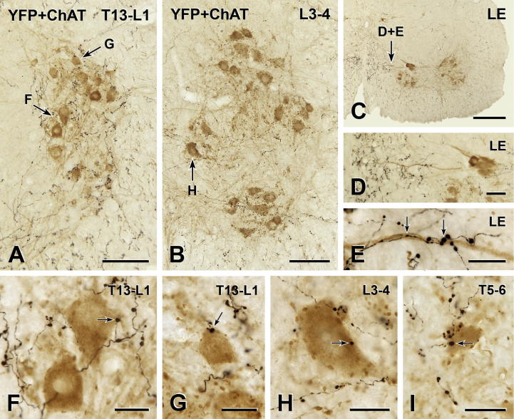
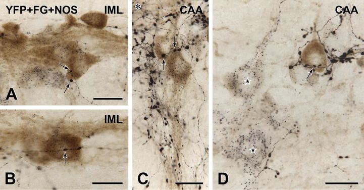
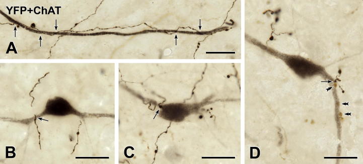
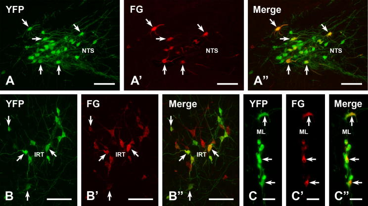
Glucagon-like peptide-1 (GLP-1) affects central autonomic neurons, including those controlling the cardiovascular system, thermogenesis, and energy balance. Preproglucagon (PPG) neurons, located mainly in the nucleus tractus solitarius (NTS) and medullary reticular formation, produce GLP-1. In transgenic mice expressing glucagon promoter-driven yellow fluorescent protein (YFP), these brainstem PPG neurons project to many central autonomic regions where GLP-1 receptors are expressed. The spinal cord also contains GLP-1 receptor mRNA but the distribution of spinal PPG axons is unknown. Here, we used two-color immunoperoxidase labeling to examine PPG innervation of spinal segments T1-S4 in YFP-PPG mice. Immunoreactivity for YFP identified spinal PPG axons and perikarya. We classified spinal neurons receiving PPG input by immunoreactivity for choline acetyltransferase (ChAT), nitric oxide synthase (NOS) and/or Fluorogold (FG) retrogradely transported from the peritoneal cavity. FG microinjected at T9 defined cell bodies that supplied spinal PPG innervation. The deep dorsal horn of lower lumbar cord contained YFP-immunoreactive neurons. Non-varicose, YFP-immunoreactive axons were prominent in the lateral funiculus, ventral white commissure and around the ventral median fissure. In T1-L2, varicose, YFP-containing axons closely apposed many ChAT-immunoreactive sympathetic preganglionic neurons (SPN) in the intermediolateral cell column (IML) and dorsal lamina X. In the sacral parasympathetic nucleus, about 10% of ChAT-immunoreactive preganglionic neurons received YFP appositions, as did occasional ChAT-positive motor neurons throughout the rostrocaudal extent of the ventral horn. YFP appositions also occurred on NOS-immunoreactive spinal interneurons and on spinal YFP-immunoreactive neurons. Injecting FG at T9 retrogradely labeled many YFP-PPG cell bodies in the medulla but none of the spinal YFP-immunoreactive neurons. These results show that brainstem PPG neurons innervate spinal autonomic and somatic motor neurons. The distributions of spinal PPG axons and spinal GLP-1 receptors correlate well. SPN receive the densest PPG innervation. Brainstem PPG neurons could directly modulate sympathetic outflow through their spinal inputs to SPN or interneurons.

Keywords: choline acetyltransferase; glucagon-like peptide-1; green fluorescent protein; nucleus of the solitary tract; parasympathetic preganglionic neurons; retrograde tracing.


Figures

Fig. 1
Fig. 1
Distribution of spinal axons showing native YFP fluorescence in segments L3-S1. (A) Schematic showing the locations of YFP-fluorescent axons and cell bodies in longitudinal sections through the lower lumbar and upper sacral spinal cord at dorsoventral levels indicated in the diagram of the transverse section, which shows L3 (modified from the Allen Brain Institute Reference Atlas; http://mousespinal.brain-map.org/imageseries/showref.html). (B) In the lower lumbar cord, YFP-fluorescent nerve cell bodies (arrows) as well as YFP-fluorescent varicose and non-varicose axons occur within the deep dorsal horn (DH). (C) In dorsal lamina X, YFP-fluorescent axons travel rostrocaudally and mediolaterally. A rostrocaudal tract of varicose YFP-fluorescent axons (stars) lies just dorsal to the central canal. Scale bars: B and C = 100 μm.
Fig. 2
Fig. 2
YFP-immunoreactive cell bodies and dendrites at the level of the lumbar enlargement (LE). Two-color immunoperoxidase staining for YFP (black) and ChAT (brown) in transverse sections through the lower lumbar spinal cord. (A) A YFP-immunoreactive cell body (black) in lamina V of the dorsal horn (DH) at spinal level L4–5. ChAT-immunoreactive somatic motor neurons (brown) occur in lamina IX in the ventral horn (VH). The arrow indicates the location of the neuron shown in B. cc, central canal. Montage of 18 micrographs. Scale bar = 250 μm. (B) Higher magnification image of the black YFP-immunoreactive neuron indicated by B in A. The boxed area is shown at higher magnification in Fig. 9A. Montage of 9 micrographs. (C and D) Other examples of black YFP-immunoreactive cell bodies and their dendrites in lamina V of the lumbar enlargement. (D) Montage of 2 micrographs. Scale bars in B–D, 50 μm. (For interpretation of the references to color in this figure legend, the reader is referred to the web version of this article.)
Fig. 3
Fig. 3
Spinal YFP-immunoreactive neurons do not project rostrally. (A) Transverse section single-stained to show FG-immunoreactivity at the center of an FG injection site (star) in thoracic segment T9. Peroxidase reaction product occurs throughout the spinal gray and white matter. Thus, it is highly likely that all axons passing through the injection site have taken up FG. Montage of 6 micrographs. Scale bar = 250 μm. (B) Two-color immunoperoxidase staining for FG (black) and YFP (brown). Four brown YFP-immunoreactive cell bodies in the lumbar enlargement (LE) lack black FG-immunoreactivity, indicating that they do not project rostrally through T9. In contrast, black puncta of FG-immunoreactivity are present in nearby cell bodies (arrows) and in black, varicose axons of undefined neurochemical phenotype. Montage of 3 micrographs. Scale bar = 50 μm. (For interpretation of the references to color in this figure legend, the reader is referred to the web version of this article.)
Fig. 4
Fig. 4
Distribution of YFP-PPG axons in thoracic spinal cord. Two-color immunoperoxidase staining for YFP (black) and ChAT (brown) in transverse sections through the thoracic spinal cord. (A–D) In spinal segments T5–6, there is a high density of rostrocaudally running black YFP-immunoreactive axons in the lateral funiculus (LF; C). There are close appositions between black YFP-immunoreactive varicosities and brown ChAT-immunoreactive sympathetic preganglionic neurons (SPN) in the intercalated nucleus (ICN; B) and brown ChAT-immunoreactive somatic motor neurons in the ventral horn (VH; D). A higher magnification image of the cell body indicated by the double arrowheads in A and D is shown in Fig. 7I. (E and F) Brown ChAT-immunoreactive SPN in the intermediolateral cell column (IML) of T3–4 receive close appositions (arrows) from black YFP-immunoreactive varicosities. (G and H) In the central autonomic area (CAA) at T9–10, black YFP-immunoreactive varicosities (black) also form appositions (arrows) on brown ChAT-immunoreactive SPN. cc, central canal. Micrographs in montages: A and E, 5; B, 6; C and D, 3; F & H, 2; G, 4. Scale bars: A and E, 100 μm; C, D and G, 50 μm; B, F and H, 20 μm. (For interpretation of the references to color in this figure legend, the reader is referred to the web version of this article.)
Fig. 5
Fig. 5
YFP-immunoreactive innervation of sympathetic preganglionic neurons (SPN). Two-color immunoperoxidase staining for YFP (black) and ChAT (brown) in horizontal sections through the spinal cord at upper thoracic (Upper T; A and B) and lower thoracic/upper lumbar (Lower T/Upper L; C) levels. At both spinal levels (B and C), black YFP-immunoreactive axons most densely innervate the central autonomic area (CAA), which lies dorsal to the central canal (cc) in lamina X. The intermediolateral cell column (IML) receives a moderately dense black YFP innervation. The lateral funiculus (LF; A and B) contains primarily black YFP-immunoreactive axons of passage that run rostrocaudally and occasional axons that travel mediolaterally (small arrows in A). The neurons indicated by arrows D–F are shown at higher magnification in D–F. Micrographs in montages: A, 3; B, 9; C, 6. (D–F) Black YFP-immunoreactive boutons from close appositions (arrows) on brown ChAT-immunoreactive SPN in the CAA. E, Montage of 2 micrographs. Scale bars: A–C, 100 μm; D–F, 20 μm. (For interpretation of the references to colour in this figure legend, the reader is referred to the web version of this article.)
Fig. 6
Fig. 6
YFP-immunoreactive innervation of parasympathetic preganglionic neurons (PPN). Two-color immunoperoxidase staining for YFP (black) and ChAT (brown) in a horizontal section through the lumbosacral (L6-S1) spinal cord. (A) The sacral parasympathetic nucleus (PN) is sparsely innervated by black YFP-immunoreactive axons. Of the 133 PPN in A, black YFP-positive axon terminals closely appose only 29 of the brown ChAT-immunoreactive PPN, some of which are indicated by arrows. Arrows B-E mark neurons that are shown at higher magnification in B–E. Black, non-varicose YFP-immunoreactive axons run rostrocaudally through the white matter of the lateral funiculus (LF). Asterisks indicate the lateral edge of the spinal cord. Montage of 6 micrographs. Scale bar: 100 μm. (B–F) Brown ChAT-immunoreactive PPN receive close appositions (arrows) from black YFP-immunoreactive varicosities. Micrographs in montages: B, 4; C, 3; D and E, 2; F, 6. Scale bars: 10 μm. (For interpretation of the references to color in this figure legend, the reader is referred to the web version of this article.)
Fig. 7
Fig. 7
YFP-immunoreactive innervation of somatic motor neurons. Two-color immunoperoxidase staining for YFP (black) and ChAT (brown) in transverse sections through the thoracic and lumbar spinal cord. (A–C) At all segmental levels, lamina IX contains a moderate number of black varicose YFP-immunoreactive axons. Micrographs in montages: A, 3; B and C, 5. Scale bars: A and B, 100 μm; C, 250 μm. (D–I) Nevertheless, only rare, brown ChAT-immunoreactive somatic motor neurons receive close appositions (arrows) from black YFP-immunoreactive varicosities. Appositions from YFP-immunoreactive boutons do occur on the dendrites of somatic motor neurons (C–E). However, in most cases, YFP-containing varicosities appose the cell bodies of somatic motor neurons (F–I). LE, lumbar enlargement. Micrographs in montages: D-F & H, 3; G, 2; I, 5. Scale bars: D, 50 μm; E–I, 20 μm. (For interpretation of the references to color in this figure legend, the reader is referred to the web version of this article.)
Fig. 8
Fig. 8
YFP-immunoreactive innervation of nitric oxide synthase (NOS)-immunoreactive spinal neurons. Triple immunoperoxidase staining for YFP (black), intraperitoneal FG (black) and NOS (brown) in the IML and CAA. Neurons containing FG-immunoreactivity send axons to the periphery and neurons without FG have axons that stay within the CNS. Hence, FG staining distinguishes between NOS-positive interneurons and NOS-positive SPN. Both NOS-containing SPN and NOS-containing spinal interneurons receive input from YFP-immunoreactive axons. (A and B) In the IML, SPN showing immunoreactivity for NOS (brown) and FG (black puncta within the cytoplasm) receive close appositions (arrows) from black YFP-immunoreactive boutons. A, Montage of 2 micrographs. Scale bars: 20 μm. (C) Similarly in the CAA, black YFP-immunoreactive varicosities closely appose (arrows) neurons that contain both brown NOS- and black FG-immunoreactivity, indicating that they are SPN. Asterisk, Ependymal cells lining the dorsal surface of the central canal are out of focus. Montage of 2 micrographs. Scale bar: 20 μm. (D) In the CAA, a varicosity from a black YFP-positive axon closely apposes (arrow) a brown NOS-positive neuron that lacks FG-immunoreactivity, indicating that it is an interneuron. Nearby neurons (stars) contain black FG-immunoreactive puncta, indicating that they are SPN. The top starred SPN is faintly immunoreactive for NOS. Montage of 5 micrographs. Scale bar: 20 μm. (For interpretation of the references to color in this figure legend, the reader is referred to the web version of this article.)
Fig. 9
Fig. 9
Innervation of spinal YFP-immunoreactive neurons. Two-color immunoperoxidase staining for YFP (black) and ChAT (brown) in transverse sections through the lumbar enlargement. (A) A YFP-immunoreactive axon forms several close appositions (arrows) on the dendrite of the YFP-immunoreactive spinal neuron shown in Fig. 2B. (B) A YFP-immunoreactive varicosity apposes (arrow) the proximal dendrite of a YFP-immunoreactive spinal neuron. (C) The cell body of a YFP-immunoreactive spinal neuron receives a close apposition (arrow) from a YFP-immunoreactive varicosity. (D) The dendrite of a YFP-positive spinal neuron receives a close apposition from a YFP-positive bouton (arrow) as well as from several ChAT-positive boutons (double arrowheads). Micrographs in montages: A, 12; D, 3. Scale bars: 20 μm. (For interpretation of the references to color in this figure legend, the reader is referred to the web version of this article.)
Fig. 10
Fig. 10
Spinally projecting YFP-PPG neurons in the brainstem. Double immunofluorescent staining for YFP (green) and FG (red) in the brainstems of YFP-PPG mice that received injections of FG at spinal segment T9. The NTS (A–A”), the IRT (B–B”) and dorsal midline (C–C”) contain YFP-immunofluorescent neurons (A–C), some of which contain FG-immunoreactivity (A’, B’ and C’). Double labeled neurons are spinally projecting YFP-PPG neurons. A”, B”, C”, merged images of micrographs showing YFP- and FG-immunoreactive neurons in each area. Scale bars: A, B, 100 μm; C, 50 μm. (For interpretation of the references to color in this figure legend, the reader is referred to the web version of this article.)

References

    1. Alhadeff A.L., Rupprecht L.E., Hayes M.R. GLP-1 neurons in the nucleus of the solitary tract project directly to the ventral tegmental area and nucleus accumbens to control for food intake. Endocrinology. 2012;153:647–658. - PMC - PubMed
    1. Anderson C.R., Edwards S.L. Intraperitoneal injections of Fluorogold reliably labels all sympathetic preganglionic neurons in the rat. J Neurosci Methods. 1994;53:137–141. - PubMed
    1. Barrera J.G., Jones K.R., Herman J.P., D’Alessio D.A., Woods S.C., Seeley R.J. Hyperphagia and increased fat accumulation in two models of chronic CNS glucagon-like peptide-1 loss of function. J Neurosci. 2011;31:3904–3913. - PMC - PubMed
    1. Buse J.B., Rosenstock J., Sesti G., Schmidt W.E., Montanya E., Brett J.H., Zychma M., Blonde L. Liraglutide once a day versus exenatide twice a day for type 2 diabetes: a 26-week randomised, parallel-group, multinational, open-label trial (LEAD-6) Lancet. 2009;374:39–47. - PubMed
    1. Cabou C., Campistron G., Marsollier N., Leloup C., Cruciani-Guglielmacci C., Penicaud L., Drucker D.J., Magnan C., Burcelin R. Brain glucagon-like peptide-1 regulates arterial blood flow, heart rate, and insulin sensitivity. Diabetes. 2008;57:2577–2587. - PMC - PubMed

Publication types

MeSH terms