. 2015 Feb;1853(2):276-84.
doi: 10.1016/j.bbamcr.2014.11.015. Epub 2014 Nov 18.

H9c2 and HL-1 cells demonstrate distinct features of energy metabolism, mitochondrial function and sensitivity to hypoxia-reoxygenation

Affiliations

H9c2 and HL-1 cells demonstrate distinct features of energy metabolism, mitochondrial function and sensitivity to hypoxia-reoxygenation

Andrey V Kuznetsov et al. Biochim Biophys Acta. 2015 Feb.

Abstract

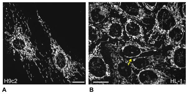
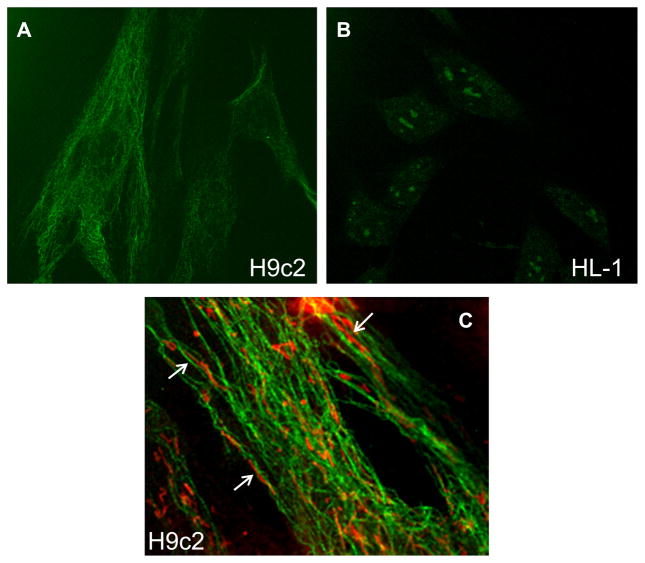
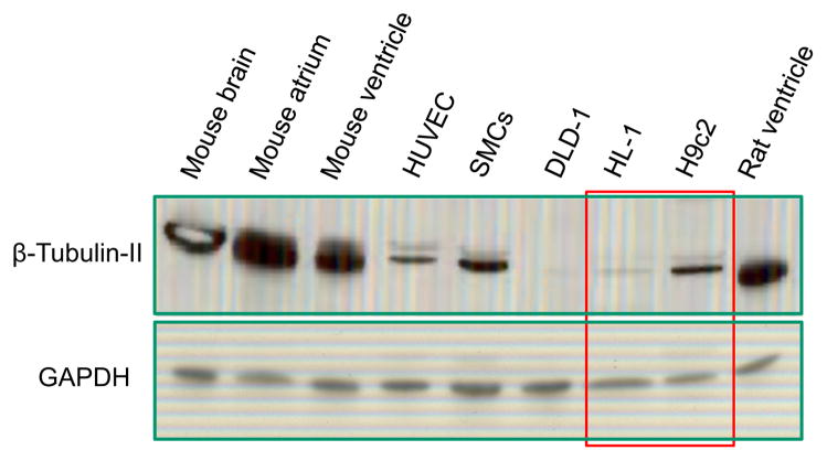
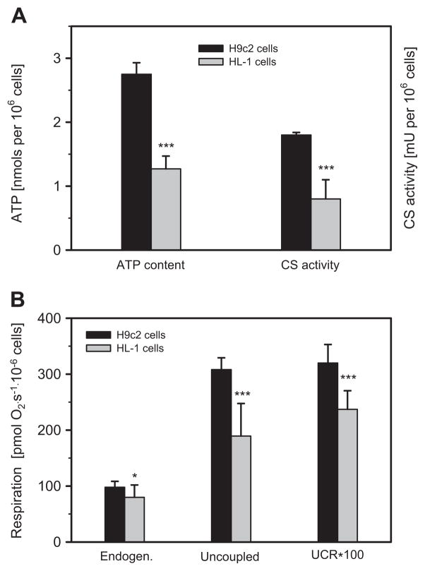
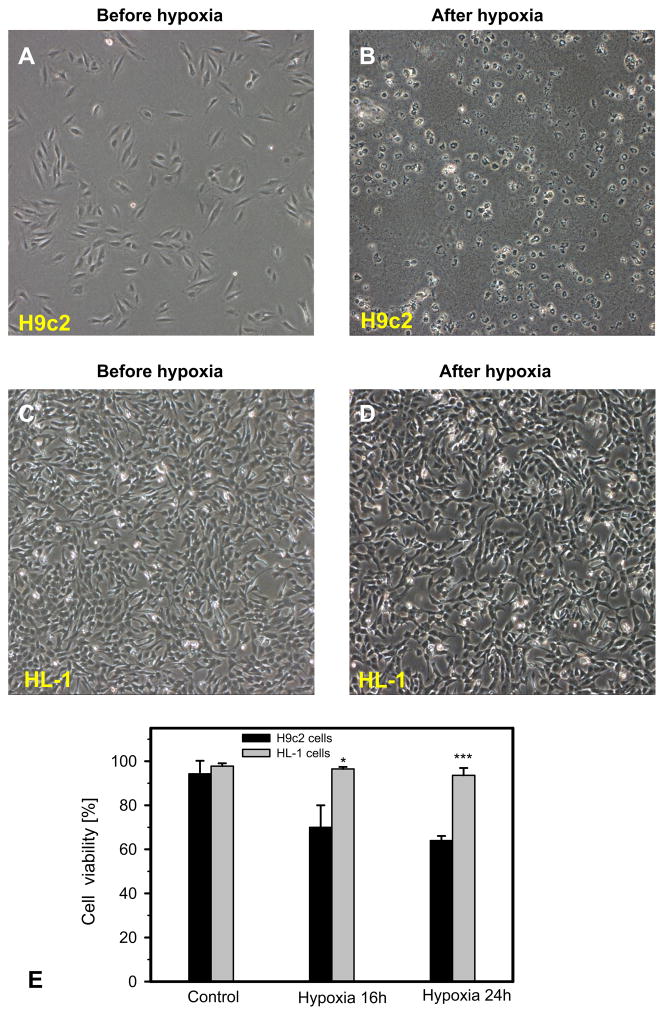
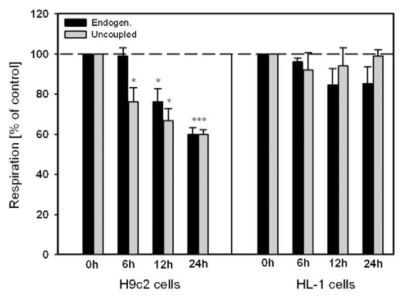
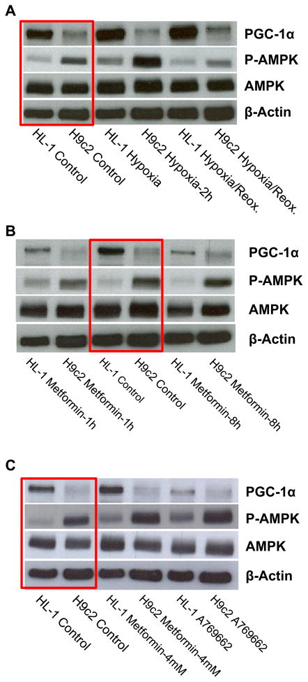
Dysfunction of cardiac energy metabolism plays a critical role in many cardiac diseases, including heart failure, myocardial infarction and ischemia-reperfusion injury and organ transplantation. The characteristics of these diseases can be elucidated in vivo, though animal-free in vitro experiments, with primary adult or neonatal cardiomyocytes, the rat ventricular H9c2 cell line or the mouse atrial HL-1 cells, providing intriguing experimental alternatives. Currently, it is not clear how H9c2 and HL-1 cells mimic the responses of primary cardiomyocytes to hypoxia and oxidative stress. In the present study, we show that H9c2 cells are more similar to primary cardiomyocytes than HL-1 cells with regard to energy metabolism patterns, such as cellular ATP levels, bioenergetics, metabolism, function and morphology of mitochondria. In contrast to HL-1, H9c2 cells possess beta-tubulin II, a mitochondrial isoform of tubulin that plays an important role in mitochondrial function and regulation. We demonstrate that H9c2 cells are significantly more sensitive to hypoxia-reoxygenation injury in terms of loss of cell viability and mitochondrial respiration, whereas HL-1 cells were more resistant to hypoxia as evidenced by their relative stability. In comparison to HL-1 cells, H9c2 cells exhibit a higher phosphorylation (activation) state of AMP-activated protein kinase, but lower peroxisome proliferator-activated receptor gamma coactivator 1-alpha levels, suggesting that each cell type is characterized by distinct regulation of mitochondrial biogenesis. Our results provide evidence that H9c2 cardiomyoblasts are more energetically similar to primary cardiomyocytes than are atrial HL-1 cells. H9c2 cells can be successfully used as an in vitro model to simulate cardiac ischemia-reperfusion injury.

Keywords: Energy metabolism; H9c2 cells; HL-1 cells; Hypoxia-reoxygenation; Mitochondria.


Conflict of interest statement

Disclosures

No confiicts of interest, financial or otherwise, are declared by the authors.

Figures

Fig. 1
Fig. 1
Different mitochondrial organization/arrangement in H9c2 and HL-1 cells. Representative confocal imaging of mitochondrial organization in H9c2 (A) and HL-1 (B) cells demonstrate punctuated/distinct mitochondria in H9c2 cells (visible also in adult primary cardiomyocytes) and more mitochondrial network in HL-1 cells more typical for various cancerous cells. Mitochondria were visualized by fluorescent confocal microscopy using mitochondrial specific probe TMRM (0.1 μM, see Methods). Bar, 20 μm.
Fig. 2
Fig. 2
Confocal imaging of beta-tubulin II in H9c2 and HL-1 cells. Cells were fixed and stained with antibodies against beta-tubulin II as described in Methods, using second antibodies Fluoro-Nanogold-anti-mouse Fab′ antibodies bound to Alexa-Fluor-488. A: Note the presence of beta-tubulin II in H9c2 cells and absence in HL-1 cells (B). C: H9c2 cells were stained with antibodies against beta-tubulin II (green) and additionally with MitoTracker red for visualization of mitochondria. In some regions, a close arrangement of mitochondrial threads and tubulin filaments can be seen (arrows).
Fig. 3
Fig. 3
Western blot analysis of beta-tubulin II in various cells and tissues. Similarly to confirming the results of fluorescent confocal imaging (see Fig. 2), the presence of beta-tubulin II in H9c2 cells and the absence in HL-1 cells were found. Note the abundant amount of beta-tubulin II in rat and mice heart and in brain. All probes contained app. 20 μg of total protein. GAPDH (“Ambion”) was used a loading control. DLD, human colorectal carcinoma cells; HUVEC, human umbilical vein endothelial cells; SMS, smooth muscle cells.
Fig. 4
Fig. 4
Biochemical, bioenergetic properties and respiratory activities are different in H9c2 and HL-1 cells. A: H9c2 cells demonstrate significantly higher level of ATP (per 106 cells) and activity of mitochondrial marker enzyme citrate synthase (CS) as compared with HL-1 cells. B: Both endogenous and uncoupled (by FCCP) respiration, as well as uncoupled control ratio (UCR) are significantly higher in H9c2 cells than in HL-1 cells (N = 3; * P < 0.05; *** P < 0.001).
Fig. 5
Fig. 5
Increased sensitivity of H9c2 cells to hypoxia compared to HL-1 cells. A–D: Representative phase contrast microscopy of H9c2 and HL-1 cells. Representative phase contrast pictures were taken before (A, C) and after (B, D) hypoxic treatment (0. 4% oxygen) for 16 h and reoxygenation for 24 h. H9c2 cells were almost completely lysed after hypoxia, whereas HL-1 substantially survived after the same hypoxic regime. HL-1 and H9c2 cells were seeded in 6-well plates and grown overnight under their corresponding conditions. E: Quantitative data of cell viability after 16 h and 24 h of hypoxia. N = 3; * P < 0.05; *** P < 0.001.
Fig. 6
Fig. 6
Mitochondrial respiratory parameters of H9c2 and HL-1 cells after hypoxia-reoxygenation. Basal and FCCP-uncoupled respiration at 6 h, 12 h and 24 h after hypoxia and standard time (2 h) of reoxygenation. In contrast to HL-1 cells (which demonstrate a stability), H9c2 cells show significant decrease (up to 60% of control in respiration rates after 24 h of hypoxia). N = 3; * P < 0.05; *** P < 0.001.
Fig. 7
Fig. 7
Comparative Western blot analysis of AMPK, P-AMPK, and PGC-1α in H9c2 and HL-1 cells. A–C: Whereas a similar level of AMPK was found in both cell lines, H9c2 cells demonstrate significantly higher amount of phospho-AMPK (activated, P-AMPK). In contrast, higher level of PGC-1α was found in HL-1 cells as compared with H9c2 cells. A: Short hypoxia (2 h) resulted in significant activation of AMPK to P-AMPK, both in H9c2 and in HL-1 cells. B: Treatment with 1 mM of activator of AMPK metformin had no effect. C: Treatment with higher (4 mM) concentration of metformin showed an activation of AMPK, more visible after treatment with specific activator A769662 (0.1 mM, 8 h).

References

    1. Aon MA, Cortassa S, Akar FG, O’Rourke B. Mitochondrial criticality: a new concept at the turning point of life or death. Biochim Biophys Acta. 2006;1762:232–240. - PMC - PubMed
    1. Balaban RS, Nemoto S, Finkel T. Mitochondria, oxidants, and aging. Cell. 2005;120:483–495. - PubMed
    1. Javadov S, Kuznetsov A. Mitochondrial permeability transition and cell death: the role of cyclophilin d. Front Physiol. 2013;4:76. - PMC - PubMed
    1. Kroemer G, Reed JC. Mitochondrial control of cell death. Nat Med. 2000;6:513–519. - PubMed
    1. Kuznetsov AV, Schneeberger S, Seiler R, Brandacher G, Mark W, Steurer W, Saks V, Usson Y, Margreiter R, Gnaiger E. Mitochondrial defects and heterogeneous cytochrome c release after cardiac cold ischemia and reperfusion. Am J Physiol Heart Circ Physiol. 2004;286:H1633–H1641. - PubMed

Publication types

LinkOut - more resources