Review
. 2014 Dec 19:194:37-48.
doi: 10.1016/j.virusres.2014.10.009. Epub 2014 Oct 17.

The avian coronavirus spike protein

Affiliations
Review

The avian coronavirus spike protein

I N Ambepitiya Wickramasinghe et al. Virus Res. .

Abstract

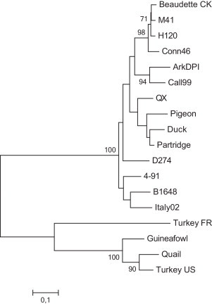
Avian coronaviruses of the genus Gammacoronavirus are represented by infectious bronchitis virus (IBV), the coronavirus of chicken. IBV causes a highly contagious disease affecting the respiratory tract and, depending on the strain, other tissues including the reproductive and urogenital tract. The control of IBV in the field is hampered by the many different strains circulating worldwide and the limited protection across strains due to serotype diversity. This diversity is believed to be due to the amino acid variation in the S1 domain of the major viral attachment protein spike. In the last years, much effort has been undertaken to address the role of the avian coronavirus spike protein in the various steps of the virus' live cycle. Various models have successfully been developed to elucidate the contribution of the spike in binding of the virus to cells, entry of cell culture cells and organ explants, and the in vivo tropism and pathogenesis. This review will give an overview of the literature on avian coronavirus spike proteins with particular focus on our recent studies on binding of recombinant soluble spike protein to chicken tissues. With this, we aim to summarize the current understanding on the avian coronavirus spike's contribution to host and tissue predilections, pathogenesis, as well as its role in therapeutic and protective interventions.

Keywords: Avian coronavirus; Binding; Gammacoronavirus; Infectious bronchitis virus IBV; Spike; Tropism.


Figures

Fig. 1
Fig. 1
Macroscopic, histological and immunohistochemical analyses of chicken trachea of mock-infected or IBV M41-infected layer chickens. Seven-day-old SPF layer chickens were oronasally infected with PBS (mock) or M41 and sacrificed at various time points after infection. (A) Longitudinally opened trachea of mock (upper) and IBV-M41 infected (lower) chicken at 7 dpi; the M41-infected trachea shows small amounts of mucoid material in the lumen and marked multifocal hyperemia of the mucosa. (B) Hematoxylin and eosin (H&E) (left) and anti-IBV S2 MAb 48.4 immunohistochemistry staining (right) of a section of the trachea of a mock-infected (7 dpi) or M41-infected (3 dpi and 7 dpi) chicken. The trachea of the M41-infected chicken at 3 dpi shows an intact epithelial lining with minimal hyperemia, while both ciliated epithelial cells and non-ciliated mucus-producing epithelial cells show marked intracytoplasmic presence of S2 antigen. At 7 dpi, the trachea has lost normal architecture due to desquamation of the ciliated and non-ciliated epithelium with replacement by a hyperplastic, more squamous non-ciliated epithelium, infiltration by large numbers of lymphocytes, marked hyperemia and in the superficial layer presence of necrotic cells. The lumen contains desquamated epithelial cells, marked numbers of heterophilic granulocytes and abundant mucoid material. Both the epithelial lining and lumen show cells containing the S2 antigen. There are no changes observed in the mock-infected chicken trachea. Scale bars represent 50 μm.
Fig. 2
Fig. 2
Infection of embryonated eggs, cell culture BHK21 cells, and primary chicken embryonic kidney cells (CEK) with IBV Beaudette and -M41. (A) Ten-day-old embryonated chicken eggs were inoculated with Beaudette and M41 and chorioallantoic membranes (CAM) were stained with anti-IBV S2 MAb 48.4. (B) BHK21 and CEK cells were inoculated with IBV Beaudette and -M41, fixed at 8 hpi and immunofluorescence was performed using the anti-IBV N protein MAb26.1. IBV antigens were present in Beaudette-inoculated BHK21, CEK and CAM, while for M41 infection was only observed in CEK and CAM. Scale bars for CAMs represent 50 μm; pictures of BHK21 and CEK cells were taken at 40× magnification.
Fig. 3
Fig. 3
Phylogenetic analysis of the amino acid sequences of the S1 subunit of the spike protein for selected avian gammacoronaviruses. Sequences were aligned using MUSCLE with default settings. The unrooted maximum likelihood tree was constructed using MEGA 6.06 with the best-fit substitution model (LG+G), with complete deletion of gaps. Bootstrap values (500 replicates) are indicated at the nodes when >70.
Fig. 4
Fig. 4
Binding of S1 proteins of different IBV strains to trachea and other clinically relevant tissues. Recombinant S1 proteins of IBV M41, H120, Beaudette, B1648 and QX were produced and attachment profiles were compared by performing spike histochemistry as previously described (Wickramasinghe et al., 2011) on trachea (A) and other chicken tissues (B). Binding of the S1 of the virulent Massachusetts M41 strain corresponded with the cells reported to be sensitive for infection with M41. Specifically, M41 S1 had a greater binding avidity than that of the attenuated vaccine strain H120 on trachea and lung (Wickramasinghe et al., 2011). For Beaudette S1 no obvious binding could be observed on chicken trachea (Wickramasinghe et al., 2011), but also not on CAM (Promkuntod et al., 2013). The S1 of the nephropathogenic B1648 did not show any appreciable staining of the trachea and the kidney, while for the S1 of QX, a strain with reported reproductive tract tropism, only mild patchy staining to the oviduct was observed. Scale bars represent 50 μm; (C) schematic representation of the ability of recombinant trimeric spike proteins to bind to a selected set of tissues and the reported ability of the corresponding IBV strains to infect these tissues; na: data not available from literature; nd: not done.
Fig. 5
Fig. 5
Binding characteristics of recombinant trimeric and dimeric IBV S1 proteins. Recombinant S1 proteins produced in frame with the GCN4 trimerization motif and the Strep-tag (‘trimers’) or in fusion with the human Fc tail (‘dimers’) were applied to chicken trachea. Using similar protein amounts for dimeric and trimeric S1 (at a concentration of 0.1 mg/ml), trimeric M41.S1 bound with higher avidity to trachea compared to dimeric M41.S1. In contrast, dimeric B1648.S1 showed limited binding to trachea, while for trimeric B1648.S1 no binding at all could be detected. Scale bars represent 50 μm.
Fig. 6
Fig. 6
IBV spike domains involved in binding. Spike histochemistry was performed using recombinant soluble spike domains. (A) The N-terminal S1 domain (M41.NTD, comprising aa 19–272 of the spike) and the C-terminal S1 domain (M41.CTD, comprising aa 273–532 of the spike) of M41 were applied to chicken trachea. The NTD was both sufficient and required for attachment to chicken tissues, and thus contained the receptor-binding domain (Promkuntod et al., 2014); (B) recombinant S1 and ectodomains of Beaudette and M41 were produced as soluble recombinant proteins and applied to BHK21 cells and chicken trachea, respectively. Binding of Beaudette spike to cell cultures was only observed for the ectodomain, while the extension of S1 with the S2 ectodomain for M41 increased the binding avidity to the trachea (similarly as observed before for the CAM; Promkuntod et al., 2013). Scale bars represent 50 μm.
Fig. 7
Fig. 7
Binding of M41.S1 is sialic acid dependent and differs between the oviduct of broiler and layer. Spike histochemistry using recombinant M41.S1 proteins was performed as described before (Wickramasinghe et al., 2011) on sections of (A) trachea of broiler and (B) oviducts of layer and broiler. Binding of M41.S1 could be inhibited by pretreating the trachea with neuraminidase from Vibrio cholerae (VCNA) and by pre-incubating the spike with the Neu5Acα2,3 specific glycan SiaLec (data not published). While M41.S1 has high binding avidity for oviduct of the layer, binding to broiler oviduct almost lacking. Scale bars represent 50 μm.
Fig. 8
Fig. 8
Beaudette spike ectodomain binding to chorioallantoic membrane tissue. Spike histochemistry was performed using recombinant wild type Beaudette spike ectodomains (Beau.ED), or the ectodomain in which the proposed heparan sulfate binding site SHRKHS (aa 686–691) was replaced by the sequence of M41 SPRRRS (Beaud.ED*M41); Beau.ED in the presence of 10 mg/ml heparin, or application of Beau.ED after treatment of tissues with 1 mU neuraminidase of Vibrio cholerae (VCNA, as described in Promkuntod et al., 2013). The proposed heparan binding sequence was not required for binding of Beaudette spike, nor could binding be blocked with heparin; binding to CAM was dependent on sialic acids. Scale bars represent 50 μm.

References

    1. Abd El Rahman S., El-Kenawy A.A., Neumann U., Herrler G., Winter C. Comparative analysis of the sialic acid binding activity and the tropism for the respiratory epithelium of four different strains of avian infectious bronchitis virus. Avian Pathol. 2009;38:41–45. - PubMed
    1. Abro S.H., Renström L.H., Ullman K., Isaksson M., Zohari S., Jansson D.S., Belák S., Baule C. Emergence of novel strains of avian infectious bronchitis virus in Sweden. Vet. Microbiol. 2012;155:237–246. - PMC - PubMed
    1. Ammayappan A., Upadhyay C., Gelb J., Jr., Vakharia V.N. Identification of sequence changes responsible for the attenuation of avian infectious bronchitis virus strain Arkansas DPI. Arch. Virol. 2009;154:495–499. - PMC - PubMed
    1. Armesto M., Cavanagh D., Britton P. The replicase gene of avian coronavirus infectious bronchitis virus is a determinant of pathogenicity. PLoS ONE. 2009;4:e7384. - PMC - PubMed
    1. Armesto M., Evans S., Cavanagh D., Abu-Median A.B., Keep S., Britton P. A recombinant avian infectious bronchitis virus expressing a heterologous spike gene belonging to the 4/91 serotype. PLoS ONE. 2011;6:e24352. - PMC - PubMed

Publication types

Substances