

Chronic methamphetamine abuse and corticostriatal deficits revealed by neuroimaging
- PMID: 25451127
- PMCID: PMC4418947
- DOI: 10.1016/j.brainres.2014.10.044
Chronic methamphetamine abuse and corticostriatal deficits revealed by neuroimaging
Abstract
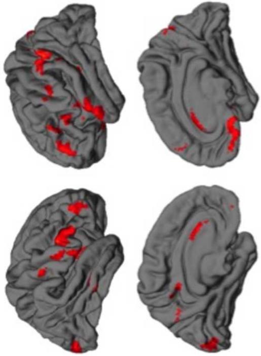
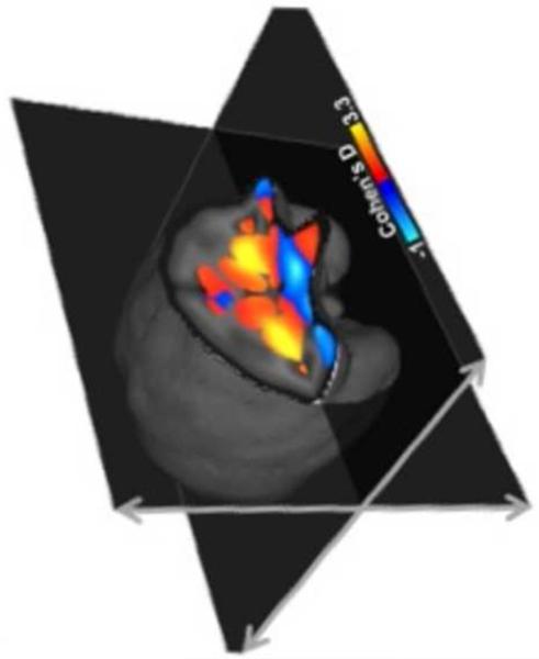
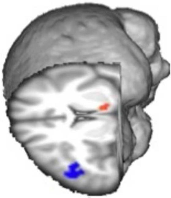
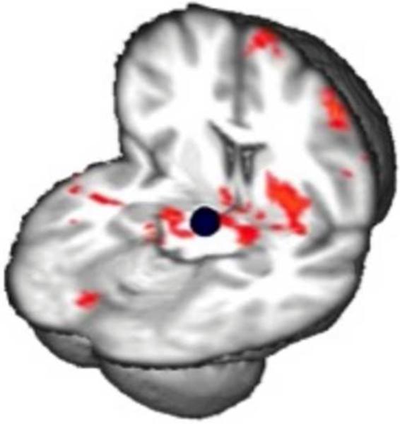
Despite aggressive efforts to contain it, methamphetamine use disorder continues to be major public health problem; and with generic behavioral therapies still the mainstay of treatment for methamphetamine abuse, rates of attrition and relapse remain high. This review summarizes the findings of structural, molecular, and functional neuroimaging studies of methamphetamine abusers, focusing on cortical and striatal abnormalities and their potential contributions to cognitive and behavioral phenotypes that can serve to promote compulsive drug use. These studies indicate that individuals with a history of chronic methamphetamine abuse often display several signs of corticostriatal dysfunction, including abnormal gray- and white-matter integrity, monoamine neurotransmitter system deficiencies, neuroinflammation, poor neuronal integrity, and aberrant patterns of brain connectivity and function, both when engaged in cognitive tasks and at rest. More importantly, many of these neural abnormalities were found to be linked with certain addiction-related phenotypes that may influence treatment response (e.g., poor self-control, cognitive inflexibility, maladaptive decision-making), raising the possibility that they may represent novel therapeutic targets.
Keywords: Addiction; Corticostriatal circuitry; Diffusion tensor imaging; Magnetic resonance imaging; Methamphetamine; Positron emission tomography.
Copyright © 2014 Elsevier B.V. All rights reserved.
Figures
References
-
- Agrawal A, Lynskey MT. Are there genetic influences on addiction: evidence from family, adoption and twin studies. Addiction. 2008;103:1069–81. - PubMed
-
- Aron AR, et al. A componential analysis of task-switching deficits associated with lesions of left and right frontal cortex. Brain. 2004;127:1561–73. - PubMed
-
- Ballard ME, et al. Low striatal dopamine D2/D3 receptor availability predicts steeper delay discounting in methamphetamine abusers; Poster session presented at the meeting of the College on Problems of Drug Dependence; San Juan, PR. Jun, 2014.
-
- Beaulieu C. In: Diffusion MRI: From Quantitiative Measurement to In Vivo Neuroanatomy. Johansen-Berg H, Behrens TE, editors. Elsevier; London: 2009. pp. 105–126.
Publication types
MeSH terms
Substances
Grants and funding
LinkOut - more resources
Full Text Sources
Other Literature Sources
Medical
