The feasibility of imaging myocardial ischemic/reperfusion injury using (99m)Tc-labeled duramycin in a porcine model
- PMID: 25451214
- PMCID: PMC4570238
- DOI: 10.1016/j.nucmedbio.2014.09.002
The feasibility of imaging myocardial ischemic/reperfusion injury using (99m)Tc-labeled duramycin in a porcine model
Abstract
When pathologically externalized, phosphatidylethanolamine (PE) is a potential surrogate marker for detecting tissue injuries. (99m)Tc-labeled duramycin is a peptide-based imaging agent that binds PE with high affinity and specificity. The goal of the current study was to investigate the clearance kinetics of (99m)Tc-labeled duramycin in a large animal model (normal pigs) and to assess its uptake in the heart using a pig model of myocardial ischemia-reperfusion injury.
Methods: The clearance and distribution of intravenously injected (99m)Tc-duramycin were characterized in sham-operated animals (n=5). In a closed chest model of myocardial ischemia, coronary occlusion was induced by balloon angioplasty (n=9). (99m)Tc-duramycin (10-15mCi) was injected intravenously at 1hour after reperfusion. SPECT/CT was acquired at 1 and 3hours after injection. Cardiac tissues were analyzed for changes associated with acute cellular injuries. Autoradiography and gamma counting were used to determine radioactivity uptake. For the remaining animals, (99m)Tc-tetrafosamin scan was performed on the second day to identify the infarct site.
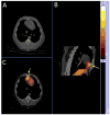
Results: Intravenously injected (99m)Tc-duramycin cleared from circulation predominantly via the renal/urinary tract with an α-phase half-life of 3.6±0.3minutes and β-phase half-life of 179.9±64.7minutes. In control animals, the ratios between normal heart and lung were 1.76±0.21, 1.66±0.22, 1.50±0.20 and 1.75±0.31 at 0.5, 1, 2 and 3hours post-injection, respectively. The ratios between normal heart and liver were 0.88±0.13, 0.80±0.13, 0.82±0.19 and 0.88±0.14. In vivo visualization of focal radioactivity uptake in the ischemic heart was attainable as early as 30min post-injection. The in vivo ischemic-to-normal uptake ratios were 3.57±0.74 and 3.69±0.91 at 1 and 3hours post-injection, respectively. Ischemic-to-lung ratios were 4.89±0.85 and 4.93±0.57; and ischemic-to-liver ratios were 2.05±0.30 to 3.23±0.78. The size of (99m)Tc-duramycin positive myocardium was qualitatively larger than the infarct size delineated by the perfusion defect in (99m)Tc-tetrafosmin uptake. This was consistent with findings from tissue analysis and autoradiography.
Conclusion: (99m)Tc-duramycin was demonstrated, in a large animal model, to have suitable clearance and biodistribution profiles for imaging. The agent has an avid target uptake and a fast background clearance. It is appropriate for imaging myocardial injury induced by ischemia/reperfusion.
Keywords: Apoptosis; Duramycin; Ischemia–reperfusion injury; Phosphatidylethanolamine.
Copyright © 2014 Elsevier Inc. All rights reserved.
Conflict of interest statement
Conflict of Interest Statement: The authors declare that they have no conflict of interest.
Figures


References
-
- Williamson P, Schlegel RA. Transbilayer phospholipid movement and the clearance of apoptotic cells. Biochim Biophys Acta. 2002;1585:53–63. - PubMed
-
- Emoto K, Toyama-Sorimachi N, Karasuyama H, et al. Exposure of phosphatidylethanolamine on the surface of apoptotic cells. Exp Cell Res. 1997;232:430–434. - PubMed
-
- Zhao M, Zhu X, Ji S, et al. 99mTc-labeled C2A domain of synaptotagmin I as a target-specific molecular probe for noninvasive imaging of acute myocardial infarction. J Nucl Med. 2006;47:1367–1374. - PubMed
-
- Hofstra L, Liem IH, Dumont EA, et al. Visualisation of cell death in vivo in patients with acute myocardial infarction. Lancet. 2000;356:209–212. - PubMed
Publication types
MeSH terms
Substances
Grants and funding
LinkOut - more resources
Full Text Sources
Other Literature Sources
Medical

