. 2015 Jan 16;290(3):1580-91.
doi: 10.1074/jbc.M114.609834. Epub 2014 Dec 1.

BRCA1-associated protein 1 (BAP1) deubiquitinase antagonizes the ubiquitin-mediated activation of FoxK2 target genes

Affiliations

BRCA1-associated protein 1 (BAP1) deubiquitinase antagonizes the ubiquitin-mediated activation of FoxK2 target genes

Yuki Okino et al. J Biol Chem. .

Abstract

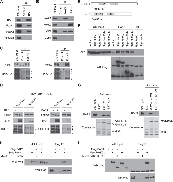
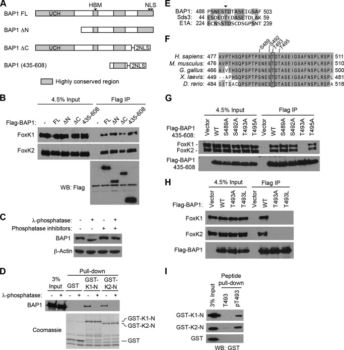
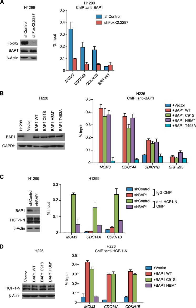
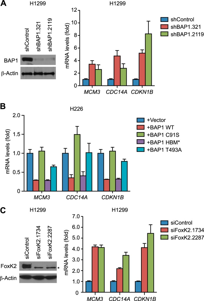
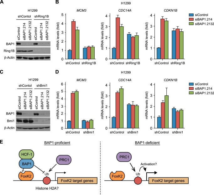
BRCA1-associated protein 1 (BAP1), which is frequently mutated in cancer, functions as a deubiquitinase (DUB) for histone H2A. Although BAP1 interacts with a transcriptional regulator, HCF-1, and transcription factors FoxK1 and FoxK2, how BAP1 controls gene expression remains unclear. This study investigates the importance of BAP1 DUB activity and the interactions with FoxK2 and HCF-1 in the regulation of FoxK2 target genes. We show that FoxK2 recruits BAP1 to the target genes through the forkhead-associated domain, which interacts with Thr(P)-493 on BAP1. BAP1, in turn, recruits HCF-1, thereby forming a ternary complex in which BAP1 bridges FoxK2 and HCF-1. BAP1 represses FoxK2 target genes, and this effect requires BAP1 DUB activity but not interaction with HCF-1. Importantly, BAP1 depletion causes up-regulation of FoxK2 target genes only in the presence of the Ring1B-Bmi1 complex, an E3 ubiquitin ligase for histone H2A, indicating an antagonizing role of BAP1 against Ring1B-Bmi1. Our findings suggest that BAP1 deficiency causes increased expression of target genes in a Ring1B-Bmi1-dependent manner.

Keywords: Deubiquitylation (Deubiquitination); Gene Expression; Phosphorylation; Tumor Suppressor Gene; Ubiquitin.


Figures

FIGURE 1.
FIGURE 1.
FoxK1 and FoxK2 form ternary complexes with BAP1 and HCF-1. A, interaction of endogenous FoxK1/K2 with BAP1 in vivo. BAP1 was immunoprecipitated (IP) with specific antibodies, and the indicated proteins were detected by Western blotting. FoxO3a is shown as a negative control. B, association of FoxK1/K2 with BAP1 in vivo. FoxK1 or FoxK2 was immunoprecipitated, and the indicated proteins were detected by Western blotting. KAP1 is shown as a negative control. C, complex formation of FoxK1/K2 with HCF-1. FoxK1 or FoxK2 was immunoprecipitated, and the indicated proteins were detected by Western blotting. D, in vivo association of FoxK1/K2 with BAP1 and HCF-1 in H226 BAP1-deficient cells. BAP1 was expressed in H226 cells, and FoxK1 or FoxK2 was immunoprecipitated with specific antibodies. The indicated proteins were detected by Western blotting. The arrowheads and asterisks indicate HCF-1 bands and nonspecific bands, respectively. E, schematic of FoxK1 and FoxK2 proteins. F, interactions between the N-terminal regions of FoxK1/K2 and BAP1. Full-length or N-terminal regions (indicated in E) of FoxK1 and FoxK2 were expressed with a FLAG tag and immunoprecipitated with anti-FLAG antibodies or control IgG. Coprecipitated endogenous BAP1 was detected by Western blotting (WB). G, in vitro interactions between BAP1 and the N-terminal regions of FoxK1/K2. GST pulldown assays were performed with the indicated GST fusion proteins mixed with 293T cell lysates. GST fusion proteins in the precipitates were visualized by Coomassie staining, and coprecipitated BAP1 was detected by Western blotting. H, coimmunoprecipitation experiments of BAP1 and the FoxK1 R127A mutant. The indicated proteins were coexpressed in 293T cells, and FLAG-BAP1 was precipitated with anti-FLAG antibodies. Myc-FoxK1 was detected by anti-Myc Western blotting. I, coimmunoprecipitation experiments of BAP1 and the FHA deletion mutant (ΔFHA) of FoxK2.
FIGURE 2.
FIGURE 2.
Phosphorylation-dependent interactions between BAP1 and FoxK1/K2. A, schematic of BAP1 and its truncation mutants. Regions highly conserved between species are shown in gray. FL, full-length; UCH, ubiquitin C-terminal hydrolase; NLS, nuclear localization signal. B, mapping of the FoxK1/K2-interacting domains of BAP1. Full-length BAP1 and its truncation mutants, depicted in A, were expressed with a FLAG tag and precipitated with anti-FLAG antibodies. Coprecipitated endogenous FoxK1 and FoxK2 proteins were detected by Western blotting (WB). IP, immunoprecipitation. C, effect of λ-phosphatase treatments on BAP1 gel mobility. Cell lysates were treated with λ-phosphatase in the presence or absence of phosphatase inhibitors, and the indicated proteins were assessed by Western blotting. D, in vitro interaction between BAP1 and the N-terminal region of FoxK1/FoxK2. GST pulldown assays were performed using the indicated GST fusion proteins, and 293T cell lysates were treated with or λ-phosphatase or left untreated. Precipitated GST fusion proteins were visualized by Coomassie staining, and coprecipitated BAP1 was detected by Western blotting. E, alignment of BAP1, Sds3, and E1A sequences surrounding the conserved Thr residue (arrowhead), which is phosphorylated in Sds3. Identical amino acids are highlighted. F, alignment of the FoxK1/K2-interacting region of BAP1 from various species. Identical and similar amino acids are highlighted with different contrasts. G, in vivo interactions between BAP1 (435–608) and FoxK1/K2. FLAG-tagged BAP1 (435–608) with various point mutations was expressed in 293T cells and immunoprecipitated with anti-FLAG antibodies. Coprecipitated endogenous FoxK1 and FoxK2 were detected by Western blotting. H, in vivo interactions between FoxK1/K2 and full-length BAP1. FLAG-tagged BAP1 proteins (wild-type, T493A, and T493L) were expressed and immunoprecipitated with anti-FLAG antibodies. Coprecipitated endogenous FoxK1/K2 was detected by Western blotting. I, peptide pulldown assays. Peptides corresponding to the 487–499 amino acid region of BAP1 were synthesized with or without phosphorylation at Thr-493 and conjugated to beads. Pulldown assays were performed by mixing beads and recombinant GST-FoxK1-N or GST-FoxK2-N proteins, and precipitated GST-fusion proteins were detected by Western blotting using anti-GST antibodies. T493, non-phosphorylated peptide; pT493, peptide phosphorylated at Thr-493.
FIGURE 3.
FIGURE 3.
Recruitment of BAP1 and HCF-1 to FoxK2 binding regions. A, recruitment of BAP1 to FoxK2 target genes. ChIP using anti-BAP1 antibodies was performed with control and FoxK2-depleted H1299 cells. Expression levels of FoxK2 and BAP1 assessed by Western blotting are shown in the left panel. B, recruitment of various BAP1 mutants to FoxK2 target genes. BAP1 mutants were expressed at endogenous levels in the H226 BAP1-deficient cell line, and ChIP was performed with anti-BAP1 antibodies. Expression levels of BAP1 assessed by Western blotting are shown in the left panel. H1299 is shown as a control for the endogenous BAP1 level. C, recruitment of HCF-1 to FoxK2 target genes. ChIP using anti-HCF-1-N antiserum was performed with control and BAP1-depleted H1299 cells. Expression levels of BAP1 and HCF-1-N assessed by Western blotting are shown in the left panel. D, ChIP analyses of HCF-1 recruitment in the H226 BAP1-deficient cell line expressing various BAP1 mutants. BAP1 mutants were expressed as in B, and ChIP was performed with anti-HCF-1-N antiserum. Expression levels of HCF-1-N were assessed by Western blotting and are shown in the left panel. β-actin and GAPDH served as loading controls in Western blotting. qPCR was done in triplicate, and mean ± S.D. is shown. In ChIP experiments, the amount of precipitated DNA at each region was expressed as a percentage of input.
FIGURE 4.
FIGURE 4.
BAP1 down-regulates FoxK2 target genes. A, expression levels of FoxK2 target genes in BAP1-depleted H1299 cells. mRNA levels of the indicated FoxK2 target genes were measured by RT-PCR in control and BAP1-depleted H1299 cells (right panel). Knockdown of BAP1 was assessed by Western blotting (left panel). β-Actin was used as a loading control. B, effects of various mutations in BAP1 on expression of FoxK2 target genes. The indicated BAP1 mutants were expressed in the H226 BAP1-deficient cell line as in Fig. 3B, and expression of the indicated genes was quantitated by RT-PCR. C, expression of FoxK2 target genes in FoxK2-depleted H1299 cells. FoxK2 was depleted with the indicated siRNAs, and FoxK2 protein levels were assessed by Western blotting (left panel). An asterisk indicates a nonspecific band. mRNA levels of the indicated genes were measured by RT-PCR (right panel). qPCR was done in triplicate, and mean ± S.D. is shown. Values were normalized to GAPDH and are shown relative to control samples.
FIGURE 5.
FIGURE 5.
Regulation of H2Aub at FoxK2 target genes by BAP1. A and B, effect of BAP1 on H2Aub levels at the FoxK2-binding regions. ChIP assays were performed using anti-H2Aub antibodies in H1299 with control or BAP1 knockdown (A) and the H226 BAP1-deficient cell line expressing the wild type or the inactive BAP1 mutant (C91S) (B). C and D, recruitment of Ring1B to FoxK2 binding regions. ChIP assays were performed using control IgG or anti-Ring1B antibodies in H1299 with control or BAP1 knockdown (C) and the H226 BAP1-deficient cell line with or without BAP1 expression (D). qPCR was done in triplicate, and mean ± S.D. is shown.
FIGURE 6.
FIGURE 6.
BAP1 suppresses PRC1-mediated activation of FoxK2 target genes. A–D, codepletion of BAP1 with Ring1B or Bmi1. BAP1 and Ring1B (A and B) or Bmi1 (C and D) were depleted individually or in combination. Verification of knockdown by Western blotting is shown in A and C. β-actin served as a loading control. Expression of the indicated FoxK2 target genes was assessed by RT-PCR and is shown in B and D. Values were normalized to GAPDH and are shown relative to the control RNAi sample. qPCR was done in triplicate, and mean ± S.D. is shown. E, model of BAP1-mediated regulation of FoxK2 target genes. See “Discussion” for details.

References

    1. Testa J. R., Cheung M., Pei J., Below J. E., Tan Y., Sementino E., Cox N. J., Dogan A. U., Pass H. I., Trusa S., Hesdorffer M., Nasu M., Powers A., Rivera Z., Comertpay S., Tanji M., Gaudino G., Yang H., Carbone M. (2011) Germline BAP1 mutations predispose to malignant mesothelioma. Nat. Genet. 43, 1022–1025 - PMC - PubMed
    1. Bott M., Brevet M., Taylor B. S., Shimizu S., Ito T., Wang L., Creaney J., Lake R. A., Zakowski M. F., Reva B., Sander C., Delsite R., Powell S., Zhou Q., Shen R., Olshen A., Rusch V., Ladanyi M. (2011) The nuclear deubiquitinase BAP1 is commonly inactivated by somatic mutations and 3p21.1 losses in malignant pleural mesothelioma. Nat. Genet. 43, 668–672 - PMC - PubMed
    1. Harbour J. W., Onken M. D., Roberson E. D., Duan S., Cao L., Worley L. A., Council M. L., Matatall K. A., Helms C., Bowcock A. M. (2010) Frequent mutation of BAP1 in metastasizing uveal melanomas. Science 330, 1410–1413 - PMC - PubMed
    1. Peña-Llopis S., Vega-Rubín-de-Celis S., Liao A., Leng N., Pavía-Jiménez A., Wang S., Yamasaki T., Zhrebker L., Sivanand S., Spence P., Kinch L., Hambuch T., Jain S., Lotan Y., Margulis V., Sagalowsky A. I., Summerour P. B., Kabbani W., Wong S. W., Grishin N., Laurent M., Xie X. J., Haudenschild C. D., Ross M. T., Bentley D. R., Kapur P., Brugarolas J. (2012) BAP1 loss defines a new class of renal cell carcinoma. Nat. Genet. 44, 751–759 - PMC - PubMed
    1. Guo G., Gui Y., Gao S., Tang A., Hu X., Huang Y., Jia W., Li Z., He M., Sun L., Song P., Sun X., Zhao X., Yang S., Liang C., Wan S., Zhou F., Chen C., Zhu J., Li X., Jian M., Zhou L., Ye R., Huang P., Chen J., Jiang T., Liu X., Wang Y., Zou J., Jiang Z., Wu R., Wu S., Fan F., Zhang Z., Liu L., Yang R., Liu X., Wu H., Yin W., Zhao X., Liu Y., Peng H., Jiang B., Feng Q., Li C., Xie J., Lu J., Kristiansen K., Li Y., Zhang X., Li S., Wang J., Yang H., Cai Z., Wang J. (2012) Frequent mutations of genes encoding ubiquitin-mediated proteolysis pathway components in clear cell renal cell carcinoma. Nat. Genet. 44, 17–19 - PubMed

Publication types

MeSH terms