. 2015 Jan 23;290(4):1979-93.
doi: 10.1074/jbc.M114.603225. Epub 2014 Dec 1.

Significantly enhanced heme retention ability of myoglobin engineered to mimic the third covalent linkage by nonaxial histidine to heme (vinyl) in synechocystis hemoglobin

Affiliations

Significantly enhanced heme retention ability of myoglobin engineered to mimic the third covalent linkage by nonaxial histidine to heme (vinyl) in synechocystis hemoglobin

Sheetal Uppal et al. J Biol Chem. .

Abstract

Heme proteins, which reversibly bind oxygen and display a particular fold originally identified in myoglobin (Mb), characterize the "hemoglobin (Hb) superfamily." The long known and widely investigated Hb superfamily, however, has been enriched by the discovery and investigation of new classes and members. Truncated Hbs typify such novel classes and exhibit a distinct two-on-two α-helical fold. The truncated Hb from the freshwater cyanobacterium Synechocystis exhibits hexacoordinate heme chemistry and bears an unusual covalent bond between the nonaxial His(117) and a heme porphyrin 2-vinyl atom, which remains tightly associated with the globin unlike any other. It seems to be the most stable Hb known to date, and His(117) is the dominant force holding the heme. Mutations of amino acid residues in the vicinity did not influence this covalent linkage. Introduction of a nonaxial His into sperm whale Mb at the topologically equivalent position and in close proximity to vinyl group significantly increased the heme stability of this prototype globin. Reversed phase chromatography, electrospray ionization-MS, and MALDI-TOF analyses confirmed the presence of covalent linkage in Mb I107H. The Mb mutant with the engineered covalent linkage was stable to denaturants and exhibited ligand binding and auto-oxidation rates similar to the wild type protein. This indeed is a novel finding and provides a new perspective to the evolution of Hbs. The successful attempt at engineering heme stability holds promise for the production of stable Hb-based blood substitute.

Keywords: Engineered Heme Affinity; Engineering Heme Stability in Myoglobin; Heme; Mimic of Third Covalent Linkage to Heme; Mutagenesis; Myoglobin; Myoglobin with Low Heme Dissociation; Protein Engineering; Protein Stability; Synechocystis Hemoglobin.


Figures

FIGURE 1.
FIGURE 1.
Side chain amino acids mutated in Synechocystis hemoglobin and their influence on the heme retention ability and protein stability. A, structural representation of Synechocystis hemoglobin (Protein Data Bank code 1RTX). The truncated helical fold (two-on-two) is represented in gray, and the heme prosthetic group is in black. Various key residues (B10, E7, E10, F7, F8, and H16) decorating the heme pocket of SynHb are displayed in color. The three histidines that are covalently associated with heme and that are unique to SynHb are shown in red. His46 and His70 directly coordinate to heme iron and constitute “hexacoordination.” The third His (His117) is covalently associated to heme vinyl group. B, comparison of the heme extraction results for Synechocystis wild type and mutant proteins. Bovine hemoglobin was used as a positive control and it released heme in the organic (top) layer (vial 1). The purified proteins used for heme extraction were as follows: vial 2, SynHbWT; vial 3, SynQ43V; vial 4, SynH46L; vial 5, SynY22L; vial 6, SynH70G; vial 7, SynH117A; vial 8, SynA69S; vial 9, SynH46L,H117A; and vial 10, myoglobin. C, comparison of pH titration profiles of SynHb wild type and mutant proteins monitored by change in the Soret wavelength against pH (2.0–11.0). SynH117A and H46L,H117A mutant proteins showed significant blue shift in the Soret wavelength at acidic pH. D, absorbance spectra of SynH117A and SynH46L,H117A at acidic pH were typical of free heme. E, changes in the Soret peak wavelength were monitored with the increase in GdmCl concentration using absorbance spectroscopy. SynH117A and H46L,H117A were significantly less stable than wtSynHb. SynH46L was also seen to be less stable. F, DSC thermograms of SynHb wild type and mutant proteins. Mb wild type protein was used as a control. The concentration of protein used was 0.12 mm in 100 mm potassium phosphate buffer, pH 7.0.
FIGURE 2.
FIGURE 2.
Selection of amino acid side chains and the strategy to introduce an additional covalent linkage in myoglobin. A, sequence alignment of Mb and SynHb generated using the ClustalW software. His117 (SynHb) aligned with Phe138 (Mb) as shown in red, indicating Phe138 to be a putative amino acid side chain for substitution. B, identification of amino acid side chain in Mb that is within 3, 4, 5, and 6 Å radius of the heme:CAB atom. These are the side chains that are most likely to form covalent linkage to vinyl heme upon substitution to His. C, structural alignment of Mb (red) and SynHb (blue) generated by using the SUPERPOSE software. Ile107 in Mb was mutated to His in silico, and the distance between heme:CAB and HisNϵ2 was calculated to be 2.54 Å, similar to that between His117Nϵ2 and heme of SynHb. The Protein Data Bank codes used are 1RTX (SynHb) and 5MBN (Mb).
FIGURE 3.
FIGURE 3.
Spectral properties, heme extraction assay, and assessment of extent of covalent linkage for myoglobin and its mutant proteins. A, absorbance spectra. B, CD spectra of Mb mutants in comparison with wild type. MRE, mean residue ellipticity. C, the proteins used for heme extraction were as follows: Mb wild type, MbF138H, MbI107H, and SynHbWT. Myoglobin released heme in the organic (top) layer. A similar result was observed for MbF138H. MbI107H did not release heme in the organic layer as also observed in SynHbWT. D, reversed phase UPLC analysis of purified Mb wild type and I107H mutant protein in BEH C18 column. The elution of the heme moiety was determined at 409 nm, whereas the protein (inset) was detected at 280 nm. An acetonitrile gradient was used to determine the elution position of free heme, Mb wild type, and MbI107H protein.
FIGURE 4.
FIGURE 4.
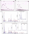
Mass spectrometric analysis of covalently bound heme in MbI107H mutant protein compared with myoglobin wild type. A, ESI-MS spectrum of intact myoglobin protein with an average molecular mass corresponding to ∼17,552.87 Da similar to the mass expected for apo-myoglobin (without heme). B, ESI-MS spectrum of intact myoglobin I107H mutant protein with an average molecular mass corresponding to ∼18,238.27 Da similar to the mass expected for the MbI107H mutant protein covalently bound with heme. C, MALDI-TOF mass spectrum of trypsin-digested myoglobin in the region of m/z = 899–3010. The inset shows the expanded view (m/z = 1922–1943 region) of the MALDI-TOF mass spectrum of the peptide fragment containing Ile at position 107 at m/z = 1927.118. D, MALDI-TOF mass spectrum of trypsin-digested myoglobin I107H mutant protein in the region of m/z = 899–3010. The left inset shows the expanded view (m/z = 1943–1954 region) of the MALDI-TOF mass spectrum of the peptide fragment containing His at position 107 at m/z = 1951.068, and the right inset shows the expanded view (m/z = 2552–2576 region) of the MALDI-TOF mass spectrum of the heme peptide fragment at m/z = 2560.109.
FIGURE 5.
FIGURE 5.
pH stability studies of myoglobin wild type and mutant proteins. A, comparison of pH titration profile of myoglobin mutant proteins (MbF138H and MbI107H) with Mb wild type protein measured by monitoring Soret peak wavelength maxima. pH titration profile of MbI107H was found to be similar to SynHb WT (inset). B–D, absorbance spectral profiles of Mb wild type (B), I107H (C), and SynHb WT (D) at pH 2.0 and pH 7.0. E, the stability of secondary structure at different pH were investigated using far UV-CD for Mb wild type and mutant proteins. CD222 nm (mdeg) values were plotted as a function of pH from 2.0 to 11.0.
FIGURE 6.
FIGURE 6.
GdmCl stability studies of myoglobin wild type and mutant proteins. A, changes in the Soret peak wavelength were monitored with increases in GdmCl concentration. B, changes in the secondary structure were monitored with increases in the GdmCl concentration using far UV-CD signal at 222 nm.
FIGURE 7.
FIGURE 7.
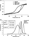
Thermal stability studies of myoglobin wild type and mutant proteins. A, changes in the secondary structure of proteins were monitored with the increase in temperature using CD spectroscopy. CD222 nm values were plotted against temperature. B, DSC thermogram of myoglobin and mutant proteins. MbI107H shows biphasic denaturation profile compared with Mb and SynHb wild type proteins, with the second peak having a higher apparent Tm than Mb wild type protein. The concentration of protein used was 0.12 mm in 100 mm potassium phosphate buffer, pH 7.0. Inset, melting temperature (apparent Tm) of Mb wild type, MbI107H, and SynHb WT obtained from differential scanning calorimetry.
FIGURE 8.
FIGURE 8.
Electronic and paramagnetic spectral characteristics of MbI107H. A and B, absorbance spectra of Mb and Mb I107H in both ferric and ferrous forms. MbI107H (B) demonstrates characteristics of high spin pentacoordinate Hb like wild type Mb (A). C, absorbance spectra of ferric and ferrous hexacoordinate Hb (HxHb; rice Hb) demonstrate characteristics typical of low spin hexacoordinate Hb. D and E, the EPR spectra of ferric Mb (D) and MbI107H (E) show the axial high spin signal. F, the EPR spectrum of ferric hexacoordinate hemoglobin (rice Hb) shows axial low spin signals.
FIGURE 9.
FIGURE 9.
Influence of mutations on auto-oxidation and hemin loss in myoglobin. A, auto-oxidation kinetics of myoglobin wild type and mutant proteins in comparison with wild type SynHb. Time courses showing the normalized changes of the ratio A581 nm/A630 nm for Mb wild type, F138H, and I107H indicated similar auto-oxidation rates. SynHb displays a much faster rate of auto-oxidation. B, time courses for hemin dissociation at pH 7.0 for SynHb, Mb, and its mutants. Normalized changes of the ratio A409 nm/A630 nm of the globins were plotted. SynHb and MbI107H were resistant to heme dissociation, whereas MbF138H had a very high rate of heme dissociation.

References

    1. Cavalier-Smith T. (2006) Cell evolution and Earth history: stasis and revolution. Philos. Trans. R Soc. Lond. B Biol. Sci. 361, 969–1006 - PMC - PubMed
    1. Falcón L. I., Magallón S., Castillo A. (2010) Dating the cyanobacterial ancestor of the chloroplast. ISME J. 4, 777–783 - PubMed
    1. Olson J. M., Blankenship R. E. (2004) Thinking about the evolution of photosynthesis. Photosynth. Res. 80, 373–386 - PubMed
    1. Hoy J. A., Kundu S., Trent J. T., 3rd, Ramaswamy S., Hargrove M. S. (2004) The crystal structure of Synechocystis hemoglobin with a covalent heme linkage. J. Biol. Chem. 279, 16535–16542 - PubMed
    1. Pesce A., Couture M., Dewilde S., Guertin M., Yamauchi K., Ascenzi P., Moens L., Bolognesi M. (2000) A novel two-over-two α-helical sandwich fold is characteristic of the truncated hemoglobin family. EMBO J. 19, 2424–2434 - PMC - PubMed

Publication types

MeSH terms

LinkOut - more resources