. 2015 Mar 10:1600:70-83.
doi: 10.1016/j.brainres.2014.10.037. Epub 2014 Oct 29.

Effects of prefrontal cortex and hippocampal NMDA NR1-subunit deletion on complex cognitive and social behaviors

Affiliations

Effects of prefrontal cortex and hippocampal NMDA NR1-subunit deletion on complex cognitive and social behaviors

Janet M Finlay et al. Brain Res. .

Abstract

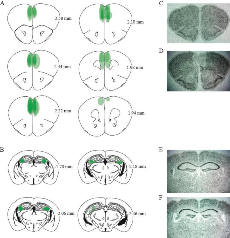
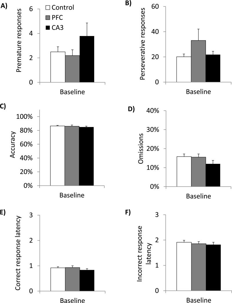
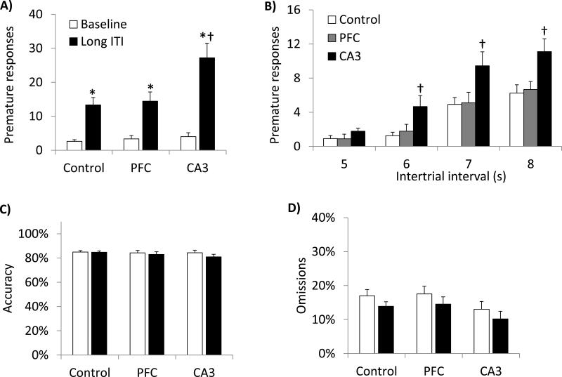
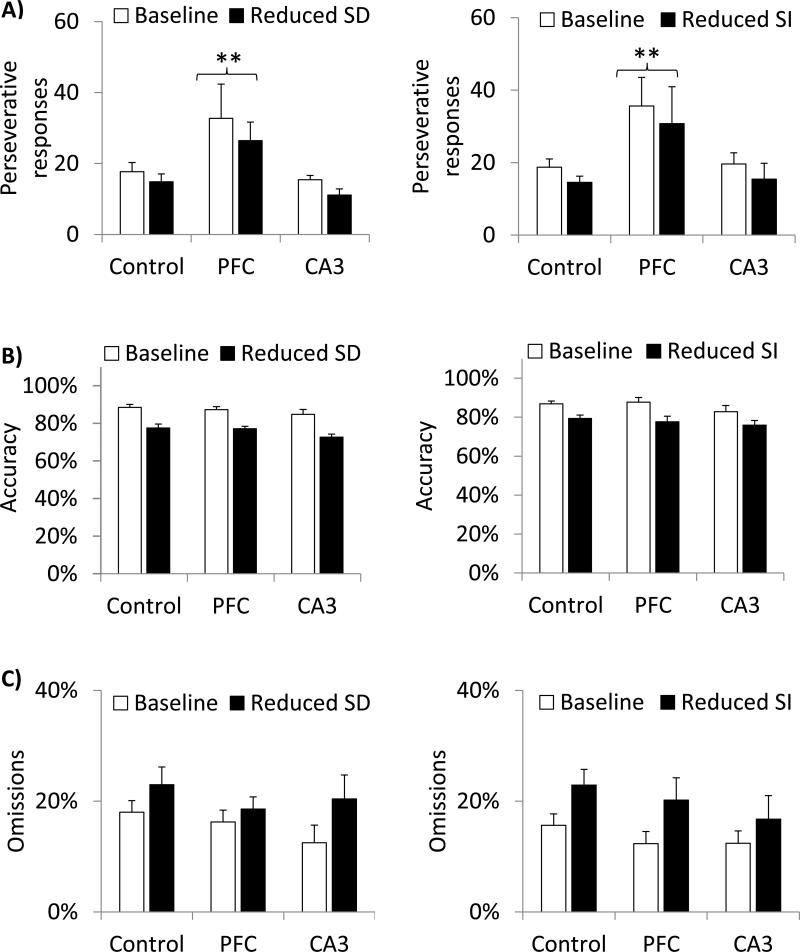
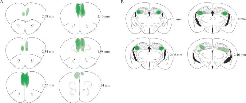
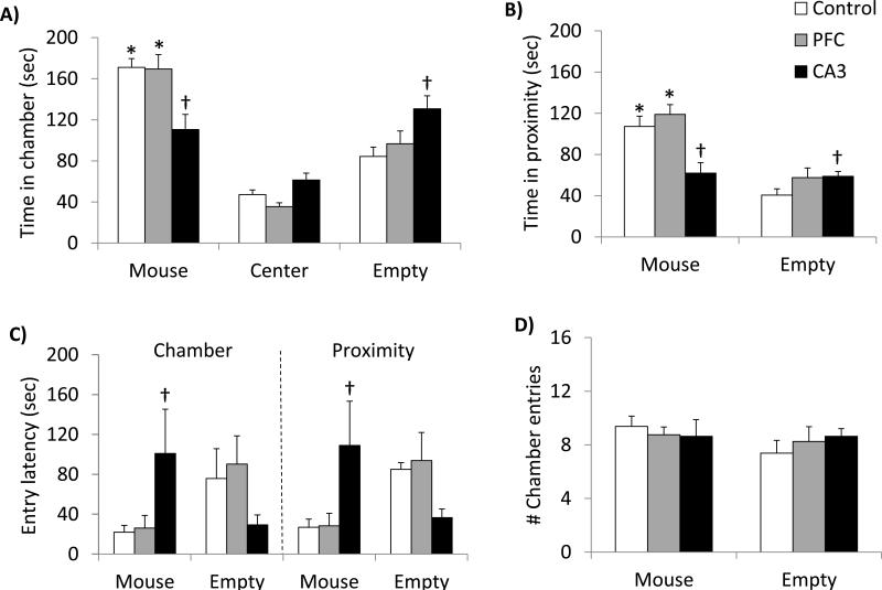
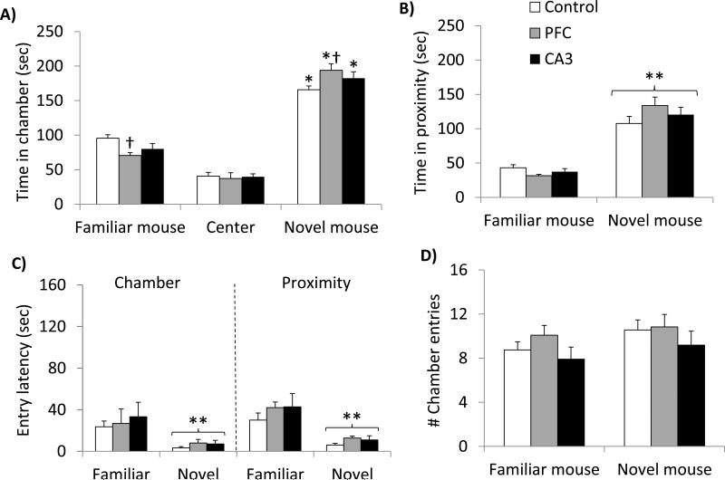
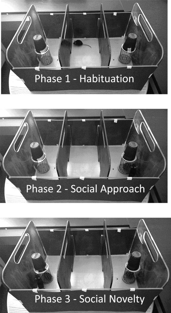
Glutamate N-methyl-D-aspartate receptors (NMDARs) in the medial prefrontal cortex (mPFC) and hippocampus may play an integral role in complex cognitive and social deficits associated with a number of psychiatric illnesses including autism, mood disorders, and schizophrenia. We used localized infusions of adeno-associated virus Cre-recombinase in adult, targeted knock-in mice with loxP sites flanking exons 11-22 of the NR1 gene to investigate the effects of chronic NMDAR dysfunction in the mPFC and CA3 hippocampus on cognitive and social behavior. A 5-choice serial reaction time task (5-CSRTT) was used to monitor aspects of cognitive function that included attention and response inhibition. Social behavior was assessed using Crowley׳s sociability and preference for social novelty protocol. Chronic NMDAR dysfunction localized to the anterior cingulate/prelimbic mPFC or dorsal CA3 hippocampus differentially affected the response inhibition and social interaction. mPFC NR1-deletion increased perseverative responding in the 5-CSRTT and enhanced preference for social novelty, whereas CA3 NR1-deletion increased premature responding in the 5-CSRTT and decreased social approach behavior. These findings suggest that mPFC and CA3 NMDARs play selective roles in regulating compulsive and impulsive behavior, respectively. Furthermore, these findings are consistent with emerging evidence that these behaviors are mediated by distinct, albeit overlapping, neural circuits. Our data also suggest that NMDARs in these regions uniquely contribute to the expression of normal social behavior. In this case, mPFC and CA3 NMDARs appear to inhibit and facilitate aspects of social interaction, respectively. The latter dissociation raises the possibility that distinct circuits contribute to the expression of social intrusiveness and impoverished social interaction.

Keywords: Attention; Hippocampus; NMDA receptor; Prefrontal cortex; Response inhibition; Social interaction.


Figures

Fig. 1
Fig. 1
NR1 gene deletion following local administration of AAV-Cre into the mPFC or CA3 hippocampus of adult fNR1 mice. The area of NR1 gene deletion induced by bilateral mPFC or CA3 infusions of AAV-Cre (0.5 μl) was determined by qualitative analysis of coronal brain sections exposed to a radiolabeled NR1-specific mRNA probe. For each mouse, grey-shaded ovals represent the visible extent of NR1 gene deletion on corresponding brain atlas images from Paxinos and Franklin (2001). A composite of shading from all mice, illustrates the overall localization of NR1 deletions in the mPFC and CA3 hippocampus (n=9/group; A & B, respectively). Numbers indicate the distance (mm) of each image from bregma. Also shown are representative radiolabeled coronal brain sections from aCSF- and LacZ-infused fNR1 control mice (C & E) and AAV-Cre-infused fNR1 mice (D & F).
Fig. 2
Fig. 2
CA3 and mPFC NR1-deletion does not significantly affect baseline performance of a 5-CSRTT task. Control mice (n=12) and mPFC and CA3 NR1-deleted mice (n=9/group) were trained on a 5-CSRTT, under baseline conditions of a 5.0 s LH, ITI, and TO and a 0.8 s SD. Each bar represents a group mean ± SEM of 8 baseline sessions (2 sessions preceding each of the 5 probe trial sessions). (A & B) Relative to controls, CA3 and mPFC NR1-deleted mice exhibited a nonsignificant trend for increased premature and perseverative responding, respectively. In contrast, mPFC and CA3 NR1-deletion did not affect baseline (C) accuracy, (D) omissions, (E) correct response latencies or (F) incorrect response latencies.
Fig. 3
Fig. 3
CA3 NR1-deletion selectively increased premature responding in a 5-CSRTT task, particularly under conditions of a variable LITI. Effects of mPFC and CA3 hippocampus NR1-deletion were examined under conditions of randomly presented variable ITIs of 5, 6, 7, and 8 s (n = 12 control, 9 mPFC and 9 CA3). Each bar represents a group mean ± SEM. (A) Relative to performance on the immediately preceding 2 days of baseline testing performed under a fixed 5 s ITI, variable LITIs differentially affected premature responses as a function of treatment condition. Control, mPFC deleted and CA3 deleted mice all exhibited increased premature responding under LITI conditions, relative to baseline conditions. However, under LITI conditions CA3 deleted mice exhibited greater premature responding than control or mPFC deleted mice. (B) To further examine the effects of individual LITIs on premature responding, the LITI data from panel A are presented as a function of the randomly presented 5, 6, 7, and 8 s ITIs used during the probe trial. CA3 deleted mice exhibited greater increases in premature responding than control mice under conditions of 6, 7, and 8s ITIs. (C) There was no effect of variable LITIs or NR1 deletion on accuracy and, (D) the LITI-induced decrease in omissions was observed across all treatment conditions. *Significantly different from baseline (within-group paired samples t-tests, p<0.05) †Significantly different from control and mPFC deleted mice (between-group independent samples t-tests, p<0.05)
Fig. 4
Fig. 4
mPFC NR1-deletion increased perseverative responding in a 5-CSRTT task. Effects of mPFC and CA3 hippocampus NR1-deletion were examined in a reduced SD probe trial consisting of randomly presented SDs of 0.2, 0.4, 0.6, and 0.8 s and a reduced SI probe trial consisting of randomly presented SIs of 30, 40, 50, 70, and 100% of baseline stimulus brightness (n = 12 control, 9 mPFC and 9 CA3). Probe trial performance was compared to that exhibited during the immediately preceding 2 days of baseline testing during which mice were tested on a fixed 0.8 s SD and 100% brightness. Each bar represents a group mean ± SEM. Relative to baseline responding, reducing the SD or SI did not differentially affect performance of control or deleted mice. (A) However, mPFC NR1-deleted mice exhibited a generalized increase in perseverative responding that was evident under baseline and manipulation conditions; this increase in reminiscent of the trend for increased perseverative responding observed under baseline conditions alone (see Figure 2B). Reducing the SD or SI did not differentially affect accuracy (B) or omissions (C) of the deleted mice. Although overall, these manipulations decreased accuracy and increased omissions. **Significantly different from control and CA3 NR1-deleted mice (pairwise comparisons collapsed across sessions for each group; post hoc LSD test, pfl0.05).
Fig. 5
Fig. 5
NR1 gene deletion following local administration of AAV-Cre into the mPFC or CA3 hippocampus of adult fNR1 mice. The area of NR1 gene deletion induced by bilateral mPFC and CA3 hippocampus infusions of AAV-Cre (0.5 μl) was determined by qualitative analysis of coronal brain sections exposed to a radiolabeled NR1-specific mRNA probe. For each mouse, grey-shaded ovals were used to indicate the visible extent of NR1 gene deletion on corresponding brain atlas images from Paxinos and Franklin (2001). A composite of shading from all mice, illustrates the overall localization of NR1 deletions in (A) the dorsal mPFC (n=12) and (B) CA3 hippocampus (n=11). Gene deletion maps for a subset of mice (n=2-3/group) tested in both social interaction and 5-CSRTTs are duplicated in Figures 1 and 10. Numbers indicate the distance (mm) of each image from bregma.
Fig. 6
Fig. 6
CA3 NR1 deletion selectively decreased social approach. During sociability testing, mice had free access to a 3-chambered box. One end chamber contained a wire retaining-cage occupied by a stimulus mouse and the other end chamber contained an empty retaining-cage. Each bar represents a group mean ± SEM (n=16 control, n=12 mPFC, and n=11 CA3). (A) CA3, but not mPFC, NR1-deletion altered the time spent in each chamber during the first 5 min of sociability testing. Control and mPFC, but not CA3, deleted mice spent more time in a chamber with a stimulus mouse than an empty chamber. Relative to controls, CA3 deleted mice spent less time in a chamber with a stimulus mouse and more time in an empty chamber. (B) CA3, but not mPFC, deletion altered the total time spent in proximity (within 3.5 cm of the outer edge of the wire retaining-cages) with a stimulus mouse and an empty cage, during the first 5 min of social approach testing. Control and mPFC, but not CA3, deleted mice spent more time in proximity with a stimulus mouse than an empty cage. Relative to controls, CA3 deleted mice spent less time in proximity with a mouse and more time in proximity with an empty cage. (C) CA3, but not mPFC, deletion altered the latency to enter a chamber and proximity zone associated with a stimulus mouse. Relative to controls, CA3 deleted mice took longer to enter a chamber and proximity zone associated with a stimulus mouse. (D) The number of chamber entries and distance traveled per chamber (data not shown) did not differ as a function of treatment condition. *Significantly different from mean time in an empty chamber or proximity zone (within-group paired samples t-tests, p<0.05). Significantly different from control mean (between-group independent samples t-tests, p<0.05)
Fig. 7
Fig. 7
mPFC NR1-deletion selectively increased preference for social novelty. Immediately following sociability testing, preference for social novelty was assessed. During social novelty testing, the “familiar” stimulus mouse introduced during sociability testing remained in the retaining-cage and a “novel” stimulus mouse was placed in the previously empty retaining-cage. Each bar represents a group mean ± SEM (n=16 control, n=12 mPFC, and n=11 CA3). (a) mPFC, but not CA3, NR1-deletion altered the time spent in each chamber during the first 5 min of social novelty testing. Control mice and mPFC and CA3 deleted mice spent more time in a chamber with a novel mouse than a familiar mouse. Relative to controls, mPFC NR1-deleted mice spent less time in a chamber with a familiar mouse and more time in a chamber with a novel mouse. (b) Time spent in proximity (within 3.5 cm of the outer edge of the wire retaining-cages) with a familiar versus novel mouse did not vary as a function of treatment condition. Overall, mice spent more time in proximity with a novel than a familiar mouse. (c) Latency to enter a chamber or proximity zone associated with a familiar versus novel mouse did not vary as a function of treatment condition. All groups entered the chamber and proximity zone associated with the new mouse more quickly than that associated with the familiar mouse. (d) The number of chamber entries and distance traveled per chamber (data not shown) did not differ as a function of treatment condition. *Significantly different from mean time spent in a chamber with a familiar mouse (within-group paired samples t-tests, p<0.05). Significantly different from control mean (between-group independent samples t-tests, p<0.05). **Significantly different from familiar mouse, collapsed across treatment condition (ANOVA main effect, p<0.05).
Fig. 8
Fig. 8
Social behavior was assessed using an apparatus and method based on previous research colleagues (Nadler et al., 2004). A single-trial test consisted of three phases. During the habituation phase (Phase 1), a test mouse was placed in and retained in the center chamber for 5 min; the end chambers were empty and not accessible to the mouse. A stimulus mouse was then placed in a retaining cage located in one of the end chambers; the empty retaining cage was also manipulated at this time but remained empty. To initiate the social approach phase (Phase 2), the guillotine doors were raised simultaneously and the test mouse was free to explore all chambers for 10 min. The test mouse was then coaxed back into the center chamber and the guillotine doors were lowered. Preference for social novelty (Phase 3) was then assessed by placing a second stimulus mouse in the previously empty retaining cage. The guillotine doors were raised and the test mouse again had free access to all chambers for 10 min.

Similar articles

Cited by

References

    1. Abela AR, Dougherty SD, Fagen ED, Hill CJ, Chudasama Y. Inhibitory control deficits in rats with ventral hippocampal lesions. Cereb Cortex. 2013;23:1396–409. - PubMed
    1. Amitai N, Semenova S, Markou A. Cognitive-disruptive effects of the psychotomimetic phencyclidine and attenuation by atypical antipsychotic medications in rats. Psychopharmacology (Berl) 2007 - PubMed
    1. Avale ME, Chabout J, Pons S, Serreau P, De Chaumont F, Olivo-Marin JC, Bourgeois JP, Maskos U, Changeux JP, Granon S. Prefrontal nicotinic receptors control novel social interaction between mice. FASEB J. 2011;25:2145–55. - PubMed
    1. Bannerman DM, Deacon RM, Offen S, Friswell J, Grubb M, Rawlins JN. Double dissociation of function within the hippocampus: spatial memory and hyponeophagia. Behav Neurosci. 2002;116:884–901. - PubMed
    1. Bari A, Dalley JW, Robbins TW. The application of the 5-choice serial reaction time task for the assessment of visual attentional processes and impulse control in rats. Nat Protoc. 2008;3:759–67. - PubMed

Publication types

Substances