. 2015 Feb;35(3):582-97.
doi: 10.1128/MCB.00775-14. Epub 2014 Dec 1.

Nedd8 regulates inflammasome-dependent caspase-1 activation

Affiliations

Nedd8 regulates inflammasome-dependent caspase-1 activation

Jesus A Segovia et al. Mol Cell Biol. 2015 Feb.

Abstract

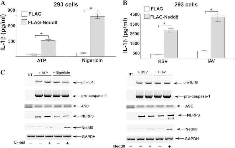
Caspase-1 is activated by the inflammasome complex to process cytokines like interleukin-1β (IL-1β). Pro-caspase-1 consists of three domains, CARD, p20, and p10. Association of pro-caspase-1 with the inflammasome results in initiation of its autocatalytic activity, culminating in self-cleavage that generates catalytically active subunits (p10 and p20). In the current study, we show that Nedd8 is required for efficient self-cleavage of pro-caspase-1 to generate its catalytically active subunits. Nedd8 silencing or treating cells with the neddylation inhibitor MLN4924 led to diminished caspase-1 processing and reduced IL-1β maturation following inflammasome activation. Coimmunoprecipitation and mass spectrometric analysis of 293 cells overexpressing pro-caspase-1 (and CARD) and Nedd8 suggested possible neddylation of caspase-1 CARD. Following inflammasome activation in primary macrophages, we observed colocalization of endogenous Nedd8 with caspase-1. Similarly, interaction of endogenous Nedd8 with caspase-1 CARD was detected in inflammasome-activated macrophages. Furthermore, enhanced autocatalytic activity of pro-caspase-1 was observed following Nedd8 overexpression in 293 cells, and such activity in inflammasome-activated macrophages was drastically diminished upon treatment of cells with MLN4924. Thus, our studies demonstrate a role of Nedd8 in regulating caspase-1 activation following inflammasome activation, presumably via augmenting autoprocessing/cleavage of pro-caspase-1 into its corresponding catalytically active subunits.


Figures

FIG 1
FIG 1
Nedd8 silencing in BMDMs reduces caspase-1 activation and IL-1β processing during inflammasome activation. (A) RT-PCR analysis of Nedd8 expression in BMDMs transfected with control siRNA or Nedd8 siRNA. (B) Western blotting results for caspase-1 p10 and mature IL-1β p17 in supernatants (supn) of Nedd8-silenced BMDMs treated with LPS followed by ATP or nigericin treatment. Actin served as a loading control (i.e., control for cell numbers). (C) RT-PCR analysis of Nedd8 expression in NR-9456 cells transfected with control siRNA or Nedd8 siRNA. (D) ELISA results for IL-1β in supernatants of Nedd8-silenced NR-9456 cells treated with LPS and nigericin. The values represent the means ± standard deviations from three independent experiments performed in triplicate. *, P < 0.05 using Student's t test. (E) Western blotting results for caspase-1 p10 in supernatants of Nedd8-silenced NR-9456 cells treated with LPS and nigericin. Actin served as a loading control (i.e., control for cell numbers). RT-PCR data (A and C) and Western blotting data (B and E) are representative of three independent experiments with similar results.
FIG 2
FIG 2
Nedd8 is required for optimal IL-1β maturation/release from RSV-infected NHBE cells, and silencing of Ubc12 in NR-9456 cells reduces IL-1β production. (A) RT-PCR analysis of Nedd8 and pro-IL-1β expression in RSV-infected NHBE cells transfected with control siRNA or Nedd8 siRNA. (B) ELISA results for IL-1β in supernatants of Nedd8-silenced NHBE cells infected with RSV for 16 h. (C) RT-PCR analysis of Ubc12 and pro-IL-1β expression in untreated (UT) and LPS-treated NR-9456 cells transfected with control siRNA or Ubc12 siRNA. (D) ELISA results for IL-1β in supernatants of Ubc12-silenced NR-9456 cells treated with LPS and ATP. The values shown in panels B and D represent the mean ± standard deviation from three independent experiments performed in triplicate. *, P < 0.05 using Student's t test. RT-PCR data (A and C) are representative of three independent experiments with similar results.
FIG 3
FIG 3
Enhanced IL-1β secretion from Nedd8-expressing 293 cells. (A and B) 293 cells were transfected with pro-IL-1β, pro-caspase-1, ASC, NLRP3, and FLAG-Nedd8 or empty FLAG vector (control). These cells were then either treated with ATP or nigericin (A) or infected with human RSV or IAV (B). IL-1β in supernatants was measured by ELISA. The values represent the means ± standard deviations from three independent experiments performed in triplicate. *, P < 0.05 using Student's t test. (C) RT-PCR analysis of gene expression in inflammasome-activated (treated with ATP or nigericin and infected with RSV or IAV) 293 cells transfected with pro-IL-1β, pro-caspase-1, ASC, NLRP3, or Nedd8. NT, nontransfected.
FIG 4
FIG 4
Inhibition of neddylation by MLN4924 (MLN) diminishes caspase-1 activation and IL-1β maturation. (A) ELISA results for IL-1β in supernatants of MLN-treated BMDMs treated with LPS and ATP or nigericin. The values represent the means ± standard deviations from three independent experiments performed in triplicate. *, P < 0.05 using Student's t test. (B) Western blotting results for caspase-1 p10 and mature IL-1β p17 in supernatants (supn) of MLN-treated BMDMs treated with LPS and ATP or nigericin. Actin served as a loading control (i.e., control for cell numbers). (C) Western blotting results for caspase-1 p10 and IL-1β p17 in the supernatants of MLN-treated THP-1 cells incubated with LPS and nigericin in the presence of DMSO (vehicle control) or MLN. Actin served as a loading control (i.e., control for cell numbers). Western blotting data (B and C) are representative of three independent experiments with similar results.
FIG 5
FIG 5
MLN4924 (MLN) administration reduces IL-1β and IL-18 production in mice. (A and B) C57BL/6 mice (n = 5 per group) were injected (i.p. route) with LPS (for 3 h), followed by i.p. injection of MLN (for 4 h) and finally i.p. administration of ATP (for 15 min). Serum collected from the mice was analyzed for IL-1β (A) and IL-18 (B) by ELISA. The values represent the means ± standard deviations. *, P < 0.05 using Student's t test. (C) C57BL/6 mice (n = 5 per group) were injected (i.p. route) with LPS (for 3 h), followed by i.p. injection of MLN (for 4 h). Serum collected from the mice was analyzed for TNF-α by ELISA. The values represent the means ± standard deviations.
FIG 6
FIG 6
Interaction of Nedd8 with caspase-1. (A) Cell lysates were collected from 293 cells expressing GFP-Nedd8 and myc-procaspase1 and immunoprecipitated (IP) with myc antibody, followed by immunoblotting (IB) with GFP antibody. Total lysate was also blotted with GFP and myc antibodies. The lower panel shows the membrane with a longer exposure. The 45-kDa and 80-kDa bands observed in Nedd8-expressing cells are indicated with arrows. (B) Cell lysates collected from 293 cells expressing GFP-Nedd8 and myc-CARD (pro-caspase-1 CARD) and immunoprecipitated with myc antibody, followed by blotting with GFP antibody. The lower panel shows the gel with a longer exposure. The 80-kDa band observed in Nedd8-expressing cells is indicated by an arrow. The data are representative of three independent experiments with similar results.
FIG 7
FIG 7
Nedd8-dependent enhanced processing of pro-caspase-1. (A) Cell lysate collected from 293 cells expressing FLAG-Nedd8 and double-tagged pro-caspase-1 (myc–caspase-1–HA; myc tag on the N terminal and HA tag on the C terminal) were immunoprecipitated (IP) with myc antibody, followed by immunoblotting (IB) with myc antibody. (B) Cell lysate collected from 293 cells expressing FLAG-Nedd8 and myc–caspase-1–HA were immunoprecipitated with HA antibody, followed by blotting with HA antibody. (C) A longer-exposed version of the blot shown in panel B. The higher-molecular-mass (approximately 53-kDa) caspase-1 was visible in Nedd8-expressing cells. (D) Western blotting results for the total lysate corresponding to the experiment shown in panels A to C. Cell lysates collected from 293 cells expressing FLAG-Nedd8 and myc–caspase-1–HA were subjected to Western blotting with either HA or FLAG antibody. The data are representative of three independent experiments with similar results.
FIG 8
FIG 8
Colocalization of endogenous Nedd8 with caspase-1 following inflammasome activation. (A) Coimmunofluorescence analysis of Nedd8 (red) and caspase-1 (green) in BMDMs following LPS treatment in the absence or presence of ATP or nigericin. (B) Coimmunofluorescence analysis of Nedd8 and caspase-1 in BMDMs following infection with RSV or IAV. Merged images (yellow) show colocalization of Nedd8 with caspase-1. Magnified versions of the merged “speck” are shown on the extreme right sides of the two panels. The images are representative of 30 viewing fields from two independent experiments with similar results.
FIG 9
FIG 9
Endogenous interaction of Nedd8 with caspase-1 CARD following inflammasome activation. (A) Cell lysates collected from BMDMs treated with LPS alone (4 h) or LPS and nigericin (LPS treatment for 4 h followed by nigericin treatment for 30 min) were immunoprecipitated (IP) with Nedd8 antibody and immunoblotted (IB) with caspase-1 CARD-specific antibody. (B) BMDMs were either untreated (UT; lane 1) or treated with LPS for 2 h (lane 2), followed by addition of MLN4924 (lane 4) or DMSO (control; lane 3) along with LPS. After 4 h, cells treated with LPS plus MLN4924 (lane 4) or LPS plus DMSO (lane 3) (total LPS treatment, 6 h) were treated with nigericin for 30 min. The cell lysates were subjected to Western blotting with anti-CARD antibody. The 17-kDa cleaved CARD fragment observed following inflammasome activation (i.e., after nigericin treatment) is indicated by a red arrow. Actin served as a loading control (i.e. control for cell numbers).

References

    1. Kostura MJ, Tocci MJ, Limjuco G, Chin J, Cameron P, Hillman AG, Chartrain NA, Schmidt JA. 1989. Identification of a monocyte specific pre-interleukin 1 beta convertase activity. Proc Natl Acad Sci U S A 86:5227–5231. doi: 10.1073/pnas.86.14.5227. - DOI - PMC - PubMed
    1. Thornberry NA, Bull HG, Calaycay JR, Chapman KT, Howard AD, Kostura MJ, Miller DK, Molineaux SM, Weidner JR, Aunins J, Elliston KO, Ayala JM, Casano FJ, Chin J, Ding GJ-F, Egger LA, Gaffney EP, Limjuco G, Palyha OC, Raju SM, Rolando AM, Salley JP, Yamin T-T, Lee TD, Shively JE, MacCross M, Mumford RA, Schmidt JA, Tocci MJ. 1992. A novel heterodimeric cysteine protease is required for interleukin-1 beta processing in monocytes. Nature 356:768–774. doi: 10.1038/356768a0. - DOI - PubMed
    1. Cerretti DP, Kozlosky CJ, Mosley B, Nelson N, Van Ness K, Greenstreet TA, March CJ, Kronheim SR, Druck T, Cannizzaro LA, et al. 1992. Molecular cloning of the interleukin-1 beta converting enzyme. Science 256:97–100. doi: 10.1126/science.1373520. - DOI - PubMed
    1. Wilson KP, Black JA, Thomson JA, Kim EE, Griffith JP, Navia MA, Murcko MA, Chambers SP, Aldape RA, Raybuck SA, Livingston DJ. 1994. Structure and mechanism of interleukin-1 beta converting enzyme. Nature 370:270–275. doi: 10.1038/370270a0. - DOI - PubMed
    1. Walker NP, Talanian RV, Brady KD, Dang LC, Bump NJ, Ferenz CR, Franklin S, Ghayur T, Hackett MC, Hammill LD, et al. 1994. Crystal structure of the cysteine protease interleukin-1 beta-converting enzyme: a (p20/p10)2 homodimer. Cell 78:343–352. doi: 10.1016/0092-8674(94)90303-4. - DOI - PubMed

Publication types

LinkOut - more resources