. 2016 Feb;75(2):430-8.
doi: 10.1136/annrheumdis-2014-205635. Epub 2014 Dec 1.

Inflammatory cytokines epigenetically regulate rheumatoid arthritis fibroblast-like synoviocyte activation by suppressing HDAC5 expression

Affiliations

Inflammatory cytokines epigenetically regulate rheumatoid arthritis fibroblast-like synoviocyte activation by suppressing HDAC5 expression

Chiara Angiolilli et al. Ann Rheum Dis. 2016 Feb.

Abstract

Objectives: Epigenetic modifications play an important role in the regulation of gene transcription and cellular function. Here, we examined if pro-inflammatory factors present in the inflamed joint of patients with rheumatoid arthritis (RA) could regulate histone deacetylase (HDAC) expression and function in fibroblast-like synoviocytes (FLS).

Methods: Protein acetylation in synovial tissue was assessed by immunohistochemistry. The mRNA levels of HDAC family members and inflammatory mediators in the synovial tissue and the changes in HDAC expression in RA FLS were measured by quantitative (q) PCR. FLS were either transfected with HDAC5 siRNA or transduced with adenoviral vector encoding wild-type HDAC5 and the effects of HDAC5 manipulation were examined by qPCR arrays, ELISA and ELISA-based assays.

Results: Synovial class I HDAC expression was associated with local expression of tumour necrosis factor (TNF) and matrix metalloproteinase-1, while class IIa HDAC5 expression was inversely associated with parameters of disease activity (erythrocyte sedimentation rate, C-reactive protein, Disease Activity Score in 28 Joints). Interleukin (IL)-1β or TNF stimulation selectively suppressed HDAC5 expression in RA FLS, which was sufficient and required for optimal IFNB, CXCL9, CXCL10 and CXCL11 induction by IL-1β, associated with increased nuclear accumulation of the transcription factor, interferon regulatory factor 1(IRF1).

Conclusions: Inflammatory cytokines suppress RA FLS HDAC5 expression, promoting nuclear localisation of IRF1 and transcription of a subset of type I interferon response genes. Our results identify HDAC5 as a novel inflammatory mediator in RA, and suggest that strategies rescuing HDAC5 expression in vivo, or the development of HDAC inhibitors not affecting HDAC5 activity, may have therapeutic applications in RA treatment.

Keywords: Chemokines; Fibroblasts; Inflammation; Rheumatoid Arthritis.


Conflict of interest statement

Competing interests None.

Figures

Figure 1
Figure 1
Expression of class I histone deacetylases (HDACs) correlates positively with tumour necrosis factor (TNF) and matrix metalloproteinase (MMP)-1 mRNA levels in rheumatoid arthritis (RA) synovial tissue, while the class IIa HDAC5 expression is negatively associated with disease activity. The mRNA expression of HDAC1–10, TNF, MMP-1 and interleukin (IL)-6 was analysed in the synovial tissue of 19 patients with RA (cohort I) by qPCR and calculated relative to the mean 18S rRNA and glyceraldehyde-3-phosphate dehydrogenase expression. Analyses of correlations between synovial expression of HDAC family members and (A) TNF, (B) MMP-1 and (C) IL-6 mRNA levels, and between (D) HDAC5 mRNA expression and the C-reactive protein (CRP) levels, erythrocyte sedimentation rate (ESR) and disease activity score in 28 joints (DAS28) were performed using Spearman’s correlation coefficient. Spearman R and p values are indicated in each graph, and circles indicate individual patient values.
Figure 2
Figure 2
Protein acetylation levels are equivalent in rheumatoid arthritis (RA), osteoarthritis (OA) and psoriatic arthritis (PsA) synovial tissue. (A) Representative photomicrographs of RA tissue sections stained with control rabbit antibody (control Ig), or with antibodies recognising acetylated lysine (acLys) and acetylated histone 3 (Lys18) (acH3). Synovial tissue sections from (B) 12 patients with RA and 12 patients with OA (cohort III), and from (C) 18 patients with RA and 12 patients with PsA (cohort IV) were stained with antibodies recognising acLys and acetylated histone 3 (Lys 18) (acH3). Integrated optical density (IOD)/mm2 values were calculated by computer-assisted digital image analysis and corrected for tissue cellularity. Differences between patient groups were analysed using the Mann–Whitney U test. (D) Synovial biopsy sections from 20 patients with RA (cohort II) were stained with antibodies against acLys and acH3 and the relationship between IOD/mm2 and patient C-reactive protein (CRP) levels was calculated using Spearman’s correlation coefficient and presented as in figure 1.
Figure 3
Figure 3
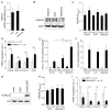
Interleukin (IL)-1β down-regulates histone deacetylase 5 (HDAC5) but not class I HDACs in rheumatoid arthritis (RA) fibroblast-like synoviocytes (FLS). (A) RA FLS (n=4) were left untreated or were stimulated with 1 ng/mL IL-1β, 10 ng/mL tumour necrosis factor (TNF) or 1 μg/mL lipopolysaccharide (LPS) for 24 h, and changes in mRNA accumulation of HDAC1–10 were analysed by quantitative (q)PCR. Data are presented as the mean±SEM mRNA expression relative to glyceraldehyde-3-phosphate dehydrogenase. (B) RA FLS were left untreated or were stimulated with 1 ng/mL IL-1β for 1–8 h and changes in HDAC5 mRNA expression analysed by qPCR and presented as above. (C) RA FLS were left untreated or were stimulated with either 1 ng/mL IL-1β (n=5) or 10 ng/mL TNF (n=4) for 24 h. Protein extracts were prepared and analysed by immunoblotting with anti-HDAC5 and anti-actin antibodies, and (D) signal intensity was subsequently quantified by densitometry analysis. (E) RA FLS (n=4) stimulated with IL-1β (1 ng/mL) or TNF (10 ng/mL) for 15 min–24 h, cell pellets were then incubated with HDAC e-N-acylated lysine substrates and HDAC activity was measured by fluorescence emission. Values for the 0 h time point were normalised to 100% and remaining values were shown as the mean±SEM percentage of HDAC activity compared with controls. Insets represent the mean±SEM fluorescence values for cells untreated or stimulated for 24 h with IL-1β or TNF. *p<0.05, **p<0.01, Friedman test followed by Dunn’s multiple comparison analysis with unstimulated cells used as reference controls.
Figure 4
Figure 4
Histone deacetylase 5 (HDAC5) regulates CXCL chemokine production in rheumatoid arthritis (RA) fibroblast-like synoviocytes (FLS). (A–F) RA FLS were left not transfected or were transfected for 48 h with 20 nM control non-targeting siRNA (scrb) or 20 nM HDAC5-specific siRNA (siHDAC5). HDAC5 knockdown efficiency was verified at the (A) mRNA and (B) protein level by quantitative (q)PCR and immunoblotting, respectively. Results in (A) are presented as the mean±SEM HDAC5 mRNA expression of three independent experiments and in (B) a representative of four independent experiments is shown. (C) Changes in RA FLS viability after HDAC5 knockdown (n=4) were analysed by 3-(4,5-dimethylthiazol-2-yl)-2,5-diphenyltetrazolium bromide (MTT) reduction assay and presented as the mean±SEM absorbance at 590 nm. (D) Transfected RA FLS (n=8) were stimulated with 1 ng/mL interleukin (IL)-1β for 4 h and mRNA expression of CXCL9, CXCL10, CXCL11 and IFNB relative to glyceraldehyde-3-phosphate dehydrogenase was determined by qPCR. Data are shown as mean±SEM fold induction compared to scrb. *p<0.05, **p<0.01, Wilcoxon matched pairs test. (E) CXCL-10 and (F) IL-6 production by control and transfected RA FLS stimulated with 1 ng/mL IL-1β for 24 h was determined by ELISA. Results are presented as the mean±SEM concentration from seven and five independent experiments for CXCL-10 and IL-6, respectively. *p<0.05, **p<0.01, Friedman test followed by Dunns’ multiple comparison analysis. (G–I) RA FLS were transduced with control GFP-encoding or wild-type HDAC5-GFP (HDAC5WT) encoding adenoviral vector for 24 h and serum-starved for another 24 h prior to further processing. (G) Overexpression of HDAC5 protein was confirmed by immunoblotting with anti-HDAC5 and anti-actin antibodies. The effects of HDAC5 overexpression on (H) cell viability and (I) mRNA expression of CXCL9, CXCL10, CXCL11 and IFNB after 4 h stimulation with 1 ng/mL IL-1β were determined as described above. Data in (H) are shown as the mean±SEM absorbance at 590 nm of four independent experiments, and in (I) as the mean±SEM relative mRNA expression of eight independent experiments. *p<0.05, **p<0.01, Wilcoxon matched pairs test.
Figure 5
Figure 5
Histone deacetylase 5 (HDAC5) regulates IRF1 transcription factor. Rheumatoid arthritis (RA) fibroblast-like synoviocytes (FLS) were transfected with 20 nM control non-targeting siRNA (scrb) or 20 nM HDAC5-specific siRNA as described in figure 4, stimulated with 1 ng/mL interleukin (IL)-1β for 4 h, and total protein lysates or nuclear extracts were prepared. (A) STAT1 phosphorylation in total cell lysates was analysed by immunoblotting with antibodies specific for phospho-STAT1 (Tyr701) and histone 3 (H3). A representative of three independent experiments is shown. (B–C) Transcription factor DNA binding assays were used to analyse DNA-binding activity of (B) STAT1 and (C) nuclear factor-κB p50 and p65 in RA FLS nuclear extracts. The data are presented as the mean±SEM absorbance at 450 nm of (B) three or (C) four independent experiments. (D–E) Interferon regulatory factor (IRF)1 nuclear retention was analysed in RA FLS nuclear fractions by (D) IRF1 ELISA or (E) immunoblotting with anti-IRF1 and anti-H3 antibodies. Results in (D) represent the mean±SEM absorbance at 450 nm of seven independent experiments and in (B) a representative of two independent experiments is shown. *p<0.05, Wilcoxon matched pairs test.

Similar articles

Cited by

References

    1. Bax M, van Heemst J, Huizinga TW, et al. Genetics of rheumatoid arthritis: what have we learned? Immunogenetics. 2011;63:459–66. - PMC - PubMed
    1. Okada Y, Wu D, Trynka G, et al. Genetics of rheumatoid arthritis contributes to biology and drug discovery. Nature. 2014;506:376–81. - PMC - PubMed
    1. Costenbader KH, Gay S, Alarcon-Riquelme ME, et al. Genes, epigenetic regulation and environmental factors: which is the most relevant in developing autoimmune diseases? Autoimmun Rev. 2012;11:604–9. - PubMed
    1. Grabiec AM, Reedquist KA. The ascent of acetylation in the epigenetics of rheumatoid arthritis. Nat Rev Rheumatol. 2013;9:311–18. - PubMed
    1. Ospelt C, Reedquist KA, Gay S, et al. Inflammatory memories: is epigenetics the missing link to persistent stromal cell activation in rheumatoid arthritis? Autoimmun Rev. 2011;10:519–24. - PubMed

Publication types

MeSH terms