. 2014 Dec 8;207(5):657-71.
doi: 10.1083/jcb.201405098. Epub 2014 Dec 1.

SOXC proteins amplify canonical WNT signaling to secure nonchondrocytic fates in skeletogenesis

Affiliations

SOXC proteins amplify canonical WNT signaling to secure nonchondrocytic fates in skeletogenesis

Pallavi Bhattaram et al. J Cell Biol. .

Abstract

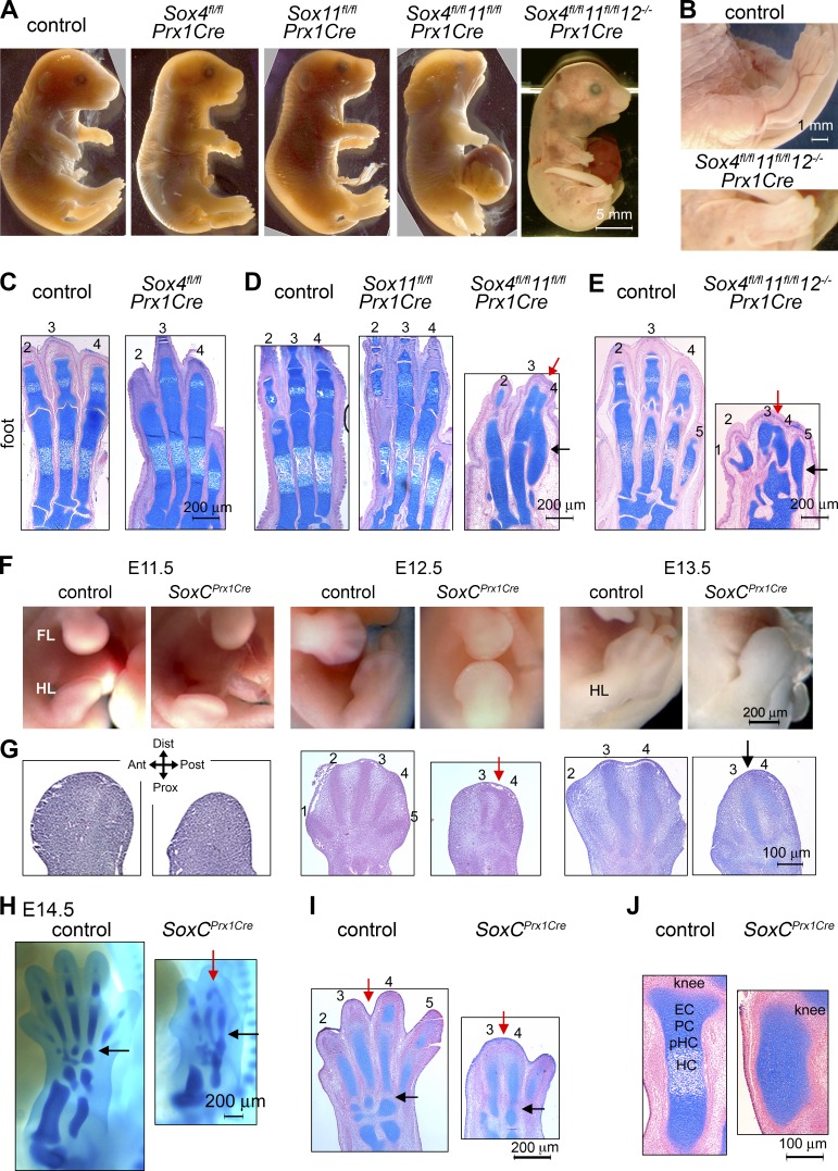
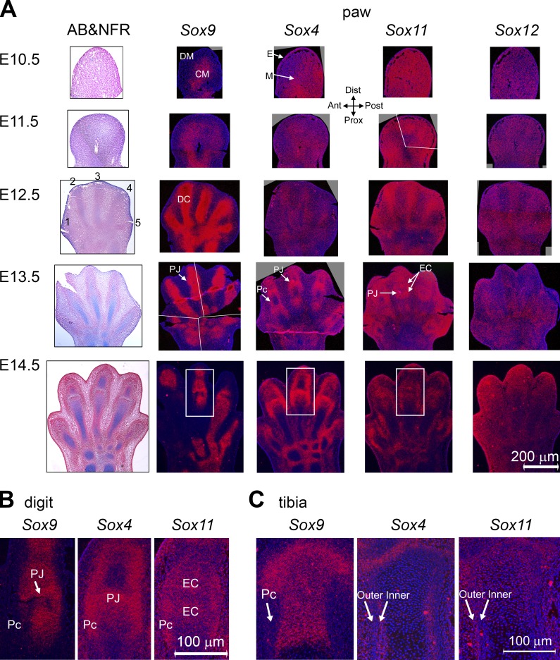
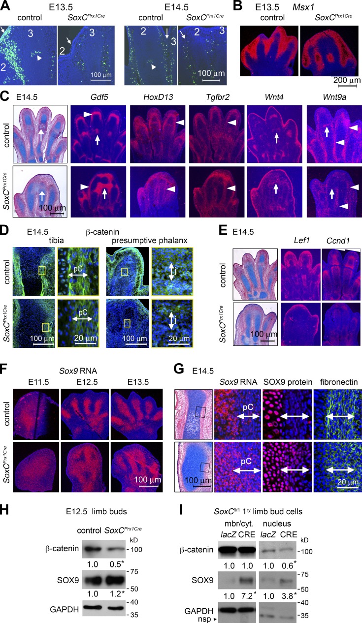
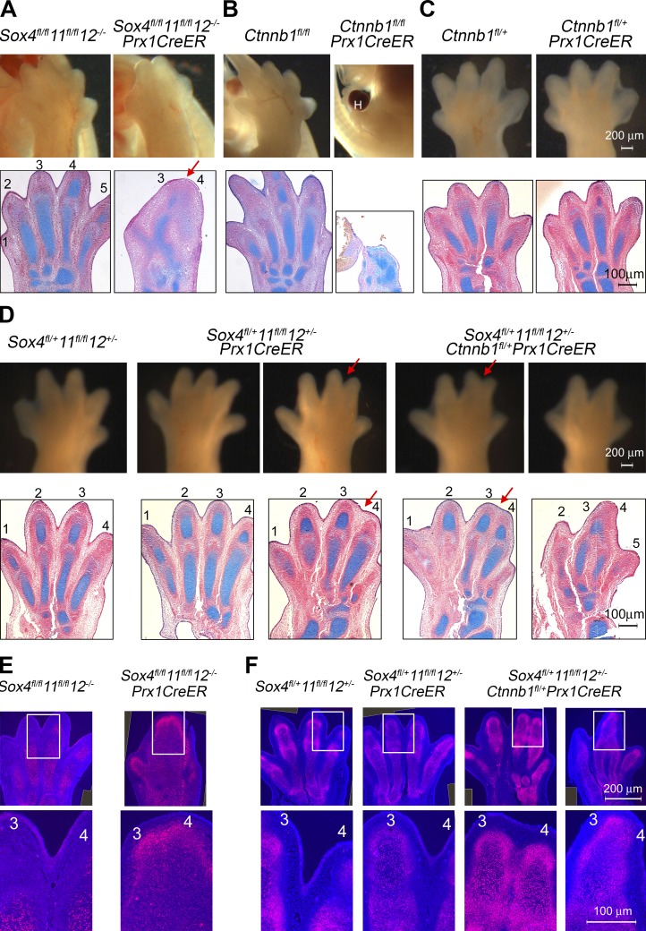
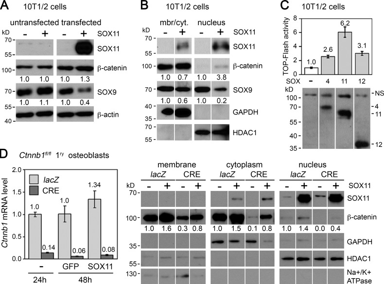
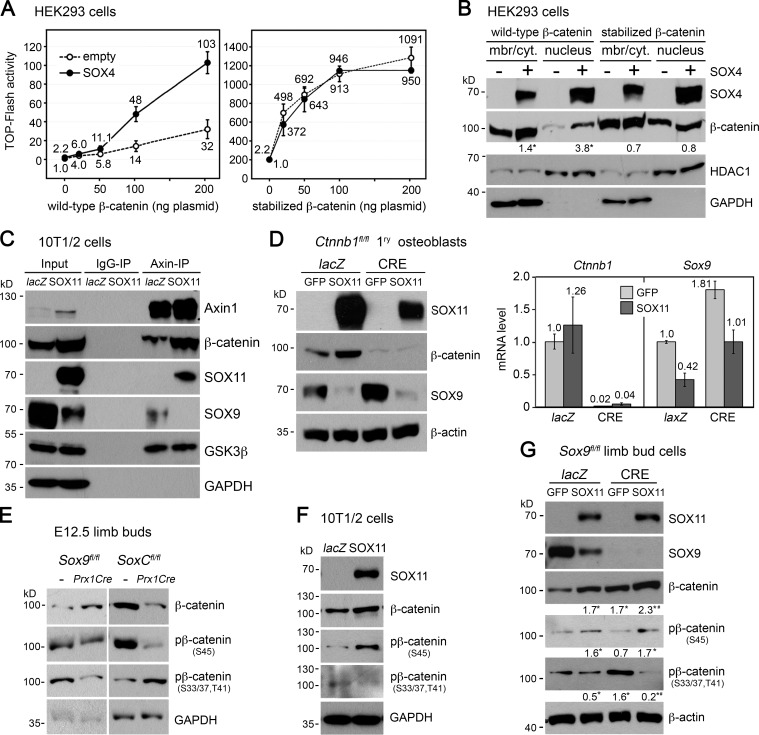
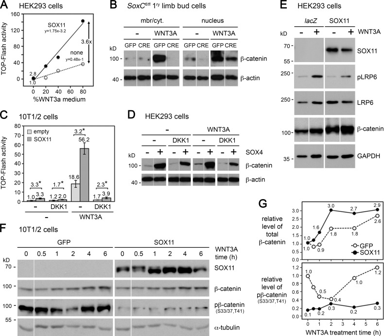
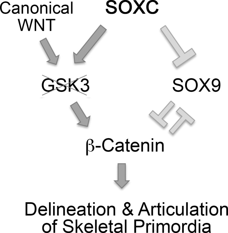
Canonical WNT signaling stabilizes β-catenin to determine cell fate in many processes from development onwards. One of its main roles in skeletogenesis is to antagonize the chondrogenic transcription factor SOX9. We here identify the SOXC proteins as potent amplifiers of this pathway. The SOXC genes, i.e., Sox4, Sox11, and Sox12, are coexpressed in skeletogenic mesenchyme, including presumptive joints and perichondrium, but not in cartilage. Their inactivation in mouse embryo limb bud caused massive cartilage fusions, as joint and perichondrium cells underwent chondrogenesis. SOXC proteins govern these cells cell autonomously. They replace SOX9 in the adenomatous polyposis coli-Axin destruction complex and therein inhibit phosphorylation of β-catenin by GSK3. This inhibition, a crucial, limiting step in canonical WNT signaling, thus becomes a constitutive event. The resulting SOXC/canonical WNT-mediated synergistic stabilization of β-catenin contributes to efficient repression of Sox9 in presumptive joint and perichondrium cells and thereby ensures proper delineation and articulation of skeletal primordia. This synergy may determine cell fate in many processes besides skeletogenesis.


Figures

Figure 1.
Figure 1.
Gross and histological analysis of SOXC mutant embryos. (A) Pictures of E18.5 control and SoxC/Prx1Cre mutants. (B) High-magnification pictures of control and triple mutant feet. (C–E) Foot coronal sections stained with Alcian blue (cartilage-specific dye) and nuclear fast red (AB&NFR). Pictures focus on metatarsals and proximal phalanges. Red and black arrows, anterior–posterior and proximal–distal fusions of cartilage primordia, respectively. (F) Pictures of E11.5–13.5 embryo limbs. FL, forelimb; HL, hind limb. (G) Hind limb coronal sections stained with hematoxylin and eosin (E11.5–12.5) or AB&NFR (E13.5). Distal/proximal and anterior/posterior axes are shown. (H–J) Alcian blue staining of E14.5 whole embryos (H) and sections of hind paws (I) and tibias (J). EC, epiphyseal cartilage; PC, proliferating chondrocytes; pHC, prehypertrophic chondrocytes; HC, hypertrophic chondrocytes. All data were verified more than three times. Each panel shows the results of a typical experiment.
Figure 2.
Figure 2.
SOXC gene expression in the limb bud. Sections of hind limbs from E10.5–14.5 wild-type embryos were stained with AB&NFR or hybridized with RNA probes (red signals) and counterstained with DAPI (blue dye), as indicated. All SOXC probes had similar length, GC content, and labeling efficiency, allowing comparison of RNA levels. CM, core mesenchyme; DC, digital condensation; DM, distal mesenchyme; E, ectoderm; EC, epiphyseal cartilage; M, mesenchyme; Pc, perichondrium; PJ, presumptive joint. (A) Coronal sections through hind limb buds. E11.5 Sox11 RNA in situ hybridization image and E13.5 Sox9 RNA in situ hybridization images are composite of several pictures. White lines mark the individual image boundaries. (B) High-magnification views of phalangeal regions boxed in A. (C) Proximal region of E14.5 tibia. All data were reproduced more than three times. Each panel shows representative results.
Figure 3.
Figure 3.
Cellular and molecular analysis of SoxCPrx1Cre limbs. (A) TUNEL assay (green) in hind limb sections from E13.5–14.5 control and SoxCPrx1Cre littermates. Counterstaining is with DAPI (blue). Arrows, mesenchyme between digits 2 and 3. Arrowheads, presumptive joints. (B and C) RNA in situ hybridization of E13.5–14.5 control and SoxCPrx1Cre hind paw sections with various probes, as indicated. Arrows, presumptive joints. Arrowheads, interdigital and perichondrium cells. (D) E14.5 hind limb sections immunostained for β-catenin (green) and counterstained with DAPI (blue). Magnified images of boxed regions are shown in side panels. j, joint; pC, perichondrium. (E) RNA in situ hybridization of E14.5 control and SoxCPrx1Cre hind paw sections with various probes, as indicated. (F) Sox9 RNA in situ hybridization of hind limb sections from E11.5–13.5 control and SoxCPrx1Cre embryos. (G, left) Coronal section of E14.5 tibias. (right) RNA in situ hybridization and immunofluorescence for SOX9 (red) and fibronectin (green). Pictures were taken at the level of the box shown in the left. (H and I) β-Catenin and SOX9 levels in whole-tissue extracts from control and SoxCPrx1Cre mutant hind limb buds (H) and in membranous/cytoplasmic and nuclear extracts from primary limb bud cells (I). Cells were isolated from E11.5 SoxCfl/fl limb buds and treated in culture with lacZ or CRE adenovirus for 24 h. Representative Western blots are shown. Fold changes in β-catenin and SOX9 levels were normalized to the levels of GAPDH and a nonspecific protein (nsp) in membrane/cytoplasm (mbr/cyt.) and nucleus fractions, respectively. *, P < 0.05; n = 3 embryo littermates (H) and experimental replicates (I). All experiments were repeated at least three times. Each panel shows the results of a representative one.
Figure 4.
Figure 4.
Genetic interaction between SoxC and Ctnnb1. (A–D) External and histological aspect of hind paws from E14.5 SoxC/Ctnnb1/Prx1CreER control, partial, and compound mutant embryos. (A) The Sox4fl/fl11fl/fl12−/−Prx1CreER paw displays anterior–posterior (red arrow) fusions of cartilage primordia. (B) The Ctnnb1fl/flPrx1CreER paw is tiny and malformed and has a large hematoma (H). (C) The Ctnnb1fl/+Prx1CreER paw is normal. (D) Most Sox4fl/+11fl/fl12+/−Prx1CreER paws are normal (left), but some develop soft-tissue syndactyly of digits 3 and 4 (red arrow, right). Most Sox4fl/+11fl/fl12+/−Ctnnb1fl/+Prx1CreER paws show soft-tissue fusions without (left) or with (right) cartilage fusions. Also see Table S1. (E and F) SOX9 immunostaining (red) in sections adjacent to those in A–D. Top images are composites of more than one original image. Bottom images are magnified pictures of boxed regions from a single image. They show SOX9 continued expression between digits 3 and 4 in SoxCPrx1CreER (E) and SoxC/Ctnnb1Prx1CreER limbs (F). Data were reproduced in at least three experiments. Each panel shows representative results.
Figure 5.
Figure 5.
Stabilization of β-catenin by SOXC proteins. (A and B) β-Catenin and SOX9 protein levels in 10T1/2 cells transfected with GFP and empty (−) or 3FLAG-SOX11 (+) expression plasmids. After 24 h, untransfected and transfected cells were sorted by flow cytometry and whole-cell extracts (A) or membranous/cytoplasmic (mbr/cyt.) and nuclear extracts (B) were made. Western blotting was performed with FLAG antibodies for SOX11 and specific antibodies for other proteins. Numbers indicate protein levels measured in representative blots and normalized with β-actin (A) or GAPDH and HDAC1 (B) levels. Thin vertical white lines added in blot pictures indicate that the order of lanes was rearranged for presentation clarity. (C) TOP-Flash reporter activity in 10T1/2 cells transfected with empty (−) or FLAG-SOXC expression plasmids. Extracts were made after 24 h. Reporter activities are presented for one representative experiment as means of triplicates with standard deviation after normalization for transfection efficiency. The Western blot shows the levels of SOXC and nonspecific (NS) proteins recognized by FLAG antibodies. (D, left) qRT-PCR assay of Ctnnb1 RNA level in Ctnnb1fl/fl osteoblasts infected with lacZ or CRE adenovirus for 24 h and with GFP or SOX11 adenovirus for the next 24 h. Data are shown as means of biological triplicates with standard deviation after normalization with Gapdh mRNA level. Please note that the slight increase in Ctnnb1 mRNA level observed in SOX11-compared with GFP-expressing cells was not statistically significant. (right) β-Catenin level in Ctnnb1fl/fl osteoblasts infected with lacZ or CRE adenovirus for 24 h and then transfected with GFP and empty (−) or SOX11 (+) expression plasmids for another 24 h. GFP-positive/lacZ and GFP-positive/CRE cells were sorted by flow cytometry. Representative Western blots are shown for subcellular fractions. Relative protein levels were quantified using GAPDH, HDAC1, and Na+/K+ ATPase as controls. All data were reproduced in at least three distinct experiments. Each panel shows the results of a representative experiment.
Figure 6.
Figure 6.
SOXC proteins inhibit β-catenin degradation in the destruction complex and repress Sox9 expression. (A) Effect of SOX4 on the activity of wild-type and CK1/GSK3-insensitive (stabilized) β-catenin. HEK293 cells were transfected with TOP-Flash and control reporters, 100 ng empty (−), or 3FLAG-SOX4 expression plasmid, and 0–200 ng of β-catenin expression plasmid. Normalized TOP-Flash activities are presented as mean values with standard deviation of triplicate cultures in a representative experiment. (B) Effect of SOX4 on the levels of wild-type and stabilized β-catenin. Representative blots of membrane/cytoplasm and nuclear extracts from HEK293 cells transfected with 200 ng of wild-type and 20 ng of stabilized β-catenin–FLAG plasmids, respectively. Levels of β-catenin were normalized to HDAC1 and GAPDH levels in nucleus and membrane/cytoplasm (mbr/cyt.) fractions, respectively. *, P < 0.05; n = 3 experimental replicates. (C) Detection of proteins binding to Axin in 10T1/2 cells infected with lacZ or 3FLAG-SOX11 adenovirus for 24 h. Cell lysates (inputs) were immunoprecipitated with nonimmune (IgG-IP) or Axin1 antibodies (Axin-IP). Western blots are shown for a representative experiment. Equal amounts of cell lysates were used for control (lacZ) and tester (SOX11) samples, as shown with GAPDH levels in inputs. SOX11 overexpression was reproducibly found to increase the level of Axin1. (D) β-Catenin–independent repression of Sox9 by SOX11. (left) Ctnnb1fl/fl osteoblasts were infected with lacZ or CRE adenovirus for 44 h and with GFP or SOX11 adenovirus for the next 24 h. Representative blots are shown for whole-cell extracts. (right) qRT-PCR assay in replicate samples. Ctnnb1 and Sox9 RNA levels, normalized to those of Gapdh, are shown as means of triplicates with standard deviation. (E) Levels of total and phosphorylated β-catenin in limb bud extracts from E12.5 SoxCPrx1Cre, Sox9Prx1Cre, and respective control littermates. β-Catenin is phosphorylated at residue S45 by CK1 and at residues S33, S37, and T41 by GSK3. Representative blots are shown. (F) Levels of total and phosphorylated β-catenin in whole extracts of 10T1/2 cells infected with lacZ or 3FLAG-SOX11 adenovirus for 24 h. Representative blots are shown. (G) Levels of SOX9 and total and phosphorylated β-catenin in whole extracts of Sox9fl/fl limb bud cells infected with lacZ or CRE adenovirus for 16 h and then with GFP or 3FLAG-SOX11 adenovirus for 24 h. Representative blots are shown. Levels of total and phosphorylated β-catenin assessed in triplicates were normalized to β-actin levels. *, P < 0.05 between lacZ/GFP controls and other samples; #, P < 0.05 between lacZ/SOX11 and Cre/SOX11 samples. Note that Sox9 depletion has opposite effects on the level of β-catenin phosphorylated by GSK3 in limb bud cells in vivo and in vitro (E and G). The differences may reflect differential effects of Sox9 deletion on cell fate or behavior in the two experimental models. All data were replicated three or more times. Each panel shows typical results.
Figure 7.
Figure 7.
SOXC proteins amplify canonical WNT/signaling by inhibiting GSK3-dependent phosphorylation of β-catenin. (A) TOP-Flash reporter activity in HEK293 cells transfected with empty (none) or FLAG-SOX11 expression plasmid and treated with various dilutions of WNT3A medium for the last 6 h of culture. Each dot represents one sample. The equations of linear fits are indicated. (B) β-Catenin level in SoxCfl/fl primary limb bud cells infected with GFP or CRE adenovirus for 24 h. Cells were treated with 20% WNT3A medium for the last 8 h. Representative blots are shown. (C) TOP-Flash reporter activity in 10T1/2 cells transfected for 24 h with empty or 3FLAG-SOX11 expression plasmid. None (−) or DKK1 protein was added 1 h before transfection, and 20% WNT3A medium was added for the last 6 h. Normalized reporter activities are presented as means with standard deviation for triplicates in a typical experiment. Fold increases caused by SOX11 are indicated. *, P < 0.05. (D) β-Catenin level in HEK293 cells transfected for 24 h with β-catenin–FLAG, empty (−), and 3FLAG-SOX4 (+) expression plasmids. DKK1 or solvent (−) was added 1 h before transfection, and 20% WNT3A medium was added for the last 6 h. Cell extracts were subjected to Western blot for β-catenin (FLAG antibody) and β-actin (loading control). Data are shown for a representative experiment. (E) Effect of SOX11 on the level and phosphorylation status of LRP6. HEK293 cells were infected with lacZ (−) or 3FLAG-SOX11 (+) adenovirus for 24 h. WNT3A medium was added for the last 8 h at 0 (−) or 20% (+). Whole-cell extracts were assayed in Western blotting. Data are shown for a representative experiment. (F) Levels of total and GSK3-phosphorylated β-catenin in whole-cell extracts from 10T1/2 cells treated with GFP or 3FLAG-SOX11 adenovirus for 20 h and then with 20% WNT3A medium for the indicated times. Data are shown for a representative experiment. (G) Quantification of protein levels detected in the Western blots shown in F. Total and phosphorylated β-catenin levels were normalized with α-tubulin levels. Each dot represents one sample. Data in each panel are representative of the results of at least three experiments. Thin vertical white lines added in blot pictures indicate that the order of lanes was rearranged for presentation clarity.
Figure 8.
Figure 8.
Model of the action of SOXC proteins in early skeletogenesis. SOXC proteins cell-autonomously block the activity of GSK3 in perichondrium and presumptive joint mesenchymal cells and thereby synergize with canonical WNT signaling. Enhanced stabilization of β-catenin along with a possible independent action of the SOXC proteins leads to Sox9 repression. SOXC proteins thereby critically contribute to the proper delineation and articulation of the cartilage primordia of the developing vertebrate skeleton.

References

    1. Akiyama H., Chaboissier M.C., Martin J.F., Schedl A., and de Crombrugghe B.. 2002. The transcription factor Sox9 has essential roles in successive steps of the chondrocyte differentiation pathway and is required for expression of Sox5 and Sox6. Genes Dev. 16:2813–2828 10.1101/gad.1017802 - DOI - PMC - PubMed
    1. Akiyama H., Lyons J.P., Mori-Akiyama Y., Yang X., Zhang R., Zhang Z., Deng J.M., Taketo M.M., Nakamura T., Behringer R.R., et al. . 2004. Interactions between Sox9 and β-catenin control chondrocyte differentiation. Genes Dev. 18:1072–1087 10.1101/gad.1171104 - DOI - PMC - PubMed
    1. Albrecht U., Eichele G., Helms J.A., and Lu H.C.. 1997. Visualization of gene expression patterns by in situ hybridization. Molecular and Cellular Methods in Developmental Toxicology. Daston G.P., editor CRC Press, Inc., Boca Raton, FL: 23–48.
    1. Baron R., and Kneissel M.. 2013. WNT signaling in bone homeostasis and disease: from human mutations to treatments. Nat. Med. 19:179–192 10.1038/nm.3074 - DOI - PubMed
    1. Bergsland M., Werme M., Malewicz M., Perlmann T., and Muhr J.. 2006. The establishment of neuronal properties is controlled by Sox4 and Sox11. Genes Dev. 20:3475–3486 10.1101/gad.403406 - DOI - PMC - PubMed

Publication types