. 2015 Jan;167(1):273-86.
doi: 10.1104/pp.114.250837. Epub 2014 Dec 1.

Iron-binding E3 ligase mediates iron response in plants by targeting basic helix-loop-helix transcription factors

Affiliations

Iron-binding E3 ligase mediates iron response in plants by targeting basic helix-loop-helix transcription factors

Devarshi Selote et al. Plant Physiol. 2015 Jan.

Abstract

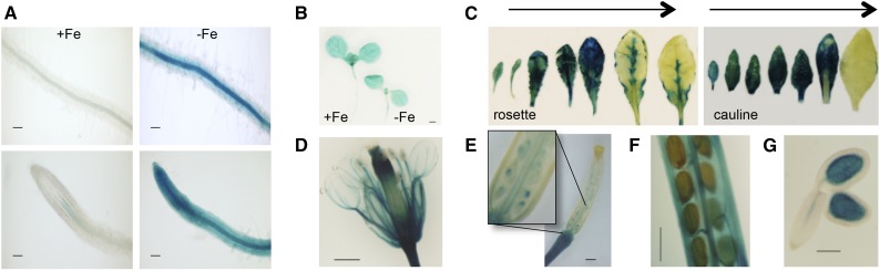
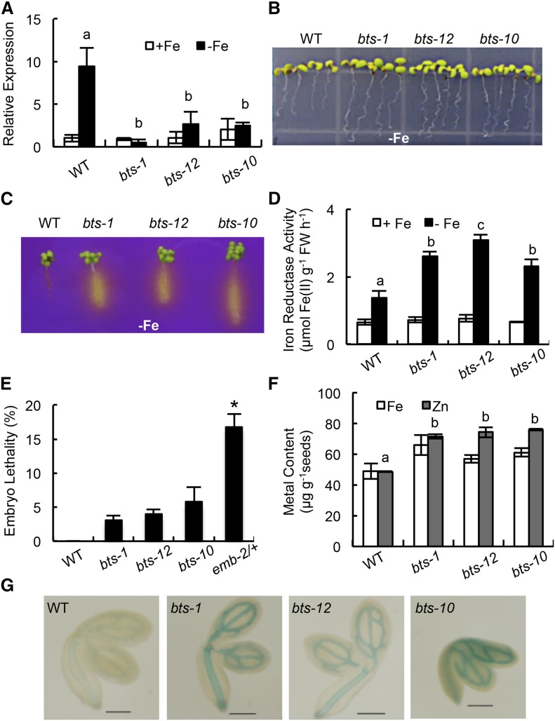
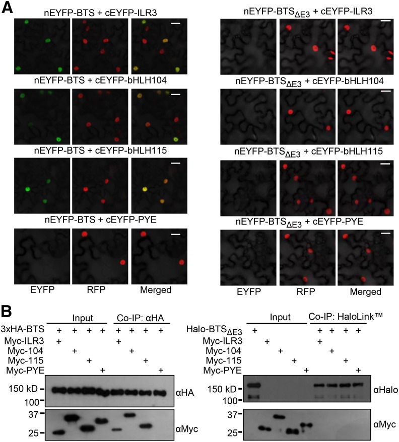
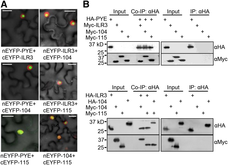
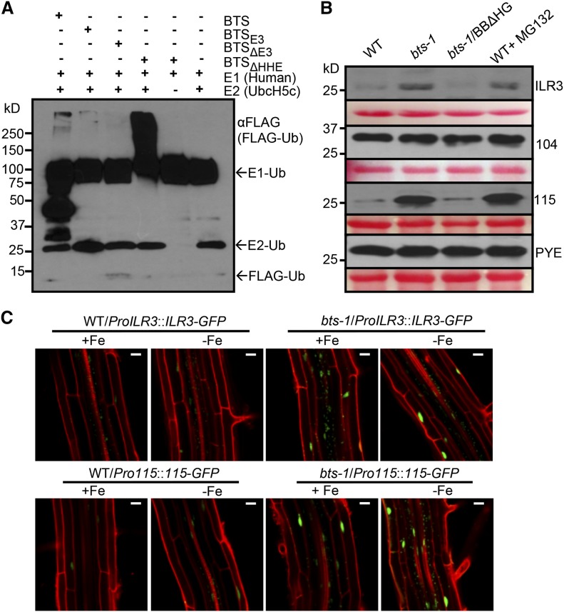
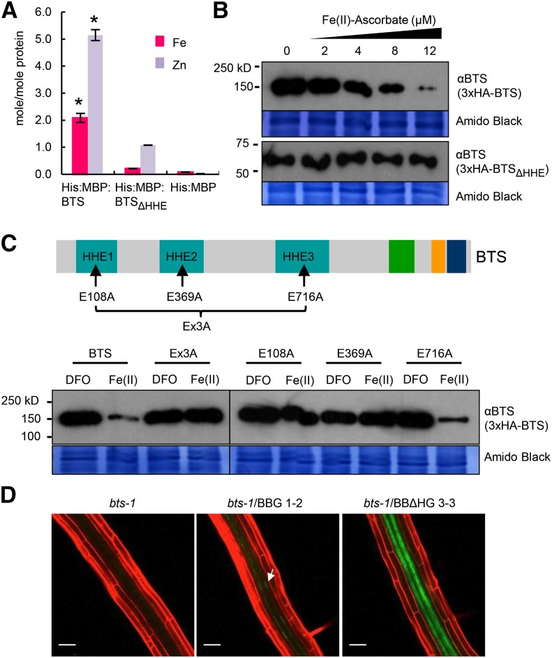
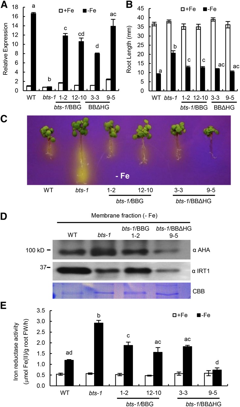
Iron uptake and metabolism are tightly regulated in both plants and animals. In Arabidopsis (Arabidopsis thaliana), BRUTUS (BTS), which contains three hemerythrin (HHE) domains and a Really Interesting New Gene (RING) domain, interacts with basic helix-loop-helix transcription factors that are capable of forming heterodimers with POPEYE (PYE), a positive regulator of the iron deficiency response. BTS has been shown to have E3 ligase capacity and to play a role in root growth, rhizosphere acidification, and iron reductase activity in response to iron deprivation. To further characterize the function of this protein, we examined the expression pattern of recombinant ProBTS::β-GLUCURONIDASE and found that it is expressed in developing embryos and other reproductive tissues, corresponding with its apparent role in reproductive growth and development. Our findings also indicate that the interactions between BTS and PYE-like (PYEL) basic helix-loop-helix transcription factors occur within the nucleus and are dependent on the presence of the RING domain. We provide evidence that BTS facilitates 26S proteasome-mediated degradation of PYEL proteins in the absence of iron. We also determined that, upon binding iron at the HHE domains, BTS is destabilized and that this destabilization relies on specific residues within the HHE domains. This study reveals an important and unique mechanism for plant iron homeostasis whereby an E3 ubiquitin ligase may posttranslationally control components of the transcriptional regulatory network involved in the iron deficiency response.


Figures

Figure 1.
Figure 1.
BTS expression throughout plant development. A and B, GUS activity in roots (A) and shoots (B) of 7-d-old wild-type ecotype Columbia (Col-0) seedlings grown on +Fe medium for 4 d and then transferred to ±Fe medium for 3 d expressing ProBTS::GUS. The results shown are representative of three independent lines. C, GUS activity in leaves from 4-week-old soil-grown plants. Arrows indicate youngest to oldest. D to G, GUS activity in flower (D), young silique (E), mature green silique (F), and mature embryo (G). Bars = 100 μm (A), 5 mm (B–F), and 2 mm (G).
Figure 2.
Figure 2.
BTS affects root iron response and embryogenesis. A, Relative BTS expression in root tissue from 7-d-old seedlings grown on +Fe medium for 4 d and then transferred to ±Fe medium for 3 d. Error bars indicate ± se of the mean (n = 4), and columns with different letters are significantly different from each other (P < 0.05). B, Root growth of 11-d-old seedlings grown on –Fe medium (4 d +Fe and 7 d –Fe). C, Rhizosphere acidification of 8-d-old seedlings grown on –Fe medium (4 d +Fe, 3 d –Fe, and 1 d bromocresol purple). Eight plants per genotype were grouped on bromocresol purple agar medium. Results shown represent four independent assays. D, Iron reductase activity of 10-d-old seedlings grown on ±Fe medium (7 d +Fe and 3 d ±Fe). Error bars indicate ± se of the mean (n = 4), and columns with different letters are significantly different from each other (P < 0.05). E, Embryo lethality in developing siliques. Error bars indicate ± se of the mean (n = 5). The asterisk indicates significant difference from the wild type (WT; P < 0.05). F, Iron and zinc content in seeds (150 mg per replicate) analyzed by ICP-OES. Error bars indicate ± se of the mean (n = 3), and columns with different letters are significantly different from each other (P < 0.05) G, Iron (Fe3+) localization in germinating seeds visualized by Perls staining. Results shown represent 10 independent assays. Bars = 2 mm. FW, Fresh weight.
Figure 3.
Figure 3.
Interaction of BTS with PYEL depends on the presence of the RING (E3 ligase) domain. A, BiFC assay showing in planta interactions of ILR3, bHLH104, bHLH115, and PYE with BTS and BTSΔE3 deletion protein in leaf epidermal cells. Indicated proteins are transiently coexpressed in red fluorescent protein (RFP)-Histone2B (nuclear marker) transgenic N. benthamiana plants. Enhanced YFP (EYFP) fluorescence indicates interaction between proteins in the nucleus. All interactions were tested using both combinations of N-/C-EYFP-fused reciprocal proteins. Results shown represent three independent assays. B, Immunoblot analysis showing Co-IP of in vitro-translated Myc-tagged ILR3, bHLH104, and bHLH115 by 3xHA-tagged BTS (full length) but not by Halo-tagged BTSΔE deletion protein. Results shown represent two independent assays. Bars = 20 μm.
Figure 4.
Figure 4.
PYE and PYEL proteins form heterodimers in the nucleus. A, BiFC assay showing in planta interactions between PYE, ILR3, bHLH104, and bHLH115 proteins in the epidermal cells. Indicated proteins are transiently coexpressed in RFP-Histone2B (nuclear marker) transgenic N. benthamiana plants. EYFP fluorescence indicates interaction between indicated proteins. All interactions were tested using both combinations of N-/C-EYFP-fused reciprocal proteins. Results shown represent three independent assays. B, Immunoblot analysis showing Co-IP of in vitro-translated HA-tagged PYE, ILR3, or bHLH104 and Myc-tagged ILR3, bHLH104, and bHLH115 proteins. Results shown represent two independent assays. Bars = 20 μm.
Figure 5.
Figure 5.
BTS targets ILR3 and bHLH115 for 26S proteasome-mediated degradation. A, E3 ligase activity of BTS and its derivatives. In vitro-translated and purified 3xHA-tagged BTS, BTSE3 domain, and BTSΔE3 and BTSΔHHE deletion proteins were subjected to in vitro ubiquitination assay containing ATP, FLAG-tagged ubiquitin (Ub), and human E1 and E2 (UbcH5c). Proteins were immunodetected using anti-FLAG antibody. Results shown represent two independent assays. B, Cell-free 26S proteasome-mediated degradation of PYEL proteins by BTS and BTSΔHHE. In vitro-translated and purified Myc-tagged ILR3, bHLH104, bHLH115, and PYE proteins were incubated with cell-free protein extracts prepared from 7-d-old seedlings of the wild type (WT), bts-1, and bts-1/ProBTS::BTSΔHHE-GFP (BBΔHG) grown on –Fe medium (4 d +Fe and 3 d –Fe). Reactions were performed with and without 160 µm MG132 (proteasome inhibitor). Proteins were immunodetected using anti-Myc antibody. Ponceau S staining indicates equal amount of cell-free protein extract loaded. C, Stability of ILR3-GFP and bHLH115-GFP in wild-type and bts-1 seedlings expressing either ProILR3::ILR3-GFP (top) or Pro-115::115-GFP (bottom) in roots. Plants were grown for 6 d on ±Fe medium (4 d +Fe and 2 d ±Fe). Results shown represent two independent assays. Bars = 20 μm.
Figure 6.
Figure 6.
BTS binds iron through HHE domains, negatively affecting BTS protein stability. A, Iron and zinc content of His:MBP fusions of BTS and HHE deleted (BTSΔHHE) proteins or His:MBP expressed in E. coli and purified. Error bars indicate ± se of the mean (n = 3 for His:MBP and His:MBP:BTS and n = 2 for His:MBP:BTSΔHHE). Asterisks indicate significant difference from BTSΔHHE and His:MBP (P < 0.05). B, In vitro translation of 3xHemagglutinin (3xHA)-tagged BTS and BTSΔHHE proteins performed in the presence of increasing concentration of ferrous iron (Fe(II)-ascorbate). Proteins were immunodetected using anti-BTS antibody. Amido Black staining indicates equal amount of wheat germ extract were loaded from in vitro protein translations. Results shown represent two independent assays. C, In vitro translation of 3xHA-tagged wild-type BTS, single amino acid substitutions (E108A, E369A, and E716A), and triple amino acid substitution (Ex3A) BTS proteins were performed in presence of ferrous iron chelator (100 μm DFO) and ferrous iron [12 μm Fe(II)-ascorbate]. Proteins were immunodetected using anti-BTS antibody. Amido Black staining indicates that equal amounts of wheat germ extract were loaded from in vitro protein translations. Results shown represent two independent assays. D, In planta stability of BTSΔHHE protein. Confocal microscopy images of roots of 7-d-old (4 d +Fe and 3 d –Fe) bts-1 seedlings expressing ProBTS::BTS-GFP (BBG, arrows) and ProBTS::BTSΔHHE-GFP (BBΔHG) stained with propidium iodide (red). Results shown represent four independent assays. Scale bars = 50 μm.
Figure 7.
Figure 7.
Complementation of bts-1 mutant by ProBTS::BTS-GFP (BBG) and ProBTS::BTSΔHHE-GFP (BBΔHG). A, Relative BTS expression in root tissue of 7-d-old seedlings grown on +Fe medium for 4 d and then transferred to ±Fe medium for 3 d. Error bars indicate ± se of the mean (n = 4), and columns with different letters are significantly different from each other (P < 0.05). B, Root length of 11-d-old seedlings grown on ±Fe medium (4 d +Fe and 7 d ±Fe). Error bars indicate ± se of the mean (n = 32), and columns with different letters are significantly different from each other (P < 0.05). C, Rhizosphere acidification of 8-d-old seedlings grown on –Fe medium (4 d +Fe, 3 d –Fe, and 1 d bromocresol purple). Eight plants per genotype were grouped on bromocresol purple agar medium. Results shown represent four independent assays. D, Levels of AHA and IRT1 proteins in membrane fractions isolated from the roots of wild-type (WT), bts-1 mutant, and BBG and BBΔHG complemented lines. Coomassie Brilliant Blue (CBB) R-250 staining shows equal loading of protein. Results shown represent two independent assays. E, Iron reductase activity of 10-d-old seedlings grown on ±Fe medium (7 d +Fe and 3 d ±Fe). Error bars indicate ± se of the mean (n = 4), and columns with different letters are significantly different from each other (P < 0.05). FW, Fresh weight.
Figure 8.
Figure 8.
Model for BTS protein stability and function. A, BTS is transcriptionally induced by iron deficiency along with bHLH transcription factors PYE and PYEL. Under low-iron conditions, BTS protein is more stable and regulates PYEL/PYE regulatory activity through its E3 ligase activity to modulate and fine-tune PYEL/PYE-mediated iron deficiency response in plants. B, Upon recovery of iron, transcriptional induction of BTS, PYE, and PYEL decreases. Elevated levels of iron are sensed through the HHE domains, altering BTS conformation and stability. This response results in the proteolysis of BTS protein, thereby further reducing the iron-responsive E3 ligase capacity of the cells.

References

    1. An YQ, Huang S, McDowell JM, McKinney EC, Meagher RB (1996) Conserved expression of the Arabidopsis ACT1 and ACT 3 actin subclass in organ primordia and mature pollen. Plant Cell 8: 15–30 - PMC - PubMed
    1. Barberon M, Zelazny E, Robert S, Conéjéro G, Curie C, Friml J, Vert G (2011) Monoubiquitin-dependent endocytosis of the iron-regulated transporter 1 (IRT1) transporter controls iron uptake in plants. Proc Natl Acad Sci USA 108: E450–E458 - PMC - PubMed
    1. Berleth T, Jurgens G (1993) The role of the monopteros gene in organising the basal body region of the Arabidopsis embryo. Development 118: 575–587
    1. Besold AN, Lee SJ, Michel SL, Sue NL, Cymet HJ (2010) Functional characterization of iron-substituted neural zinc finger factor 1: metal and DNA binding. J Biol Inorg Chem 15: 583–590 - PubMed
    1. Cabiscol E, Piulats E, Echave P, Herrero E, Ros J (2000) Oxidative stress promotes specific protein damage in Saccharomyces cerevisiae. J Biol Chem 275: 27393–27398 - PubMed

Publication types

MeSH terms