

Positron Emission Tomography with [(18)F]FLT Revealed Sevoflurane-Induced Inhibition of Neural Progenitor Cell Expansion in vivo
- PMID: 25452743
- PMCID: PMC4233913
- DOI: 10.3389/fneur.2014.00234
Positron Emission Tomography with [(18)F]FLT Revealed Sevoflurane-Induced Inhibition of Neural Progenitor Cell Expansion in vivo
Abstract
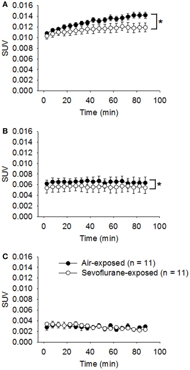
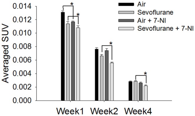
Neural progenitor cell expansion is critical for normal brain development and an appropriate response to injury. During the brain growth spurt, exposures to general anesthetics, which either block the N-methyl-d-aspartate receptor or enhance the γ-aminobutyric acid receptor type A can disturb neuronal transduction. This effect can be detrimental to brain development. Until now, the effects of anesthetic exposure on neural progenitor cell expansion in vivo had seldom been reported. Here, minimally invasive micro positron emission tomography (microPET) coupled with 3'-deoxy-3' [(18)F] fluoro-l-thymidine ([(18)F]FLT) was utilized to assess the effects of sevoflurane exposure on neural progenitor cell proliferation. FLT, a thymidine analog, is taken up by proliferating cells and phosphorylated in the cytoplasm, leading to its intracellular trapping. Intracellular retention of [(18)F]FLT, thus, represents an observable in vivo marker of cell proliferation. Here, postnatal day 7 rats (n = 11/group) were exposed to 2.5% sevoflurane or room air for 9 h. For up to 2 weeks following the exposure, standard uptake values (SUVs) for [(18)F]-FLT in the hippocampal formation were significantly attenuated in the sevoflurane-exposed rats (p < 0.0001), suggesting decreased uptake and retention of [(18)F]FLT (decreased proliferation) in these regions. Four weeks following exposure, SUVs for [(18)F]FLT were comparable in the sevoflurane-exposed rats and in controls. Co-administration of 7-nitroindazole (30 mg/kg, n = 5), a selective inhibitor of neuronal nitric oxide synthase, significantly attenuated the SUVs for [(18)F]FLT in both the air-exposed (p = 0.00006) and sevoflurane-exposed rats (p = 0.0427) in the first week following the exposure. These findings suggested that microPET in couple with [(18)F]FLT as cell proliferation marker could be used as a non-invasive modality to monitor the sevoflurane-induced inhibition of neural progenitor cell proliferation in vivo.
Keywords: [18F]FLT; neural progenitor cell; positron emission tomography; proliferation; sevoflurane.
Figures

Similar articles
-
In Vivo Monitoring of Sevoflurane-induced Adverse Effects in Neonatal Nonhuman Primates Using Small-animal Positron Emission Tomography.Anesthesiology. 2016 Jul;125(1):133-46. doi: 10.1097/ALN.0000000000001154. Anesthesiology. 2016. PMID: 27183169
-
3'-deoxy-3'-[18F]fluorothymidine as a new marker for monitoring tumor response to antiproliferative therapy in vivo with positron emission tomography.Cancer Res. 2003 Jul 1;63(13):3791-8. Cancer Res. 2003. PMID: 12839975
-
Glioma proliferation as assessed by 3'-fluoro-3'-deoxy-L-thymidine positron emission tomography in patients with newly diagnosed high-grade glioma.Clin Cancer Res. 2008 Apr 1;14(7):2049-55. doi: 10.1158/1078-0432.CCR-07-1553. Clin Cancer Res. 2008. PMID: 18381944
-
Molecular PET and PET/CT imaging of tumour cell proliferation using F-18 fluoro-L-thymidine: a comprehensive evaluation.Nucl Med Commun. 2009 Dec;30(12):908-17. doi: 10.1097/MNM.0b013e32832ee93b. Nucl Med Commun. 2009. PMID: 19794320 Review.
-
PET imaging of proliferation with pyrimidines.J Nucl Med. 2013 Jun;54(6):903-12. doi: 10.2967/jnumed.112.112201. Epub 2013 May 14. J Nucl Med. 2013. PMID: 23674576 Review.
Cited by
-
Isoflurane and Sevoflurane Induce Cognitive Impairment in Neonatal Rats by Inhibiting Neural Stem Cell Development Through Microglial Activation, Neuroinflammation, and Suppression of VEGFR2 Signaling Pathway.Neurotox Res. 2022 Jun;40(3):775-790. doi: 10.1007/s12640-022-00511-9. Epub 2022 Apr 26. Neurotox Res. 2022. PMID: 35471722 Free PMC article.
-
Potential Adverse Effects of Prolonged Sevoflurane Exposure on Developing Monkey Brain: From Abnormal Lipid Metabolism to Neuronal Damage.Toxicol Sci. 2015 Oct;147(2):562-72. doi: 10.1093/toxsci/kfv150. Epub 2015 Jul 23. Toxicol Sci. 2015. PMID: 26206149 Free PMC article.
-
Collapsin Response Mediator Protein-2 Ameliorates Sevoflurane-Mediated Neurocyte Injury by Targeting PI3K-mTOR-S6K Pathway.Med Sci Monit. 2018 Jul 18;24:4982-4991. doi: 10.12659/MSM.909056. Med Sci Monit. 2018. PMID: 30018280 Free PMC article.
References
LinkOut - more resources
Full Text Sources
Other Literature Sources