. 2014 Nov 12:5:605.
doi: 10.3389/fmicb.2014.00605. eCollection 2014.

Benthic protists and fungi of Mediterranean deep hypsersaline anoxic basin redoxcline sediments

Affiliations

Benthic protists and fungi of Mediterranean deep hypsersaline anoxic basin redoxcline sediments

Joan M Bernhard et al. Front Microbiol. .

Abstract

Some of the most extreme marine habitats known are the Mediterranean deep hypersaline anoxic basins (DHABs; water depth ∼3500 m). Brines of DHABs are nearly saturated with salt, leading many to suspect they are uninhabitable for eukaryotes. While diverse bacterial and protistan communities are reported from some DHAB water-column haloclines and brines, the existence and activity of benthic DHAB protists have rarely been explored. Here, we report findings regarding protists and fungi recovered from sediments of three DHAB (Discovery, Urania, L' Atalante) haloclines, and compare these to communities from sediments underlying normoxic waters of typical Mediterranean salinity. Halocline sediments, where the redoxcline impinges the seafloor, were studied from all three DHABs. Microscopic cell counts suggested that halocline sediments supported denser protist populations than those in adjacent control sediments. Pyrosequencing analysis based on ribosomal RNA detected eukaryotic ribotypes in the halocline sediments from each of the three DHABs, most of which were fungi. Sequences affiliated with Ustilaginomycotina Basidiomycota were the most abundant eukaryotic signatures detected. Benthic communities in these DHABs appeared to differ, as expected, due to differing brine chemistries. Microscopy indicated that only a low proportion of protists appeared to bear associated putative symbionts. In a considerable number of cases, when prokaryotes were associated with a protist, DAPI staining did not reveal presence of any nuclei, suggesting that at least some protists were carcasses inhabited by prokaryotic scavengers.

Keywords: DHABs; L’ Atalante; Urania; discovery; diversity; eukaryote; rRNA.


Figures
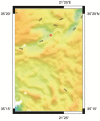
FIGURE 1
FIGURE 1
Bathymetric map of L’ Atalante DHAB generated by Seabeam. Red asterisk = approximate sample site.
FIGURE 2
FIGURE 2
Underwater photographs showing small portion of redoxcline impinging the seafloor in Urania (A), Discovery (B,D), and L’ Atalante (C) DHABs, eastern Mediterranean. Note red laser spots on seafloor in (C); dots are spaced 10 cm apart. N = normoxic control region; UH = upper halocline zone; MH = mid-halocline zone; LH = lower halocline zone; B = brine. Injector cores emplaced in Discovery upper and mid-halocline (D) illustrate the ability to sample different zones. All images © Woods Hole Oceanographic Institution.
FIGURE 3
FIGURE 3
Depth profiles of the oxygen concentrations in overlying waters and sediments of pushcores collected for this study, presented by DHAB and general habitat. From left to right: Normoxic (control;A); Upper Halocline(B); Lower Halocline(C).
FIGURE 4
FIGURE 4
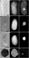
Light micrographs. (A,B) Thecate foraminifer from 611 c3 (L’ Atalante control), paired images showing DIC (A) and epifluorescence (B) of DAPI staining. Note the nucleus and lack of associated prokaryotes (B) in the well-vacuolated cytoplasm (A). This specimen was considered living at the time of fixation. (C,D) protist carcass from 609-610 cL (Discovery lower halocline), paired images showing DIC (C) and epifluorescence (D) of DAPI staining. Note the lack of a DAPI labeled nucleus and presence of a few prokaryotes. This specimen was considered dead at the time of fixation. Scales = 10 μm.
FIGURE 5
FIGURE 5
Light micrographs of ciliates from Urania halocline. (A,C) Possible armophorean, Metopus-like ciliate from 608 c11. (A,B) Paired images showing DIC (A) and epifluorescence (B) of DAPI staining. Box in (B) shows area depicted in (C). Note the nucleus (B) and endobionts (B,C). (D,E) Possible karyorelictid or hypotrich/strichothrich ciliate from 607 c10. DIC image (D) of large endobiont-bearing ciliate, somewhat masked by debris, shown in (E; epifluorescence of DAPI staining at slightly higher magnification). Note nucleus. (F) Ciliate from 607 c10 shown in double exposure of DIC and DAPI epifluorescence. Note macro- and micronucleus. Scales: B = 50 μm; C = 10 μm; D, E = 25 μm; F = 10 μm.
FIGURE 6
FIGURE 6
Light micrographs of protists from Discovery DHAB. (A–C) Ciliate from 609 c14 (mid-halocline). (A–C) Paired images showing DIC (A,B) and epifluorescence (C) of DAPI staining. Note the nucleus (C) and lack of abundant prokaryotic associates (C). (D,E) Paired images showing DIC (D) and epifluorescence (E) of protist from 610 c9 (lower halocline). Note lack of obvious single large nucleus and presence of possible endobionts or parasites/scavengers. Scale bars = 20 μm.
FIGURE 7
FIGURE 7
Light micrographs of ciliates from L’ Atalante DHAB. (A,C) Ciliate from 611 c5 (upper halocline); paired images showing DIC (A) and epifluorescence (B,C) of DAPI staining. Note the nucleus in (A); DAPI staining of the nucleus also occurred (not shown). Also note the abundant endobionts (B,C), mostly at protist periphery. (D,E) Specimen from 611 c10 (mid-halocline); paired images showing DIC (D) and epifluorescence (E) of DAPI staining. Note the nucleus and lack of prokaryotic associates. (F,G) Specimen from 611 c10; paired images showing DIC (F) and epifluorescence (G) of DAPI staining. Note the nucleus (somewhat out of the plane of view in G) and abundant endobiont associates. (H–K) Specimen from 611 c10; DIC image (H) along with paired images (I–K) showing darkfield (I) and epifluorescence (J,K) of DAPI staining. Note the nucleus (* in I), ectobionts (arrows) and aggregated endobiont associates (arrowheads). Scales: C,G = 100 μm; E = 25 μm; H,K = 50 μm.
FIGURE 8
FIGURE 8
Light micrographs of protists incubated in both CellTracker Green and DAPI. (A–C) Specimen from 607-608 cB (Urania control); paired images showing DIC (A) and epifluorescence (B,C) of CellTracker Green labeling (B) and DAPI (C) staining. Note the fluorescence of the cytoplasm, the nucleus (C), and absence of abundant associated prokaryotes. (D–F) Ciliate from 607-608 cE (Urania halocline); paired images showing DIC (D) and epifluorescence (E,F) of CellTracker Green labeling (E) and DAPI (F) staining. Note fluorescence of the cytoplasm, the macronucleus, and micronucleus (F) and lack of prokaryotic associates. (G–I) Ciliate from 609-610 cH (Discovery upper halocline); paired images showing DIC (G) and epifluorescence (H,I) of CellTracker Green labeling (H) and DAPI (I) staining. Note the lack of prokaryotic associates. (J–L) Unidentified protist from 609-610 cL (Discovery lower halocline). Note the nucleus and scattered bacteria on exterior. Scales: A,G = 10 μm, D = 100 μm, J = 50 μm.
FIGURE 9
FIGURE 9
Number of protists per unit cubic centimeter of sediment (in situ), presented for each habitat. Error bars represent the SD resultant from counts of triplicate subsamples from each sample. Urania control = 607-08 cB; Urania halocline = 607-08 cE; Discovery upper halocline = 610 c14; Discovery mid-halocline = 609-10 cL; L’ Atalante upper halocline = 611 c10; L’ Atalante lower halocline = 611 c17.
FIGURE 10
FIGURE 10
Relative abundance (%) of rRNA signatures of unicellular eukaryotic groups of the DHAB sediments based on V4 SSU rRNA pyrotags clustered at 97% similarity. Taxonomic assignments were performed using BLAST implemented in QIIME against SILVA 111 database.
FIGURE 11
FIGURE 11
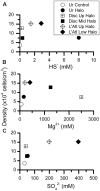
Abundances of protists per cubic centimeter, calculated from microscopic counts, presented with respect to inferred chemical parameter in overlying waters. Chemical constituent data from van der Wielen et al. (2005).

References

    1. Alexander E., Stock A., Breiner H. W., Behnke A., Bunge J., Yakimov M. M., et al. (2009). Microbial eukaryotes in the hypersaline anoxic L’ Atalante deep-sea basin. Environ. Microbiol. 11 360–381 10.1111/j.1462-2920.2008.01777.x - DOI - PubMed
    1. Alongi D. M. (1987). The distribution and composition of deep-sea microbenthos in a bathyal region of the Western Coral Sea. Deep Sea Res. Part A Oceanogr. Res. Pap. 34 1245–1254 10.1016/0198-0149(87)90074-4 - DOI
    1. Amend A. (2014). From dandruff to deep-sea vents: Malassezia-like Fungi are ecologically hyper-diverse. PLoS Pathog. 10:e1004277 10.1371/journal.ppat.1004277 - DOI - PMC - PubMed
    1. Bernhard J. M. (2003). Potential symbionts in bathyal foraminifera. Science 299 861–861 10.1126/science.1077314 - DOI - PubMed
    1. Bernhard J. M., Barry J. P., Buck K. R., Starczak V. R. (2009). Impact of intentionally injected carbon dioxide hydrate on deep-sea benthic foraminiferal survival. Global Change Biol. 15 2078–2088 10.1111/j.1365-2486.2008.01822.x - DOI

LinkOut - more resources