. 2014 Dec 16;111(50):17983-8.
doi: 10.1073/pnas.1421047111. Epub 2014 Dec 1.

Life-long in vivo cell-lineage tracing shows that no oogenesis originates from putative germline stem cells in adult mice

Affiliations

Life-long in vivo cell-lineage tracing shows that no oogenesis originates from putative germline stem cells in adult mice

Hua Zhang et al. Proc Natl Acad Sci U S A. .

Abstract

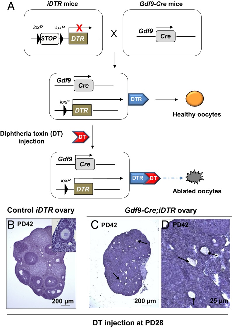
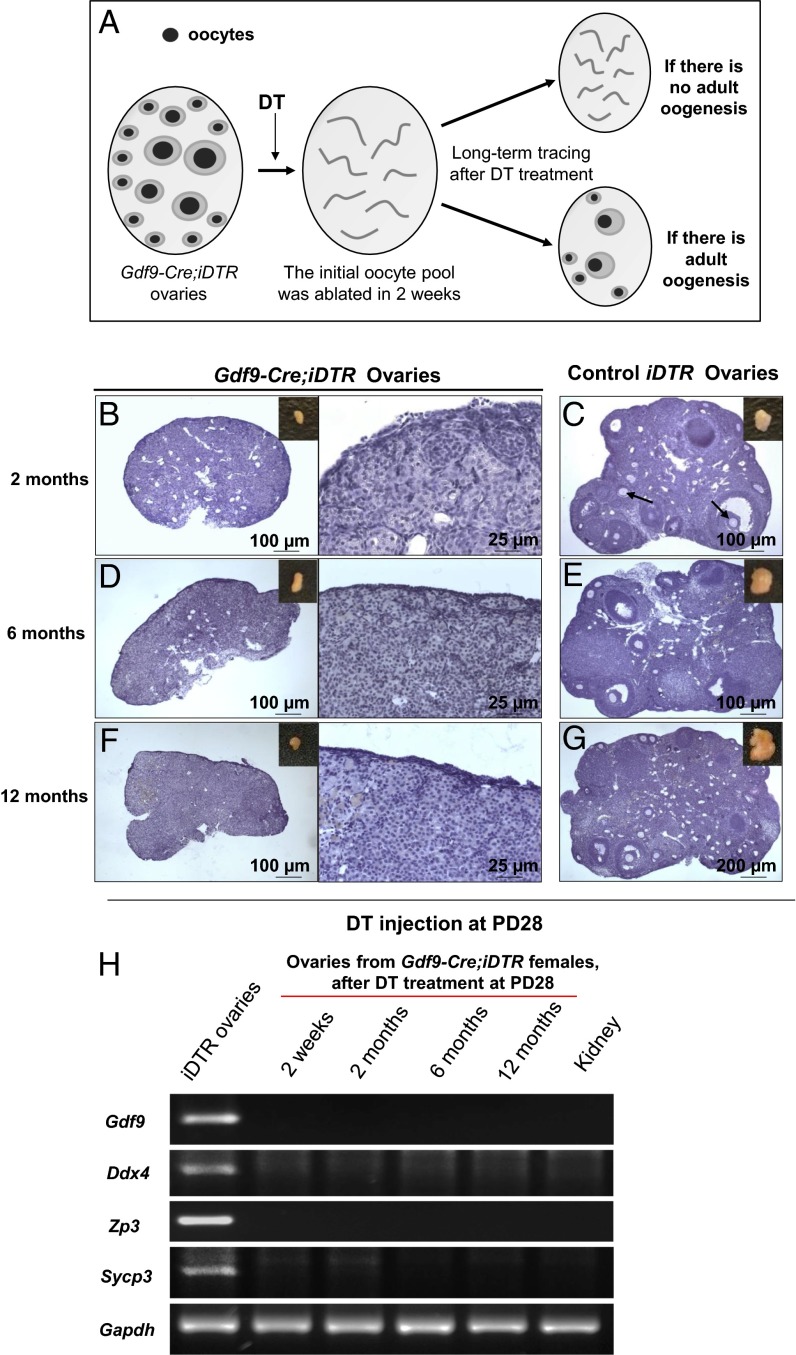
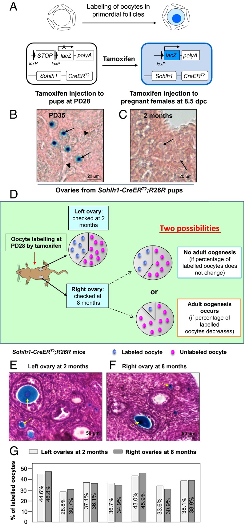
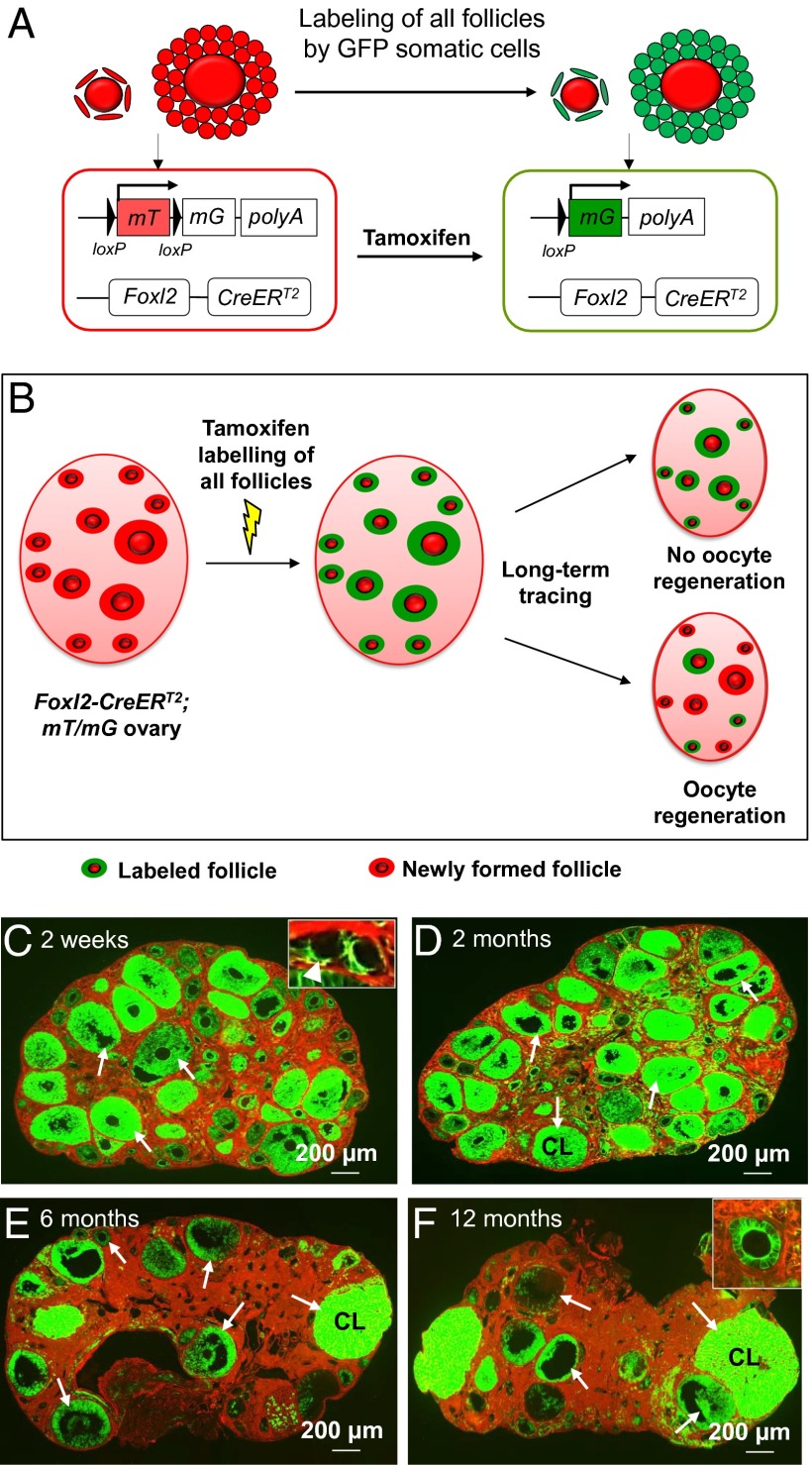
Whether or not oocyte regeneration occurs in adult life has been the subject of much debate. In this study, we have traced germ-cell lineages over the life spans of three genetically modified mouse models and provide direct evidence that oogenesis does not originate from any germline stem cells (GSCs) in adult mice. By selective ablation of all existing oocytes in a Gdf9-Cre;iDTR mouse model, we have demonstrated that no new germ cells were ever regenerated under pathological conditions. By in vivo tracing of oocytes and follicles in the Sohlh1-CreER(T2);R26R and Foxl2-CreER(T2);mT/mG mouse models, respectively, we have shown that the initial pool of oocytes is the only source of germ cells throughout the life span of the mice and that no adult oogenesis ever occurs under physiological conditions. Our findings clearly show that there are no GSCs that contribute to adult oogenesis in mice and that the initial pool of oocytes formed in early life is the only source of germ cells throughout the entire reproductive life span.

Keywords: follicle tracing; genetically modified mouse models; life-long observations; no postnatal oocyte regeneration; oocyte tracing.


Conflict of interest statement

The authors declare no conflict of interest.

Figures

Fig. 1.
Fig. 1.
Specific and efficient ablation of oocytes in the Gdf9-Cre;iDTR mouse ovaries. (A) Illustration of the diphtheria toxin (DT)-induced ablation of oocytes in Gdf9-Cre;iDTR mouse ovaries. In Gdf9-expressing oocytes, the Cre recombinase removes the loxP-flanked STOP sequence and allows the expression of the DTR. Upon DT administration, the Gdf9-expressing oocytes are selectively ablated. (BD) All existing oocytes were ablated in Gdf9-Cre;iDTR mouse ovaries after DT injection. DT was given to PD28 females for 5 consecutive days, and ovaries were examined 2 wk later. Compared with the normal ovarian development in iDTR females (B), Gdf9-Cre;iDTR ovaries demonstrated a complete loss of all oocytes (C and D). Only follicle structures without oocytes or with oocyte debris were observed (C and D, arrows) (n = 6).
Fig. 2.
Fig. 2.
No oocyte regeneration occurred in oocyte-ablated Gdf9-Cre;iDTR mouse ovaries. (A) Schema for long-term tracing of oocyte regeneration in Gdf9-Cre;iDTR mouse ovaries. Neogenic oocytes and follicles should be visible in the ovaries of DT-injected mice if there is oocyte regeneration during the 12-mo tracing study. (B, D, and F) No live oocytes or follicles were observed in Gdf9-Cre;iDTR ovaries at 2 mo (n = 6) (B), 6 mo (n = 6) (D), or 12 mo (n = 6) (F) after DT administration. (C, E, and G) As controls, the ovarian morphology of iDTR females was normal at 2 mo (n = 4) (C), 6 mo (n = 6) (E), and 12 mo (n = 6) (G) after DT administration. (H) Comparative gene expression profiling of germ-cell markers in oocyte-ablated Gdf9-Cre;iDTR ovaries. Semiquantitative RT-PCR analysis showed that none of the germ-cell markers studied, including Gdf9, Ddx4, Zp3 and Sycp3, were expressed in the Gdf9-Cre;iDTR ovaries from 2 wk to 12 mo after DT administration. An RNA sample from the mouse kidney was used as a negative control, and Gapdh was used as the internal loading control. All experiments were repeated at least three times, and representative images are shown.
Fig. 3.
Fig. 3.
The developing oocytes in later reproductive life originated from immature oocytes that were labeled in early life in Sohlh1-CreERT2;R26R females. (A) Illustration of the tamoxifen-induced labeling of oocytes in primordial follicles of the Sohlh1-CreERT2;R26R ovary. CreERT2 recombinase is expressed in Sohlh1-expressing oocytes of primordial follicles. Upon tamoxifen administration, the CreERT2 recombinase removes the STOP sequence and turns on the expression of lacZ (the gene encoding β-galactosidase), and this generates a blue color in the oocyte cytoplasm after β-galactosidase staining. (B) Labeling oocytes of primordial follicles by postnatal tamoxifen injection. Tamoxifen was given to Sohlh1-CreERT2;R26R females for 7 consecutive days at PD28, and ovaries were collected for analysis at PD35 (n = 6). The labeled oocytes were visualized by their blue color after β-galactosidase staining (arrows) whereas the unlabeled oocytes showed only background color (arrowheads). (C) No PGCs were labeled by embryonic tamoxifen injection. R26R females were plugged with Sohlh1-CreERT2 males, and tamoxifen was given to pregnant females from 8.5 to 12.5 dpc. No labeled oocytes were observed at 2 mo in the ovaries of Sohlh1-CreERT2;R26R pups that were born from the tamoxifen-injected females (n = 6). (D) Schema for long-term tracing of oocyte regeneration in Sohlh1-CreERT2;R26R mouse ovaries. The ratio of labeled-to-unlabeled oocytes should remain the same if there is no adult oogenesis but should decrease if oocyte neogenesis occurs. (E and F) Labeling the existing oocytes in the adult mouse ovary. The labeled oocytes (identified by the blue dots) can be observed in both the left ovary at 2 mo (E) and the right ovary at 8 mo (F) after tamoxifen injection. Some labeled oocytes had entered into the growing phase (arrows). (G) The ratio of labeled-to-unlabeled oocytes remained the same in each mouse at 2 mo (left ovaries) (n = 7) and at 8 mo (right ovaries) (n = 7) after tamoxifen injection. The similarity in the proportion of labeled oocytes indicates that no oocyte neogenesis occurred in the adult mouse ovaries.
Fig. 4.
Fig. 4.
Long-term tracing of follicle populations by labeling the follicular somatic cells in adult Foxl2-CreERT2;mT/mG ovaries. (A) Illustration of the tamoxifen-induced labeling of follicles in Foxl2-CreERT2;mT/mG mice. In Foxl2-expressing follicular somatic cells, the CreERT2 recombinase is not active and the cells express mT, a red fluorescent protein. Upon tamoxifen injection, the CreERT2 recombinase deletes the mT region and switches on the expression of mG, a green fluorescent protein. Thus, the Foxl2-expressing follicular somatic cells were labeled with green fluorescence. (B) Schema for long-term tracing of oocyte regeneration in Foxl2-CreERT2;mT/mG mouse ovaries. All follicular somatic cells were labeled with the green fluorescent mG at PD28. Follicles with only red (mT) somatic cells would be observed if oocyte regeneration occurred during the tracing study. (CF) The mice were given an i.p. injection of 80 mg⋅kg−1 BW (∼2 mg per mouse) tamoxifen per day for 7 consecutive days starting at PD28. The mice were killed at 2 wk (n = 6) (C), 2 mo (n = 6) (D), 6 mo (n = 6) (E), and 12 mo (n = 6) (F) after the injection. Ovaries were collected and all sections were carefully checked under the microscope, and a follicle or a corpus luteum was considered to be labeled if green fluorescent granulosa or luteal cells were observed. At 2 wk after tamoxifen injection, all follicles, including the dormant primordial follicles in the cortex (C, Inset, arrowhead) and growing follicles in the medulla (C, arrows), were labeled with green fluorescence in their somatic cells. All follicles (DF, arrows) or corpora lutea (DF, CL) were labeled with green fluorescence in their somatic cells, and no follicles with only red somatic cells were found in mice killed at 2 mo (D), 6 mo (E), or 12 mo (F) after tamoxifen injection.

Comment in

References

    1. Zuckerman S. The number of oocytes in the mature ovary. Recent Prog Horm Res. 1951;6:63–109.
    1. Johnson J, Canning J, Kaneko T, Pru JK, Tilly JL. Germline stem cells and follicular renewal in the postnatal mammalian ovary. Nature. 2004;428(6979):145–150. - PubMed
    1. Johnson J, et al. Oocyte generation in adult mammalian ovaries by putative germ cells in bone marrow and peripheral blood. Cell. 2005;122(2):303–315. - PMC - PubMed
    1. White YA, et al. Oocyte formation by mitotically active germ cells purified from ovaries of reproductive-age women. Nat Med. 2012;18(3):413–421. - PMC - PubMed
    1. Zou K, et al. Production of offspring from a germline stem cell line derived from neonatal ovaries. Nat Cell Biol. 2009;11(5):631–636. - PubMed

Publication types