. 2015 Jan 6;112(1):100-5.
doi: 10.1073/pnas.1413764111. Epub 2014 Dec 1.

Vibrio effector protein VopQ inhibits fusion of V-ATPase-containing membranes

Affiliations

Vibrio effector protein VopQ inhibits fusion of V-ATPase-containing membranes

Anju Sreelatha et al. Proc Natl Acad Sci U S A. .

Abstract

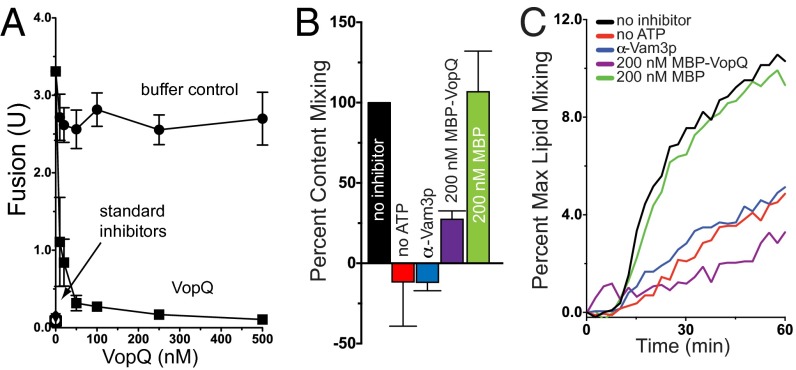
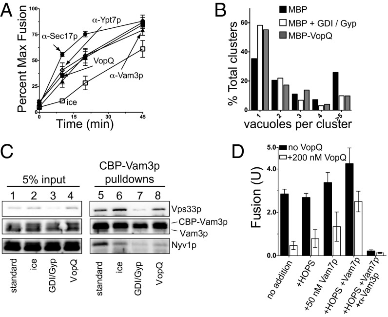
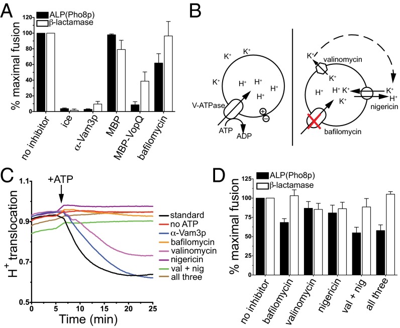
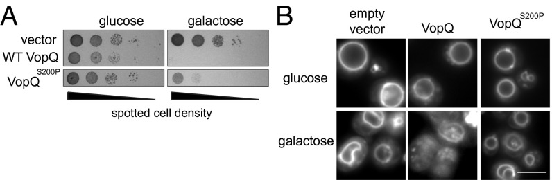
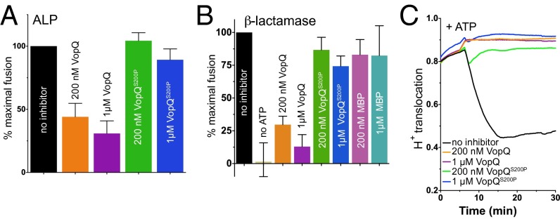
Vesicle fusion governs many important biological processes, and imbalances in the regulation of membrane fusion can lead to a variety of diseases such as diabetes and neurological disorders. Here we show that the Vibrio parahaemolyticus effector protein VopQ is a potent inhibitor of membrane fusion based on an in vitro yeast vacuole fusion model. Previously, we demonstrated that VopQ binds to the V(o) domain of the conserved V-type H(+)-ATPase (V-ATPase) found on acidic compartments such as the yeast vacuole. VopQ forms a nonspecific, voltage-gated membrane channel of 18 Å resulting in neutralization of these compartments. We now present data showing that VopQ inhibits yeast vacuole fusion. Furthermore, we identified a unique mutation in VopQ that delineates its two functions, deacidification and inhibition of membrane fusion. The use of VopQ as a membrane fusion inhibitor in this manner now provides convincing evidence that vacuole fusion occurs independently of luminal acidification in vitro.

Keywords: SNARE; Vibrio parahaemolyticus; vesicle fusion; vp1680; yeast vacuole.


Conflict of interest statement

The authors declare no conflict of interest.

Figures

Fig. 1.
Fig. 1.
VopQ inhibits yeast homotypic vacuole fusion. (A) ALP vacuole fusion reactions in the absence or presence of increasing concentrations of recombinant His6-VopQ. Standard fusion inhibitors were α-Vam3p, α-Sec17p, 1 µM Gdi1p, Gyp1-46, or ice incubation. Error bars indicate SD from the mean; n = 4. (B and C) The β-lactamase content-mixing assay (B) was performed in parallel with the Rh-PE dequenching lipid-mixing assay (C) in the absence of ATP or including α-Vam3p, MBP-VopQ, or MBP. Data for the content-mixing assay are reported as mean ± SD, n = 3. The Rh-PE dequenching plot is a representative curve measured from the same three experiments used for the content-mixing assay.
Fig. 2.
Fig. 2.
VopQ inhibits trans-SNARE complex formation. (A) At 0-, 10-, 20-, and 45-min intervals, ALP vacuole fusion reaction was incubated on ice, or one of the following fusion inhibitors was added: α-Sec17p, α-Ypt7p, α-Vam3p, or 200 nM VopQ. After 90 min, fusion was measured and compared with the fusion of an uninhibited fusion reaction. Error bars indicate SD from the mean; n = 3. (B) The quantitative docking assay was performed in the presence of 500 nM MBP, 2.8 µM Gdi1p and 11.4 µM Gyp1-46, or 500 nM MBP-VopQ. At least 130 clusters from 10 random fields were scored for each condition. (C) Trans-SNARE assays were performed without or with the following inhibitors: ice incubation, 1 µM Gdi1p/1 µM Gyp1-46, or 200 nM VopQ. (D) ALP vacuole fusion reactions were performed in the absence or presence of 200 nM MBP-VopQ. Recombinant 50 nM Vam7p SNARE, 20 nM HOPS complex, or α-Vam3p was added where indicated.
Fig. 3.
Fig. 3.
A pH gradient is not required for vacuole fusion. (A) ALP and β-lactamase fusion reactions were performed in parallel, using standard inhibitors or 200 nM MBP, 200 nM MBP-VopQ, or 500 nM bafilomycin. Maximal fusion was defined as the extent of fusion of the no-inhibitor reaction; error bars indicate SD from the mean; n = 3. (B) Schematic diagram of the mechanism of action of the ionophores used. (C) Proton translocation activity of vacuoles was assayed in the absence or presence of ATP, α-Vam3p, 500 nM bafilomycin, 1 µM valinomycin, 1 µM nigericin, or combinations of ionophores and bafilomycin. Curves are representative of three independent experiments. (D) ALP and β-lactamase fusion reactions were performed in parallel, using a combination of ionophores at the concentrations used in C. Maximal fusion was defined as the no-inhibitor reaction. Error bars indicate SD from the mean; n = 3.
Fig. 4.
Fig. 4.
VopQS200P does not induce vacuole fragmentation. (A) Serial dilutions of yeast strain BY4742 harboring pRS413-Gal1, pRS413-Gal1-VopQ, or pRS413-Gal1-VopQS200P in Complete Supplement Mixture (CSM) medium lacking histidine, supplemented with either 2% glucose or 2% galactose. (B) Vacuolar morphology of the BY4742 strain harboring pRS413-Gal1, pRS413-Gal1-VopQ, or pRS413-Gal1-VopQS200P was visualized via FM4-64 staining. (Scale bar, 5 μm.)
Fig. 5.
Fig. 5.
VopQS200P deacidifies the vacuole but does not inhibit fusion. (A) ALP vacuole fusion reactions or (B) β-lactamase fusion reactions were performed using standard inhibitors, MBP-VopQ, or MBP-VopQS200P. (C) Proton translocation activity was measured in the presence of MBP-VopQ or MBP-VopQS200P when proteins were added before the addition of ATP.

Comment in

References

    1. Hiesinger PR, et al. The v-ATPase V0 subunit a1 is required for a late step in synaptic vesicle exocytosis in Drosophila. Cell. 2005;121(4):607–620. - PMC - PubMed
    1. Wickner W. Membrane fusion: Five lipids, four SNAREs, three chaperones, two nucleotides, and a Rab, all dancing in a ring on yeast vacuoles. Annu Rev Cell Dev Biol. 2010;26:115–136. - PubMed
    1. Armstrong J. Yeast vacuoles: More than a model lysosome. Trends Cell Biol. 2010;20(10):580–585. - PubMed
    1. Strasser B, Iwaszkiewicz J, Michielin O, Mayer A. The V-ATPase proteolipid cylinder promotes the lipid-mixing stage of SNARE-dependent fusion of yeast vacuoles. EMBO J. 2011;30(20):4126–4141. - PMC - PubMed
    1. Bayer MJ, Reese C, Buhler S, Peters C, Mayer A. Vacuole membrane fusion: V0 functions after trans-SNARE pairing and is coupled to the Ca2+-releasing channel. J Cell Biol. 2003;162(2):211–222. - PMC - PubMed

Publication types

MeSH terms

LinkOut - more resources